
Who Invented MRI? The Complete History of Magnetic Resonance Imaging and the Scientists Who Created It
Who invented MRI? Discover how Lauterbur, Mansfield, Damadian, Rabi & Bloch created magnetic resonance imaging — a complete history of MRI invention. ✏️
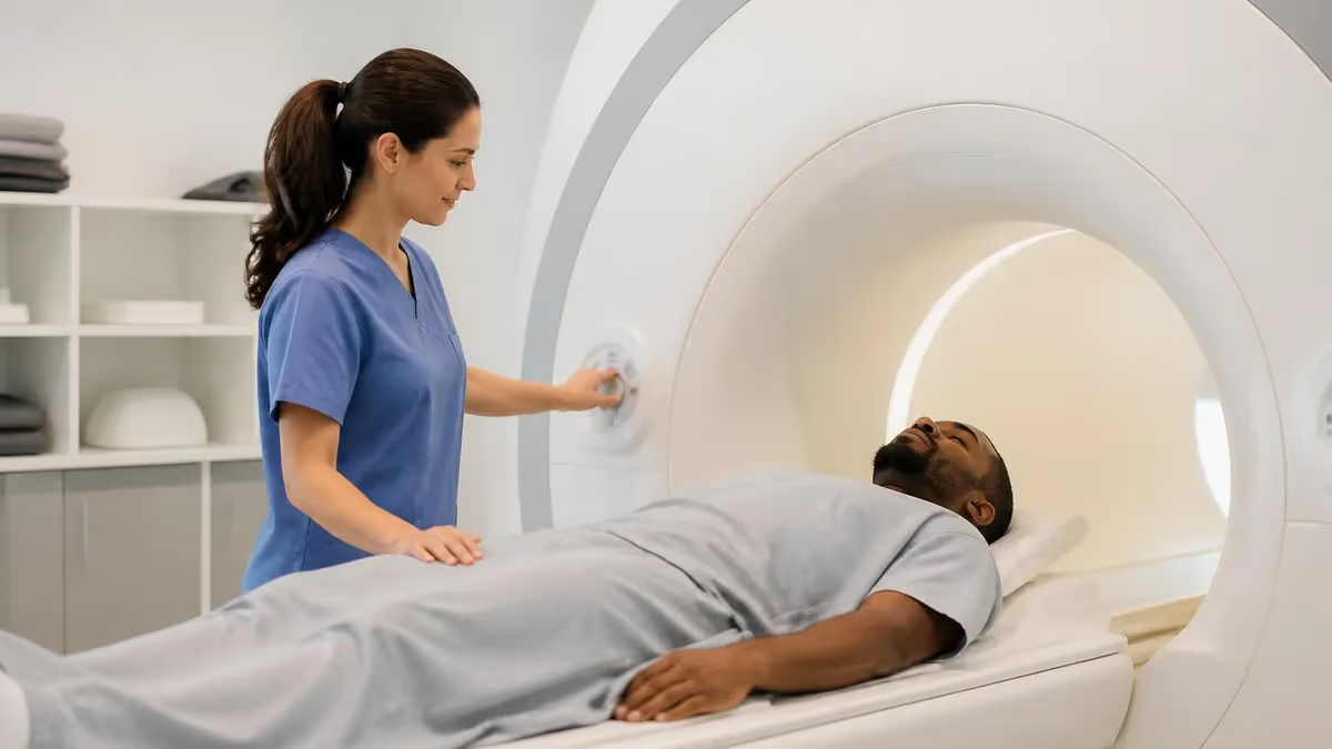
The question of who invented MRI has one of the most contested answers in modern medical history. Magnetic resonance imaging — the technology that allows physicians to see inside the human body without radiation — was not the work of a single genius but a chain of brilliant scientists building on each other's discoveries across nearly five decades. The names most central to the story are Isidor Rabi, Felix Bloch, Edward Purcell, Raymond Damadian, Paul Lauterbur, and Sir Peter Mansfield, each contributing a piece of the puzzle that ultimately revolutionized diagnostic medicine and earned multiple Nobel Prizes.
To understand who invented MRI, you have to start with the physics. In 1938, Columbia University physicist Isidor Rabi demonstrated nuclear magnetic resonance in molecular beams, work that earned him the 1944 Nobel Prize in Physics. Rabi showed that atomic nuclei placed in a magnetic field would absorb and re-emit electromagnetic radiation at specific frequencies. Without this foundational discovery, no MRI scanner could ever exist. His experiments turned an abstract quantum property into a measurable physical phenomenon that later scientists would harness for imaging.
Less than a decade later, in 1946, Felix Bloch at Stanford and Edward Purcell at Harvard independently extended Rabi's work to liquids and solids. The two physicists shared the 1952 Nobel Prize for developing nuclear magnetic resonance spectroscopy — a technique that became standard in chemistry labs worldwide for analyzing molecular structure. For roughly twenty years afterward, NMR remained a chemistry tool, not a medical one. Nobody seriously believed it could be used to peer inside a living human being, and the equipment was the size of a small room with no obvious clinical application.
That changed in 1971 when Raymond Damadian, a physician at the State University of New York Downstate Medical Center, published a landmark paper in Science demonstrating that cancerous tissue in rats had different NMR relaxation times than healthy tissue. Damadian's insight — that NMR could distinguish diseased from normal tissue — opened the door to medical imaging. He filed a patent in 1972 titled "Apparatus and Method for Detecting Cancer in Tissue," and in 1977 built the first whole-body MRI scanner, nicknamed "Indomitable," which now sits in the Smithsonian Institution.
However, Damadian's machine detected signals but did not produce true cross-sectional images. The breakthrough that made modern MRI possible came from Paul Lauterbur at Stony Brook University, who in 1973 introduced magnetic field gradients — a method of spatially encoding NMR signals to construct two-dimensional images. Sir Peter Mansfield at the University of Nottingham then developed the mathematical framework and echo-planar imaging techniques that allowed scans to be acquired in seconds rather than hours. Lauterbur and Mansfield shared the 2003 Nobel Prize in Physiology or Medicine.
For more historical context on these breakthrough decades, see the history of MRI, which traces the technology's evolution from laboratory curiosity to indispensable clinical tool. The omission of Damadian from the 2003 Nobel Prize remains one of the most controversial decisions in the prize's history, sparking full-page newspaper advertisements and intense debate within the scientific community about credit attribution.
Today, more than 40,000 MRI scanners operate worldwide, performing over 150 million scans annually. The technology has transformed neurology, oncology, orthopedics, and cardiology. But behind every modern scanner stands a lineage of physicists, chemists, and physicians whose combined work answers the question of who invented MRI — not one person, but a constellation of minds whose discoveries built upon each other across generations to give medicine one of its most powerful diagnostic tools.
MRI Invention by the Numbers
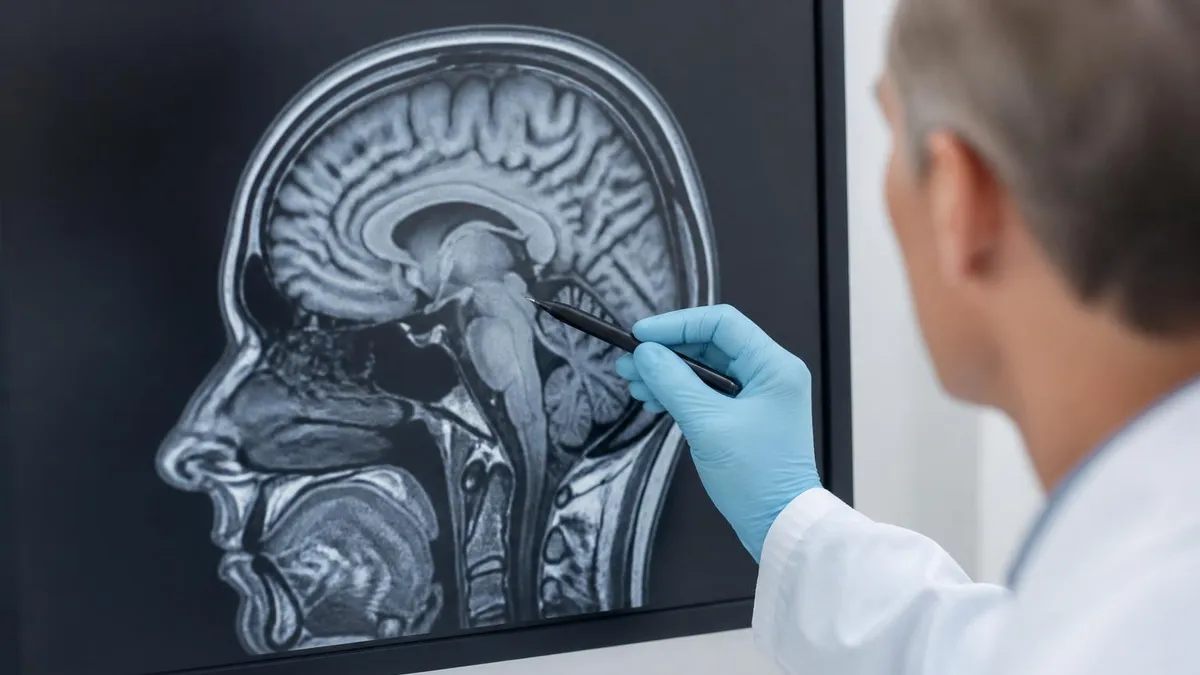
The Key Inventors of MRI
Isidor Rabi (1938)
Felix Bloch & Edward Purcell (1946)
Raymond Damadian (1971)
Paul Lauterbur (1973)
Sir Peter Mansfield (1977)
The story of who invented MRI cannot be told without going back to the late 1930s and the discovery of nuclear magnetic resonance itself. Isidor Isaac Rabi, a Polish-born physicist who fled antisemitism in Europe and built his career at Columbia University, was studying the magnetic properties of atomic nuclei using molecular beam techniques. In 1938, his team showed that nuclei placed in a strong magnetic field would resonate — absorbing radio frequency energy and re-emitting it — at characteristic frequencies determined by the nucleus type and the field strength.
Rabi's 1938 experiments were not aimed at medicine. He was pursuing fundamental physics, trying to understand the quantum mechanical behavior of atomic nuclei. Yet his work proved that nuclear spins could be manipulated and measured using radio waves, an observation so significant that it earned him the 1944 Nobel Prize in Physics. The award citation specifically credited his "resonance method for recording the magnetic properties of atomic nuclei." Without this seminal discovery, the entire chain of MRI development would have been impossible — every modern scanner relies directly on Rabi's resonance principle.
The next major leap came in 1946, when two physicists working independently on opposite American coasts achieved the same breakthrough. Felix Bloch, a Swiss-born professor at Stanford University, and Edward Purcell, working at Harvard, separately demonstrated that NMR could be observed in bulk matter — in liquids and solids, not just molecular beams. Bloch detected the proton signal in liquid water, while Purcell measured it in paraffin wax. Both publications appeared in Physical Review within weeks of each other, and the discoveries earned them a shared 1952 Nobel Prize in Physics.
For the next two decades, NMR remained almost exclusively a chemistry tool. Chemists used it to identify molecular structures, study reaction kinetics, and analyze unknown compounds. The pharmaceutical and petrochemical industries adopted NMR spectroscopy as a standard quality-control technique. Companies like Varian Associates and Bruker built commercial NMR spectrometers that became staples of academic and industrial laboratories. But the idea of using NMR on humans seemed absurd — the machines were small, signals were weak, and there was no obvious medical question NMR could answer.
The chemistry-to-medicine bridge began forming slowly through the 1960s as researchers explored NMR in increasingly complex biological samples. Scientists studied NMR signals in protein solutions, then tissue extracts, then small biological specimens. Each study revealed that biological tissues had distinct NMR characteristics, but nobody yet connected these observations to clinical diagnosis. The dominant view among physicists and physicians alike was that NMR was too cumbersome, too expensive, and too signal-poor to ever scan a living patient meaningfully.
That conventional wisdom would soon be shattered by an outsider — a young physician with limited NMR experience but extraordinary determination. Raymond Damadian, working in Brooklyn, was about to ask a question that no established NMR scientist had seriously pursued: could NMR distinguish cancerous tissue from normal tissue in a way that might detect tumors before they became visible by other methods? For deeper background on these foundational decades, see the comprehensive history of MRI documentation.
What's remarkable about this early period is how disconnected the scientific community was from the medical possibilities of NMR. Bloch and Purcell themselves never seriously pursued biological applications. Rabi remained focused on fundamental physics throughout his career. The physicists who created NMR did not invent MRI — that required a completely different mindset, one that saw the technology not as a chemistry instrument but as a potential window into human disease, willing to imagine what most established experts dismissed as impossible.
MRI Practice Test Questions
Prepare for the MRI - Magnetic Resonance Imaging exam with our free practice test modules. Each quiz covers key topics to help you pass on your first try.
MRI Knowledge
MRI Exam Questions covering Knowledge. Master MRI Test concepts for certification prep.
MRI Physics
Free MRI Practice Test featuring Physics. Improve your MRI Exam score with mock test prep.
MRI Anatomy and Pathology
MRI Test Prep for MRI Anatomy and Pathology. Practice MRI Quiz questions and boost your score.
MRI Anatomy and Positioning
MRI Questions and Answers on MRI Anatomy and Positioning. Free MRI practice for exam readiness.
MRI Contrast Agents
Free MRI Quiz on MRI Contrast Agents. MRI Exam prep questions with detailed explanations.
MRI Patient Care and Positioning
MRI Practice Questions for MRI Patient Care and Positioning. Build confidence for your MRI certification exam.
The Damadian vs Lauterbur Controversy
Raymond Damadian believed strongly that he was the true inventor of MRI, citing his 1971 Science paper showing NMR could distinguish cancerous from healthy tissue. He filed the first MRI patent in 1972, built the first whole-body human scanner in 1977, and founded FONAR Corporation — the first MRI manufacturer. Damadian's supporters argue that without his foundational insight that NMR could detect disease, no one would have pursued medical imaging applications at all.
When the 2003 Nobel Committee awarded the Prize in Physiology or Medicine to Lauterbur and Mansfield without including Damadian, he was deeply upset. He took out full-page advertisements in The New York Times, Washington Post, and Los Angeles Times protesting the decision under the headline "The Shameful Wrong That Must Be Righted." The exclusion remains one of the most publicly contested Nobel decisions ever made and continues to generate scholarly debate about credit attribution in collaborative science.
The Multi-Inventor Reality of MRI: Strengths and Limitations
- +Multiple Nobel Prizes recognize the breadth of contributions to NMR and MRI
- +Collaborative discovery process led to faster technological refinement
- +Different specialists (physicists, chemists, physicians) brought complementary expertise
- +Patent disputes pushed competing companies to innovate aggressively
- +Damadian's medical focus accelerated clinical adoption
- +Lauterbur's gradient method became the universal MRI standard
- +Mansfield's mathematics made real-time imaging possible
- −Credit disputes overshadow legitimate contributions of all parties
- −Damadian's exclusion from the 2003 Nobel remains controversial
- −Patent litigation slowed commercial development in the 1980s
- −Public confusion about who actually invented MRI persists today
- −Early scanners were prohibitively expensive due to licensing disputes
- −Lesser-known contributors like Richard Ernst rarely receive recognition
- −Historical narratives often oversimplify the multi-decade discovery process
Key MRI Invention Milestones to Remember
- ✓1938 — Isidor Rabi demonstrates nuclear magnetic resonance in molecular beams at Columbia University
- ✓1944 — Rabi awarded the Nobel Prize in Physics for his NMR discovery
- ✓1946 — Felix Bloch and Edward Purcell independently extend NMR to liquids and solids
- ✓1952 — Bloch and Purcell share the Nobel Prize in Physics for NMR spectroscopy
- ✓1971 — Raymond Damadian publishes Science paper showing NMR detects cancer in rat tissue
- ✓1972 — Damadian files the first MRI patent, titled apparatus for detecting cancer
- ✓1973 — Paul Lauterbur introduces magnetic field gradients for spatial encoding in Nature
- ✓1977 — Damadian completes the first whole-body human MRI scan with Indomitable
- ✓1977 — Peter Mansfield develops echo-planar imaging for ultra-fast scans
- ✓1980 — First commercial MRI scanners enter clinical practice worldwide
- ✓2003 — Lauterbur and Mansfield receive the Nobel Prize in Physiology or Medicine
- ✓2026 — Over 40,000 MRI scanners operate globally, performing 150 million scans annually
Six scientists, three Nobel Prizes, one revolutionary technology
The question of who invented MRI has no single answer because the technology required contributions across three distinct domains: the fundamental physics of nuclear resonance (Rabi, Bloch, Purcell), the medical application to disease detection (Damadian), and the mathematical encoding that produced actual images (Lauterbur, Mansfield). Together they transformed a quantum mechanical curiosity into one of the most powerful diagnostic tools in modern medicine — a journey spanning 65 years from theory to clinical practice.
Moving MRI from laboratory prototype to clinical workhorse required overcoming enormous engineering, financial, and regulatory hurdles. When Raymond Damadian's Indomitable scanner produced the first human MRI image on July 3, 1977, the scan took nearly five hours to complete and produced a single grainy cross-section of his graduate student Larry Minkoff's chest. The machine itself was assembled from scrap parts in a Brooklyn laboratory, with a magnet wound from miles of superconducting wire that Damadian and his team installed by hand over many months of painstaking work.
By the early 1980s, commercial MRI development was racing forward at companies like FONAR (founded by Damadian in 1978), General Electric, Siemens, Philips, and Picker International. The first FDA-approved clinical MRI scanner reached the market in 1984, manufactured by FONAR, and competing systems followed within months. Early scanners cost between $1.5 and $3 million per unit, plus installation costs that often doubled the total expense due to the need for radiofrequency shielding and dedicated buildings with reinforced floors capable of supporting the multi-ton magnets.
Patent litigation defined much of the 1980s and 1990s commercial landscape. Damadian's foundational 1972 patent led to years of courtroom battles, most famously his 1997 lawsuit against General Electric. After a jury trial, GE was ordered to pay FONAR $128.7 million for patent infringement — at the time, the largest patent verdict ever against a Fortune 500 company. The legal battles were bitter, expensive, and shaped how MRI manufacturers approached intellectual property for decades, but they also helped establish Damadian's legacy as a foundational inventor regardless of his Nobel exclusion.
Clinical adoption accelerated dramatically through the 1990s as scanner technology improved and prices gradually decreased. Neurology was the first specialty to embrace MRI widely because of the technology's unmatched ability to visualize soft tissue and detect conditions like multiple sclerosis, brain tumors, and stroke. Orthopedics followed quickly, with MRI becoming the gold standard for diagnosing knee ligament tears, rotator cuff injuries, and spinal disc problems. Cardiology, abdominal imaging, and breast cancer screening came later as specialized coils and pulse sequences were developed for each application.
The development of functional MRI (fMRI) in the early 1990s opened entirely new medical and research frontiers. Seiji Ogawa at AT&T Bell Laboratories discovered the BOLD (blood oxygen level dependent) contrast mechanism in 1990, allowing scientists to map brain activity in real time. This breakthrough transformed neuroscience research and clinical neurology, enabling surgeons to plan operations around critical brain regions and researchers to study cognition, emotion, and disease in unprecedented detail across thousands of studies published each year.
Modern MRI scanners are technological marvels that would astonish the original inventors. Today's 3 Tesla machines produce images with sub-millimeter resolution in under 30 minutes. Specialized techniques like diffusion tensor imaging map white matter tracts in the brain. MR spectroscopy measures specific metabolites in tissue. MR angiography images blood vessels without contrast injection in many cases. To understand the broader development of these techniques, see what an MRI test involves and how the scan procedure has evolved.
The economic and human impact of MRI invention is difficult to overstate. The global MRI market exceeds $7 billion annually and is projected to surpass $10 billion by 2030. More importantly, MRI has revolutionized patient care across every medical specialty, with no ionizing radiation exposure, making it safer than CT scans for repeat imaging. From its invention in scattered laboratories to its current status as essential medical infrastructure, MRI represents one of the most successful translations of fundamental physics into life-saving clinical technology in the history of medicine.
Many people incorrectly believe MRI was invented by a single person. In reality, modern MRI exists because of contributions from at least six major scientists working across nearly 40 years. The 2003 Nobel Prize recognized Lauterbur and Mansfield specifically for the imaging breakthrough, but the underlying physics and the medical application came from earlier and parallel work. Understanding this collaborative history is essential for anyone studying MRI physics or preparing for radiologic technology board exams.
The legacy of MRI's inventors extends far beyond the patents and Nobel medals — it lives in every modern scanner and every patient diagnosis made possible by their work. Today's MRI technology incorporates innovations from countless researchers building on the foundations laid by Rabi, Bloch, Purcell, Damadian, Lauterbur, and Mansfield. Modern superconducting magnets routinely operate at 1.5 or 3 Tesla, with research scanners reaching 7 Tesla or higher. These field strengths would have seemed impossible to the pioneers of NMR, whose original experiments used magnets thousands of times weaker than today's clinical systems.
One of the most important modern developments has been the parallel imaging revolution that began in the late 1990s. Techniques like SENSE (Sensitivity Encoding) and GRAPPA (Generalized Autocalibrating Partially Parallel Acquisition) use multiple receiver coils simultaneously to accelerate image acquisition by factors of two to eight times. These methods directly extend Mansfield's echo-planar work but achieve scan speeds he could not have imagined. A complete brain MRI that took an hour in 1990 can now be completed in 10–15 minutes with vastly superior image quality and diagnostic detail.
Artificial intelligence has become the latest frontier in MRI evolution, with deep learning algorithms now used to denoise images, reconstruct undersampled k-space data, and even predict diagnoses directly from raw scanner data. Companies like Subtle Medical, GE Healthcare, and Siemens have FDA-cleared AI tools that improve image quality while reducing scan times by 30% to 60%. These advances stand on the mathematical shoulders of Lauterbur and Mansfield while extending their impact in ways the original inventors could never have predicted in the 1970s.
The diagnostic applications of MRI continue to expand into new clinical territories. MR-guided focused ultrasound now allows non-invasive brain surgery for essential tremor and Parkinson's disease. Cardiac MRI has become the gold standard for evaluating myocardial viability, congenital heart disease, and inflammatory conditions like myocarditis. Whole-body MRI screening for cancer detection is gaining traction among health-conscious patients, though its cost-effectiveness for general populations remains actively debated by medical experts and policy researchers worldwide.
One often-overlooked legacy of MRI's invention is its safety record. Unlike CT and X-ray imaging, MRI uses no ionizing radiation, making it the preferred imaging modality for children, pregnant women, and patients requiring repeated scans. The technology's safety profile reflects the careful engineering and physics that Rabi, Bloch, and their successors built into the fundamental NMR principles. However, MRI is not entirely without risk — strong magnetic fields create hazards from ferromagnetic objects, and certain implants remain contraindicated for scanning under specific conditions.
The educational and research infrastructure built around MRI represents another enduring achievement of its inventors. Dozens of university programs train MRI physicists, radiologic technologists, and clinical radiologists each year. Professional societies like the International Society for Magnetic Resonance in Medicine (ISMRM), founded in 1994, host annual meetings with thousands of attendees presenting new research. For more context on the abbreviation and terminology, see MRI medical abbreviation and its formal definitions in clinical practice.
Perhaps most importantly, the multi-inventor nature of MRI offers a powerful lesson about modern scientific discovery. Major technological breakthroughs rarely emerge from solitary geniuses; they require communities of researchers contributing different expertise across decades. The story of who invented MRI is ultimately a story about how physics, chemistry, medicine, and engineering converged through cooperation and competition to create something none of the individual contributors could have achieved alone — a tool that has changed medicine forever and continues to save lives in hospitals around the world every single day.
For students, technologists, and clinicians preparing to learn or work with MRI, understanding the history of who invented MRI is more than trivia — it's essential context for grasping how the technology actually works. The fundamental physics of nuclear magnetic resonance, first measured by Rabi and refined by Bloch and Purcell, remains the operational basis of every scan. When a technologist places a patient in the bore and selects pulse sequences, they are directly applying principles discovered in the 1930s and 1940s, with imaging techniques developed in the 1970s by Lauterbur and Mansfield.
If you're studying for the ARRT MRI registry exam or the ARMRIT certification, expect questions about MRI history alongside physics, safety, and clinical procedures. The major exam categories include MRI physics, instrumentation, pulse sequences, imaging procedures, and patient care. Knowing the chronological development of the technology helps you remember why certain pulse sequences exist and what problems they were designed to solve. For example, echo-planar imaging exists because Mansfield needed a way to acquire data faster than conventional spin-echo techniques could ever achieve.
When studying MRI physics, focus on the core concepts that link directly to the inventors' work. The Larmor frequency — the resonance condition Rabi measured — determines the radiofrequency pulses needed to excite specific nuclei. T1 and T2 relaxation times, which Damadian showed differed between tumors and normal tissue, remain the basis of most clinical contrast. Magnetic field gradients, Lauterbur's contribution, control slice selection and spatial encoding. Understanding these connections turns abstract physics into intuitive concepts grounded in historical context that makes them easier to memorize for exams.
Practical clinical applications of MRI continue to evolve, and staying current is part of any technologist's career. Newer techniques like diffusion-weighted imaging, susceptibility-weighted imaging, and arterial spin labeling are now standard in many imaging centers, especially for neurological and oncological studies. Familiarity with these advanced sequences sets apart competent technologists from exceptional ones who can adapt to specialized protocols. Most certification bodies now expect candidates to understand at least the basic principles behind these modern techniques and their clinical indications.
Safety knowledge is non-negotiable for anyone working with MRI scanners. The strong static magnetic fields used in clinical MRI — typically 1.5 or 3 Tesla — pose serious risks if not properly managed by trained staff. Ferromagnetic objects can become deadly projectiles when accidentally brought into Zone IV. Implanted devices like pacemakers, cochlear implants, and certain aneurysm clips must be carefully screened before scanning. Patients with claustrophobia, kidney disease (relevant to gadolinium contrast), or metallic foreign bodies need careful protocol planning before being scanned.
For ongoing professional development, take advantage of free practice resources to reinforce your learning. Working through MRI practice questions regularly is one of the most effective study strategies, helping you identify knowledge gaps and reinforce key concepts before exam day. Combine question practice with reading classic MRI textbooks like Bushong's MRI: Physical and Biological Principles, Westbrook's MRI in Practice, and the ACR MRI Safety guidelines. These foundational texts trace the same historical threads we've covered while diving deep into technical details.
Finally, never lose sight of why MRI matters beyond exams and certifications. Every scan you perform or interpret traces back to the brilliance and persistence of the scientists who invented MRI. From Rabi's molecular beams in 1938 to Damadian's persistence in the face of skepticism, from Lauterbur's gradient insight to Mansfield's mathematical genius, each contribution made it possible for modern clinicians to diagnose disease without surgery or radiation. Carrying that history into daily practice honors the inventors' legacy and reminds us that every patient benefits from decades of patient, creative scientific work.
MRI Questions and Answers
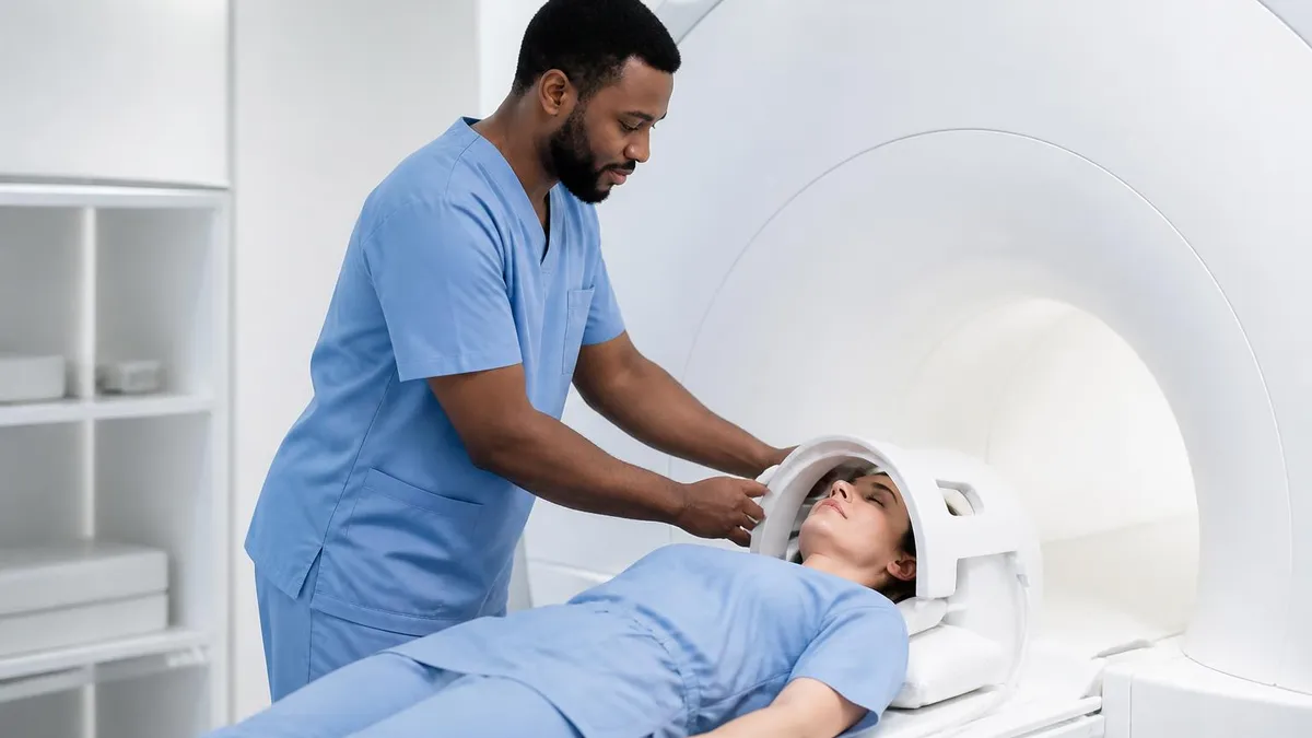
What Is an MRI Test? How Magnetic Resonance Imaging Scans Diagnose Disease in 2026
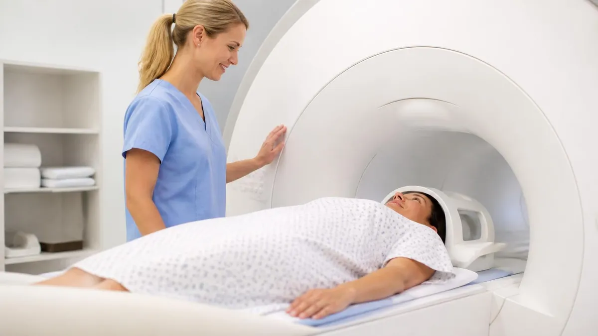
MRI Medical Abbreviation: What MRI Stands For and Why It Matters
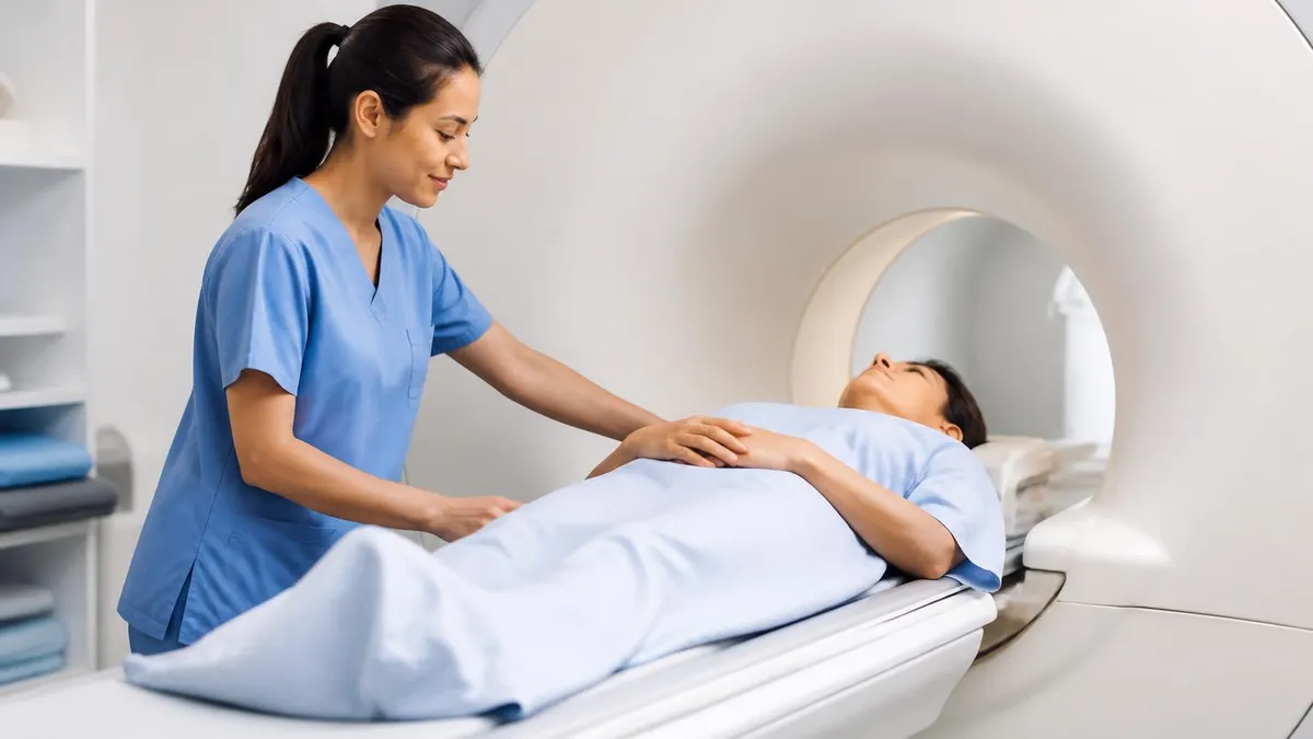
The History of MRI: From Discovery to Modern Medicine

Knee MRI Images: A Complete Guide to Reading, Understanding, and Interpreting Knee Scans
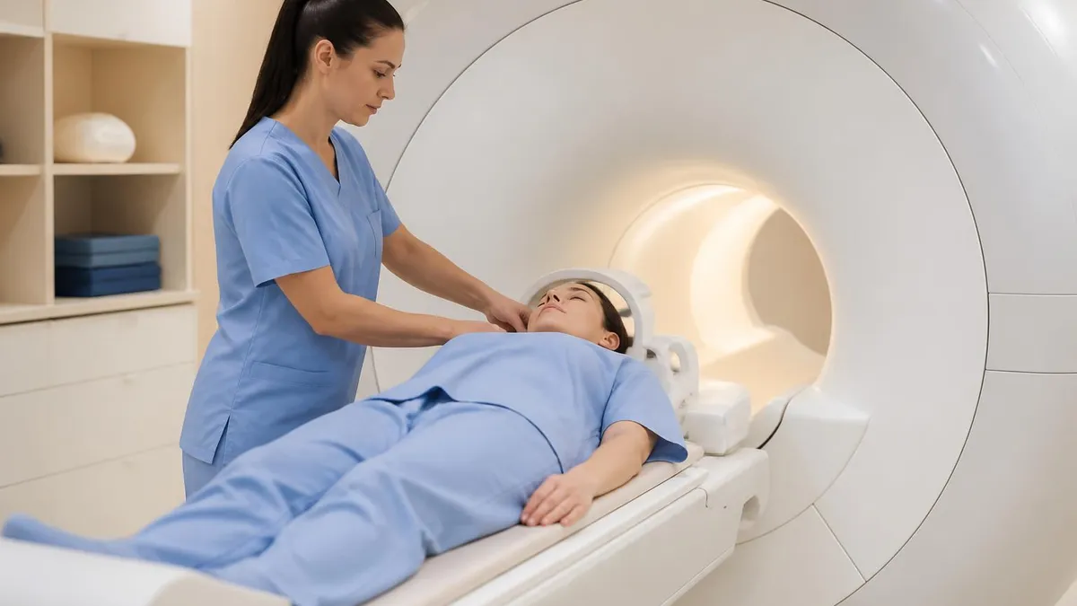
Noise of MRI Machine: Why MRI Scanners Are So Loud and What to Expect
About the Author

Medical Laboratory Scientist & Clinical Certification Expert
Johns Hopkins UniversityDr. Sandra Kim holds a PhD in Clinical Laboratory Science from Johns Hopkins University and is certified as a Medical Technologist (MT) and Medical Laboratory Scientist (MLS) through ASCP. With 16 years of clinical laboratory experience spanning hematology, microbiology, and molecular diagnostics, she prepares candidates for ASCP board exams, MLT, MLS, and specialist certification tests.
Join the Discussion
Connect with other students preparing for this exam. Share tips, ask questions, and get advice from people who have been there.
View discussion (8 replies)