Open MRI Machine: How It Works, When to Use, and Realistic Tradeoffs 2026 July
Open MRI machine explained: ✍🏼 design differences from closed MRI, ideal patient situations, image quality tradeoffs, and how to choose between options.
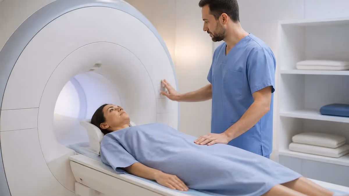
Open MRI machine refers to magnetic resonance imaging equipment designed with open sides rather than the traditional cylindrical tube of closed MRI. The open design accommodates patients who can't tolerate the enclosed environment of closed MRI due to claustrophobia, anxiety, body size limitations, or other reasons. Open MRI uses magnets positioned above and below the patient (like a giant sandwich) rather than around them. Understanding when open MRI is appropriate, what tradeoffs it involves compared to closed MRI, and how to make informed decisions helps patients navigate imaging choices effectively.
The major tradeoff for open MRI is image quality. Open MRI machines typically operate at lower magnetic field strength (0.2 to 1.0 Tesla) compared to closed MRI (typically 1.5 to 3.0 Tesla). Lower field strength produces lower image resolution, less detailed anatomical visualization, and longer scan times. For many imaging needs, open MRI provides adequate diagnostic information; for some specific situations requiring high-resolution detail, closed MRI is necessary regardless of patient preference. The tradeoff is real and clinically meaningful but doesn't make open MRI inferior — it makes it appropriate for some situations and inappropriate for others.
Patient comfort with open MRI is substantially better than closed MRI for many people. Less feeling of confinement. Better visibility of surroundings. Easier accommodation of larger body sizes (open MRI typically handles patients up to 500+ pounds vs closed MRI weight limits often 350 pounds). Better tolerance for patients with claustrophobia or anxiety. Better accommodation for pediatric patients who can have parent visible during scan. Each comfort advantage matters substantially for specific patient populations. Many patients who couldn't complete closed MRI complete open MRI successfully.
This guide covers open MRI machines comprehensively: how they work and differ from closed MRI, when open MRI is appropriate, image quality tradeoffs, finding open MRI facilities, and how to make informed decisions when both options are available. Whether you're scheduled for MRI and considering options or generally curious about imaging technology, you'll find practical guidance here.
Design: Magnets above and below patient (open sides) vs closed cylinder
Field strength: 0.2-1.0 Tesla typical (vs 1.5-3.0 Tesla closed)
Image quality: Lower resolution than closed MRI
Best for: Claustrophobic patients, larger patients, pediatric, anxious patients
Limitations: Some imaging requires high-resolution closed MRI
For how MRI works specifically, magnetic resonance imaging uses powerful magnets and radio waves to create detailed images of internal body structures. The patient's body's hydrogen atoms align with the magnetic field. Radio waves disturb this alignment; sensors detect the energy released as atoms realign. Computer processing constructs detailed images from these signals. Different tissue types produce different signal characteristics, supporting differentiation in images. The technology is remarkable; understanding the basics helps patients understand what's happening during scans. The magnetic resonance imaging resources cover MRI fundamentals.
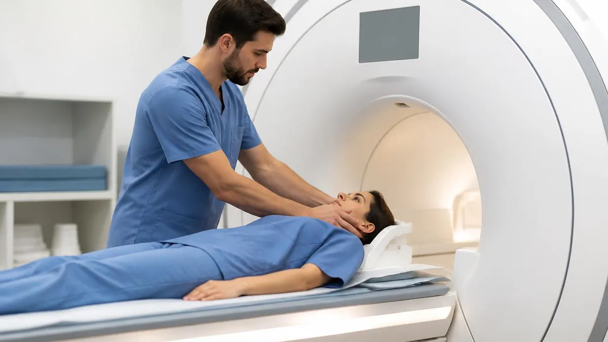
For closed vs open MRI specifically, the design difference affects multiple factors. Closed MRI: cylindrical tube design with magnetic field generated by surrounding magnet. Higher field strength supports better imaging. More confining experience for patient. Open MRI: magnets above and below with sides open. Lower field strength. Less confining experience. The choice between designs typically depends on patient tolerance and clinical imaging needs. Both are legitimate MRI technologies serving different purposes.
For Tesla strength specifically, this measurement of magnetic field strength affects image quality substantially. 0.2-0.7 Tesla — many older open MRI machines; lower image quality. 1.0 Tesla — newer high-field open MRI; better image quality. 1.5 Tesla — standard closed MRI; good image quality for most needs. 3.0 Tesla — high-field closed MRI; highest image quality for detailed imaging. Higher Tesla isn't always better — 3.0 T can produce some artifact issues that 1.5 T avoids; specific clinical needs determine appropriate Tesla. The open MRI resources cover open MRI specifically.
For patient situations where open MRI specifically helps, several patterns emerge. Severe claustrophobia making closed MRI unbearable. Large body size exceeding closed MRI weight limits. Pediatric patients requiring parental presence during scan. Anxiety disorders making enclosed spaces problematic. Some specific physical conditions making confinement difficult. Each situation suggests open MRI may produce better completion than closed MRI, even with image quality tradeoffs. Completed lower-quality scan provides more diagnostic value than incomplete higher-quality scan attempt.
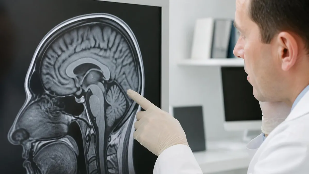
For when closed MRI is necessary specifically, several clinical situations require closed MRI. Detailed neurological imaging often requires high-resolution closed MRI. Some cardiac imaging requires specific closed MRI capabilities. Detailed musculoskeletal imaging for some conditions needs closed MRI resolution. Functional MRI (fMRI) typically requires closed MRI. Specific advanced imaging techniques require closed MRI. When ordering physician specifies closed MRI for clinical reason, alternative open MRI may not produce adequate diagnostic information. Discuss with ordering physician if open MRI is preferred.
Open vs Closed MRI Comparison
Better patient comfort. Accommodates larger patients (often up to 500+ lbs vs 350 lb limits on closed). Reduces claustrophobia issues. Allows parent presence for pediatric patients. Lower scan completion failure rates from anxiety. More accessible for anxious or claustrophobic patients. Good imaging for many common indications.
Lower magnetic field strength reduces image resolution. Longer scan times often needed. Some imaging requires closed MRI for adequate detail. Specific advanced techniques (fMRI, some specialized imaging) typically require closed. Less common in some areas (limiting accessibility). Higher cost in some markets despite older technology.
Higher magnetic field strength produces better image resolution. Faster scan times typically. More clinical sites available. Better for detailed neurological, cardiac, and specialized imaging. More flexible for various clinical needs. Standard for many specific imaging types. Generally lower cost per scan.
Confined environment causes claustrophobia issues for ~10-20% of patients. Weight limits (350 lbs typical) exclude some larger patients. Anxiety can prevent scan completion. Less accommodation for special needs. Some patients require sedation to complete. Pediatric patients often need anesthesia for full closed MRI.
For finding open MRI facilities specifically, several approaches work. Ask referring physician's office if open MRI is option for your scan. Imaging facility directories often note open vs closed availability. Major hospitals and imaging centers often offer both. Independent imaging centers sometimes specialize in open MRI. Online searches with location and "open MRI" identify nearby options. Insurance provider lists indicate covered facilities. Calling facilities directly to verify open MRI availability and specific Tesla strength provides accurate information. Plan ahead — open MRI may require more travel than closed MRI.
For insurance coverage specifically, both open and closed MRI typically covered when medically necessary. Some insurance plans require justification for specific imaging type. If physician orders open MRI for specific clinical reason (claustrophobia, body size), insurance typically covers. If patient requests open MRI when closed is medically appropriate, coverage varies. Some plans require trying closed MRI first before approving open MRI alternative. Check coverage with insurance before scheduling to prevent unexpected costs. The MRI cost resources cover related cost information.
For typical MRI scan experience specifically, several elements matter. Pre-scan: removing metal objects (jewelry, certain clothing items, dental work in some cases). Lying still on scan bed during examination. Hearing loud noises during scan (earplugs or headphones provided). Holding breath for some sequences. Scan duration typically 30-60 minutes for most studies. Some patients receive contrast dye injection. Post-scan: standard discharge unless contrast or sedation used. Each element is similar between open and closed MRI; specific differences relate to environment rather than fundamental scan process.
For pediatric imaging specifically, MRI in children has unique considerations. Younger children often need anesthesia for closed MRI to remain still. Open MRI may allow some children to complete scan without anesthesia given better tolerance of environment. Sedation has its own risks; avoiding sedation through better-tolerated open MRI may be preferable. Each child's specific situation guides decisions. Discussion between pediatric radiologist, anesthesiologist (if needed), and family about options produces best outcomes. The MRI safety resources cover safety considerations.
For pregnancy specifically, MRI generally considered safe during pregnancy when medically necessary. No ionizing radiation (unlike CT/x-ray). Specific contrast agents avoided during pregnancy. Closed MRI is well-tolerated by most pregnant patients despite environment. Open MRI can be alternative if closed environment is concerning during pregnancy. Discuss with both ordering physician and obstetrician before scheduling. Most decisions about MRI during pregnancy weigh benefits of imaging information against minimal theoretical risks of MRI exposure during fetal development.
Open MRI by Patient Situation
Patients with claustrophobia:
- Open MRI value: Substantially better tolerance vs closed MRI
- Alternative: Sedation for closed MRI (additional risks and recovery)
- Common solution: Open MRI when image quality adequate for diagnosis
- Imaging needs: Verify open MRI provides adequate detail for specific imaging required
- Communication: Discuss anxiety with imaging facility for additional support
For wide-bore closed MRI specifically, this represents middle ground between traditional closed MRI and open MRI. Wide-bore (typically 70 cm bore vs traditional 60 cm) provides more space for patients. Same magnetic field strength (1.5 T or 3.0 T) as standard closed MRI. Better tolerance for some patients vs traditional closed MRI without sacrificing image quality. Wide-bore is increasingly common as alternative to both traditional closed and open MRI. Many facilities now have wide-bore as standard rather than offering both narrow closed and open. Discuss bore options with imaging facility for best fit.
For high-field open MRI specifically, technological advances have produced higher-Tesla open MRI machines (1.0 T or higher). High-field open MRI produces better image quality than older 0.2-0.7 T open MRI while maintaining open design comfort advantages. Availability is growing but still limited compared to traditional MRI options. Cost is typically higher than older open MRI. The technology represents partial resolution of the traditional open vs closed tradeoff. Specific availability varies by region. The MRI machine resources cover MRI equipment broadly.
For specific imaging studies and open MRI suitability specifically, several patterns matter. Brain MRI for stroke or major findings — typically benefits from closed MRI's higher resolution; open MRI sometimes adequate. Spine MRI for various indications — open MRI often adequate; closed MRI for detailed conditions. Musculoskeletal MRI (knee, shoulder, etc.) — open MRI often adequate for many indications; specific situations need closed MRI. Cardiac MRI — typically requires closed MRI. Each clinical indication has typical patterns guiding open vs closed appropriateness.
For radiologist preferences specifically, individual radiologists have preferences about imaging studies. Some specialize in interpreting open MRI imaging; others primarily work with closed MRI. Image interpretation skill matters; same scan interpreted by experienced vs inexperienced radiologist produces different diagnostic results. When choosing between facilities for imaging, radiologist expertise matters alongside equipment quality. Specifically asking who will read your scan provides useful information about expected interpretation quality.
For follow-up imaging consistency specifically, several considerations matter. When imaging is for follow-up of previous condition, using same MRI type as previous scan supports better comparison. Comparing 0.5 T open MRI to subsequent 3.0 T closed MRI complicates comparison due to different image characteristics. When possible, follow-up imaging should match previous imaging for consistency. When changing types (initial closed, follow-up open or vice versa), expect interpretation challenges from non-comparable imaging.
Open MRI image quality may not be adequate for specific clinical needs. Before choosing open MRI based on patient preference, discuss with ordering physician whether open MRI adequacy for the specific clinical question. Some imaging requires closed MRI for adequate diagnostic information; using open MRI in those situations may produce inadequate results requiring repeat closed MRI later. The convenience of open MRI doesn't help if scan must be repeated due to inadequate quality. Verify with physician before scheduling — they understand specific clinical needs and can advise on whether open MRI is acceptable alternative. Don't assume open MRI is always adequate substitute for closed MRI.
For sedation alternatives to open MRI specifically, several options handle anxiety without changing imaging type. Light sedation (oral medication like Valium or Ativan) reduces anxiety enough for many patients to tolerate closed MRI. Moderate sedation (anesthesiologist-administered) for patients needing more support. General anesthesia for patients unable to tolerate any awake scan. Each option has tradeoffs in cost, recovery time, and risk. Sedation can preserve closed MRI image quality while addressing patient tolerance issues. The decision between sedation for closed MRI vs open MRI depends on specific patient situation and clinical needs.
For comfort measures during MRI specifically, several practices help all MRI patients. Music or audio entertainment during scan (provided through compatible headphones). Communication with technologist between sequences. Mirror inside bore to see outside (in some closed MRI machines). Positioning aids for comfort during long scans. Stop button for patient to halt scan if absolutely necessary. Each comfort measure helps both open and closed MRI experience. Discussing comfort needs with imaging facility before scan supports better tolerance.
For preparing for MRI specifically, several practices support successful scan completion. Eat light meal before scan to avoid discomfort. Arrive early to allow registration and preparation. Wear metal-free clothing or change into provided gown. Remove all metal objects (jewelry, dental work in some cases, hair accessories). Discuss any medical implants or metal in body with technologist (some are MRI-incompatible). Take prescribed anti-anxiety medication if recommended. Use bathroom before starting. Each preparation supports better scan experience. The MRI with contrast resources cover contrast considerations.
For after the scan specifically, several outcomes follow. Standard scans without sedation or contrast — discharge immediately, normal activities. Contrast scans — typically observe briefly for reaction; normal activities afterward. Sedated scans — recovery period and specific aftercare instructions. Results — typically delivered within 1-3 days through ordering physician. Most patients have unremarkable post-scan course. Following any specific instructions about contrast hydration or sedation recovery matters for safety. Plan ride home if sedation used; can drive yourself for standard scans.
Looking forward, MRI technology continues advancing. Higher-field open MRI (1.0 T+) becoming more common. Wider-bore closed MRI providing comfort without quality sacrifice. Faster scan techniques reducing patient time in machine. AI-assisted image processing improving image quality from any source. Patient comfort considerations increasingly integrated into MRI design. The traditional binary choice between open and closed MRI is becoming less stark as technology evolves to provide better options across the comfort-quality tradeoff.
For specialty MRI services specifically, several niche services exist beyond standard open vs closed. Stand-up or upright MRI allows imaging in weight-bearing positions for some musculoskeletal indications. Extremity MRI scans only specific body parts (knee, foot, etc.) with smaller dedicated machines accommodating arms or legs while patient sits. Each specialty has specific clinical indications. Most general MRI needs are met by standard open or closed MRI; specialty options serve specific situations.
For coverage maps of open MRI specifically, geographic distribution varies. Major metropolitan areas typically have open MRI options. Smaller cities and rural areas may have limited or no open MRI access. Some patients travel substantially for open MRI access. The geographic limitation affects practical access to open MRI option. When open MRI is necessary, planning for travel may be required. Major medical centers often have both options.
For clinical decision-making between options specifically, the key principle is matching imaging to clinical needs while accommodating patient constraints. Imaging that requires closed MRI quality should use closed MRI even with patient discomfort; sedation can address discomfort. Imaging with quality flexibility can use open MRI if patient prefers. Neither type is universally better — they serve different situations. Open communication between patient, ordering physician, and imaging facility produces best decisions.
For technological evolution specifically, MRI continues developing. Faster scan techniques reduce time in machine. Higher-field open MRI improves image quality of comfort option. Wide-bore closed MRI provides comfort of open with quality of closed. AI-assisted reconstruction improves images from any MRI type. Patient comfort design improvements throughout. Future MRI may resolve traditional tradeoffs more completely than current options. Today's choices reflect current technology; future evolution will continue improving available options.
Open MRI Quick Facts
Open MRI Decision
- +Substantially better patient comfort than closed MRI
- +Accommodates larger patients (500+ lbs typical)
- +Reduces claustrophobia issues that prevent closed MRI completion
- +Allows parent presence for pediatric patients
- +Better tolerance for anxious patients without requiring sedation
- −Lower magnetic field strength produces lower image resolution
- −Some imaging requires closed MRI for adequate diagnostic detail
- −Longer scan times for some imaging
- −Less common than closed MRI — limited geographic availability
- −Sometimes higher cost despite lower technology level
MRI Questions and Answers
About the Author
Medical Laboratory Scientist & Clinical Certification Expert
Johns Hopkins UniversityDr. Sandra Kim holds a PhD in Clinical Laboratory Science from Johns Hopkins University and is certified as a Medical Technologist (MT) and Medical Laboratory Scientist (MLS) through ASCP. With 16 years of clinical laboratory experience spanning hematology, microbiology, and molecular diagnostics, she prepares candidates for ASCP board exams, MLT, MLS, and specialist certification tests.
Join the Discussion
Connect with other students preparing for this exam. Share tips, ask questions, and get advice from people who have been there.
View discussion (6 replies)