Nevro HF10 Spinal Cord Stimulator MRI Safety: Complete Guide for Patients and Technologists
Nevro HF10 spinal cord stimulator MRI safety: conditions, scan rules, patient prep, and technologist screening protocols for safe imaging.
Understanding nevro hf10 spinal cord stimulator MRI safety is essential for anyone scheduling magnetic resonance imaging with an implanted neurostimulator. The Nevro Senza system, which delivers 10 kHz high-frequency therapy, is conditionally approved for both 1.5T and 3T MRI scans when specific manufacturer guidelines are followed precisely. Misunderstanding these conditions can lead to canceled appointments, device damage, tissue heating, or in rare cases serious patient injury. This guide explains exactly what patients, referring physicians, and MRI technologists need to verify before, during, and after the scan.
The HF10 platform earned full-body MR conditional labeling after extensive bench testing, animal modeling, and clinical evaluation. Unlike older spinal cord stimulators that were strictly MR unsafe or only head-scan compatible, the Senza II, Senza Omnia, and HFX iQ generators allow imaging of any anatomy when the system is intact, properly programmed into MRI mode, and the scanner operates within published specific absorption rate (SAR) limits. Knowing which generator model the patient has implanted is therefore the first checkpoint of every safety workflow.
Patients often arrive at the imaging center without their patient identification card, without recent device interrogation reports, and without a clear sense of whether their stimulator is even on or off. Technologists must therefore build a redundant verification process: chart review, patient interview, physical inspection of the remote control, and direct confirmation with the implanting pain management clinic when uncertainty exists. A 15-minute delay to confirm device status is always preferable to scanning an unverified implant.
The regulatory framework also matters. The FDA cleared HF10 MR conditional labeling under specific phantom-tested conditions, and deviating from those conditions voids the safety claim. That means a 3T scan with body transmit coil at 4.0 W/kg whole-body SAR is outside labeling even though the same anatomy at 2.0 W/kg would be acceptable. Technologists must read the current Nevro MRI guidelines document, which is periodically updated, rather than relying on memory or outdated printouts taped to the console.
Patient experience during the scan is another important consideration. Even with a fully compliant setup, patients may feel mild warmth, vibration, or paresthesia at the lead site. These sensations are typically benign and self-limited, but technologists must counsel patients to use the squeeze-ball alert immediately if discomfort intensifies. A well-prepared patient who understands what is normal versus what is not is far less likely to terminate the scan prematurely, which preserves diagnostic quality and reduces repeat-imaging costs.
This article walks through generator models, scan conditions, the pre-scan checklist, what to do mid-scan, and post-scan device verification. We also cover common questions about lead fractures, partial systems, recharging timing, and what happens if a patient is scanned in error. For broader context on how stimulator artifact can affect spinal imaging interpretation, you may also want to review Common MRI Findings: Brain, Spine and Joints Guide to understand how implant artifacts present alongside true pathology.
Whether you are a patient preparing for a knee MRI, a technologist screening a new referral, or a radiologist reading a post-implant lumbar study, the practical rules below will help you complete the exam safely and obtain images of diagnostic quality. The protocols are conservative by design, and following them protects both the patient and the device investment that took weeks to trial and titrate.
HF10 MRI Safety by the Numbers
Generator Models and MR Conditional Status
The Senza II implantable pulse generator is full-body MR conditional at 1.5T and 3T when programmed into MRI mode. It supports both head and body transmit coils within published SAR limits.
The Omnia generator supports multiple waveforms including 10 kHz HF10 therapy. It carries the same full-body MR conditional labeling and requires identical pre-scan programming and verification steps.
The HFX iQ is Nevro's AI-enabled generator with sensor-driven programming. MR conditional labeling matches earlier generators, but firmware must be current and the device placed into MRI mode via the clinician programmer or patient remote.
External trial stimulators worn during the 5-7 day evaluation period are MR unsafe. Patients in the trial phase must not undergo MRI until the external generator and percutaneous leads are removed.
The Nevro HF10 MR conditional labeling specifies exact scanner conditions that must be met for every exam. The static magnetic field strength must be either 1.5 tesla or 3.0 tesla in a horizontal closed-bore scanner. Open MRI systems, extremity-only scanners with vertical fields, and any field strength outside the approved range are explicitly excluded. Even a 1.0T scanner, which might intuitively seem safer due to lower field strength, is not validated and therefore not permitted under current labeling for HF10 systems.
Specific absorption rate, or SAR, is the most commonly misunderstood condition. The whole-body averaged SAR must remain at or below 2.0 W/kg in normal operating mode for the duration of the scan. First-level controlled mode, which allows SAR up to 4.0 W/kg, is not approved and must not be selected. Technologists should verify the scanner is set to normal mode before each sequence, since some protocols default to first-level when patient weight is entered or when high-resolution sequences are selected.
Gradient slew rate and dB/dt limits also apply. The maximum gradient slew rate must not exceed 200 T/m/s per axis, which is well within standard clinical scanner capabilities but worth confirming on advanced research systems. Maximum spatial gradient field strength at the implant location is capped at 40 T/m, again a parameter rarely exceeded in routine imaging but relevant for ultrafast diffusion or echo-planar sequences. Documentation of these parameters in the scan log protects both the facility and the patient.
Coil selection is another critical decision point. Transmit body coils, transmit head coils, and transmit-receive extremity coils are all acceptable provided the SAR limit is respected. Local transmit coils placed directly over the implant or leads should be avoided when alternative coil configurations exist. For lumbar spine imaging in a patient with thoracic leads, this usually means using the integrated body coil for transmit and a phased-array spine coil for receive, which is the standard configuration anyway.
Active scan time per imaging session is limited to 30 minutes of cumulative radiofrequency exposure. Cumulative means the running total of sequence durations, not wall-clock time in the bore. If a patient needs both a brain and a lumbar study, the technologist must track total RF-on time across both exams and pause for a cooling period if approaching the limit. Many facilities split such combined studies across two separate appointment days to avoid any ambiguity. For deeper context on what these multi-region studies can reveal, see What MRI Can Detect: Conditions & Diagnostic Capabilities.
The implanted system itself must be intact and functional. Broken leads, partially implanted systems with abandoned hardware, and any evidence of lead migration or fracture on prior imaging change the safety analysis. In these cases the scan is not automatically contraindicated, but the Nevro MRI guidelines require additional review, often including a phone consult with Nevro technical support. Documenting the conversation and the recommendation in the patient chart is a defensive best practice.
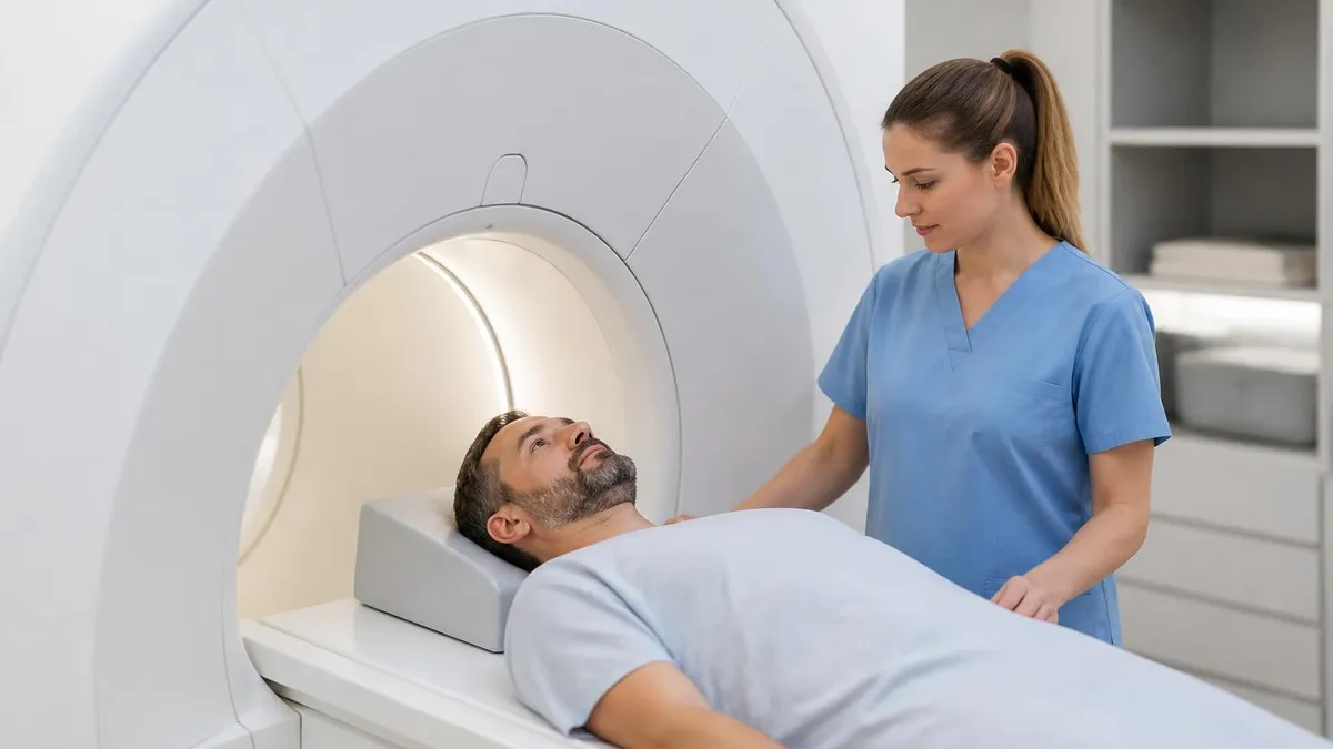
Finally, the device must be placed into MRI mode before the patient enters the scan room. MRI mode disables stimulation output and locks the generator into a safe configuration. This is accomplished via the patient remote control or, for older systems, the clinician programmer. Confirmation that MRI mode is active should be verbal, visual on the remote screen, and ideally documented with a screenshot in the patient record before transferring the patient to the magnet.
Pre-Scan Verification Workflow for HF10 MRI Safety
Begin with documentation review before the patient arrives. Confirm the implanted generator model, implant date, lead positions, and current programming from the most recent pain management visit. Pull the patient implant card if scanned into the EMR, and verify the device serial number matches the chart entry. Any mismatch between recorded model and the patient's recollection requires resolution before scheduling proceeds.
Check for prior MRI exams since implantation. A patient who has been safely scanned at your facility before is lower risk than a first post-implant scan, since lead integrity has been functionally tested. Note whether prior exams used 1.5T or 3T fields and any reported issues such as paresthesia, warmth, or post-scan reprogramming needs. Pattern recognition across exams catches subtle device drift early.
Scanning an HF10 Patient: Benefits and Risks to Weigh
- +Full-body MR conditional approval at both 1.5T and 3T field strengths
- +Standard diagnostic imaging quality achievable with proper sequence selection
- +No need to explant the device for medically necessary MRI exams
- +MRI mode programming is reversible and quickly restored to therapy
- +Patients can continue receiving HF10 pain relief indefinitely without imaging trade-offs
- +Most diagnostic questions can be answered within the 30-minute RF time budget
- −Lead and IPG artifact may obscure anatomy directly adjacent to hardware
- −Requires additional pre-scan screening time and clinic coordination
- −Patient must bring functional remote and have adequate battery charge
- −First-level SAR mode is unavailable, limiting some high-resolution sequences
- −Therapy is paused during MRI mode, which may temporarily increase pain
- −Post-scan reprogramming visit occasionally needed if device exits MRI mode incorrectly
Patient Preparation Checklist for Nevro HF10 MRI Safety
- ✓Confirm generator model is Senza II, Senza Omnia, or HFX iQ (not a trial system)
- ✓Verify implant date is at least six weeks before the scheduled scan
- ✓Charge the IPG to above 25% capacity the night before the exam
- ✓Bring the patient remote control to every MRI appointment
- ✓Bring the patient implant identification card or a photograph of it
- ✓Avoid eating a heavy meal within two hours of the scan to reduce motion
- ✓Wear metal-free clothing or change into a facility gown before screening
- ✓Practice using MRI mode on the remote with the implanting clinic in advance
- ✓Notify the technologist immediately of any new pain, swelling, or device issues
- ✓Plan for a follow-up with pain management within two weeks to verify programming
MRI mode disables therapy — but does not turn off the device
When the HF10 generator is placed into MRI mode, stimulation output stops but the device itself remains powered and continues to consume battery. Patients sometimes confuse this with the device being off and panic when paresthesia disappears. Counsel patients clearly that this is expected, temporary, and fully reversible with a single remote-control action after the exam ends.
Once the patient is positioned in the scanner and MRI mode is confirmed active, the technologist's job shifts to monitoring rather than verifying. The intercom should be tested before the first sequence, the squeeze-ball alert placed firmly in the patient's hand, and the patient instructed to use it for any new warmth, twitching, vibration, or sharp pain at the implant or lead sites. These sensations are uncommon when SAR is within limits, but they can occur, and early notification allows the technologist to pause sequences and reassess before harm develops.
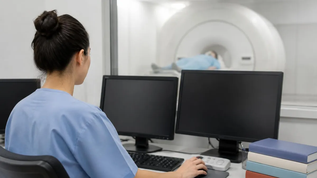
Monitor the SAR display on the scanner console for every sequence. If a planned sequence pushes whole-body SAR above 2.0 W/kg, the technologist must modify parameters before running it. Common adjustments include increasing TR, reducing the number of slices, lowering flip angle, or switching from spin-echo to gradient-echo techniques. Documentation of any parameter changes in the technologist worksheet helps the radiologist understand why a substitute sequence appears in the final study and supports billing accuracy.
Patient communication between sequences is essential. Brief check-ins of two or three seconds — "How are you doing in there, any new sensations?" — keep the patient engaged and provide multiple opportunities to catch developing problems early. Patients who feel ignored often hesitate to use the squeeze ball for fear of being a bother, which delays useful feedback and increases the likelihood of an aborted exam. A short scripted check between every sequence is a low-cost, high-value safety habit.
If the patient reports new sensation, pause the sequence immediately and ask clarifying questions through the intercom. Distinguish positional discomfort, which is common during long exams, from new paresthesia, warmth, or pain at the implant site, which requires immediate sequence termination. When in doubt, end the active scan, verify the patient remote still shows MRI mode, remove the patient from the bore, and reassess. A non-diagnostic exam is preferable to an injury, and most concerning sensations resolve quickly outside the magnet.
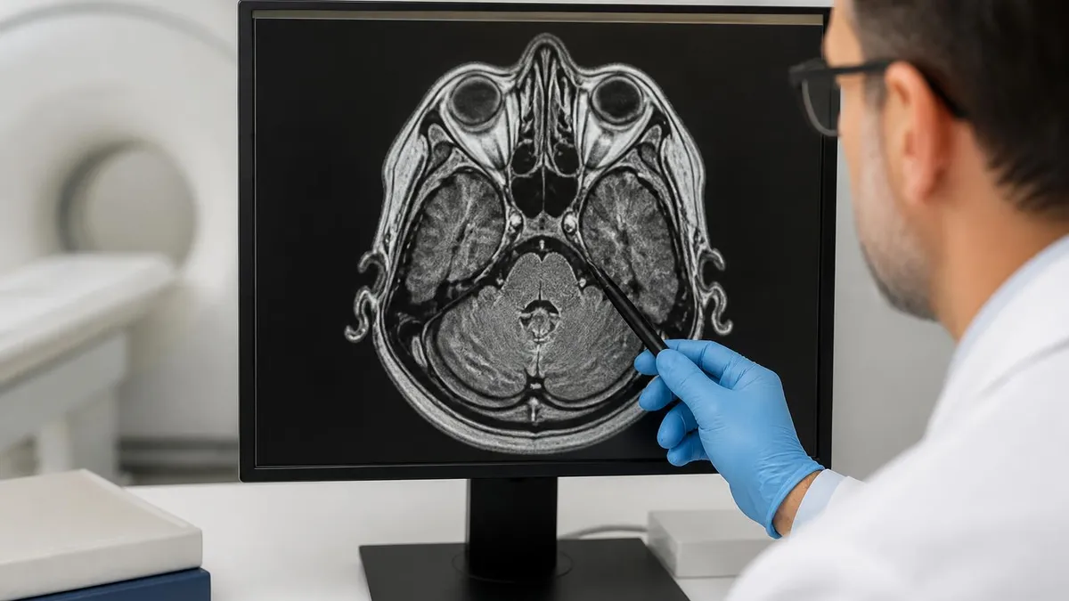
Image artifact from the IPG and leads is expected and predictable. The titanium generator case produces a signal void of about 4 to 6 centimeters in diameter on most sequences, while the leads create thin linear voids tracking along the epidural space. Radiologists familiar with stimulator artifact can usually interpret around it, but ordering providers benefit from a note in the report identifying the device and the expected artifact pattern. This avoids confusion with pathology and supports clean communication of the diagnostic findings.
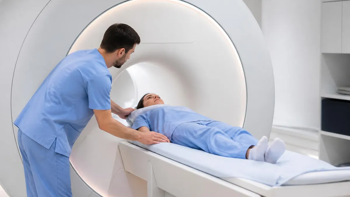
After the final sequence ends, the patient should remain still while the technologist enters the scan room and verbally confirms they feel well. Help the patient sit up slowly, and only then provide the remote control to switch the device back to therapy mode. Switching modes inside the bore or immediately upon sitting up is unnecessary and increases the chance of remote drop or fumble. A calm, stepwise exit from the scanner protects both the patient and the device.
Document the entire encounter thoroughly. The technologist worksheet should record generator model, serial number, pre-scan mode status, post-scan mode status, maximum SAR reached, total RF active time, any reported sensations, and the time MRI mode was deactivated. This documentation supports continuity of care, defends the facility in the rare event of an adverse outcome, and creates a reference record for the next time the patient needs imaging at your center.
The percutaneous trial system worn for 5-7 days before permanent implant is MR unsafe. Scanning a patient with a trial system risks lead displacement, severe burns, and serious tissue injury. Always confirm the patient has the permanent implanted pulse generator before proceeding, and reschedule the MRI if any uncertainty exists about which system is in place.
Troubleshooting begins with a clear understanding of what is and is not a contraindication. A patient with a single fractured lead and an intact remaining lead may still be eligible for imaging if Nevro technical support reviews the case and approves the scan in writing. A patient with abandoned hardware from a prior explanted system requires careful evaluation, since older fragments may not carry MR conditional labeling even when the current Nevro device does. Documenting these complex cases meticulously protects everyone involved.
One common edge case involves the recently implanted patient. Nevro guidance recommends waiting six weeks after implantation before non-urgent MRI to allow tissue ingrowth around the leads, which stabilizes lead position and reduces migration risk during scanning. For urgent imaging within that window, the referring physician and the implanting team should consult before scheduling, and the risk-benefit discussion should be documented in the chart with patient consent.
Another scenario worth planning for is the patient who arrives without their remote. Without the remote, MRI mode usually cannot be activated, and the exam must be rescheduled. A facility-stocked loaner remote from Nevro is not currently available, so educating patients about the absolute requirement to bring their remote is the only practical solution. Some centers add a confirmation reminder to appointment confirmation calls 24 hours before the scan to reduce no-remote arrivals. For a closer look at the imaging careers that handle these workflows, see How to Become an MRI Technician: Schools, Certification, Salary.
Battery depletion mid-scan is rare but worth understanding. If the IPG battery falls below the minimum threshold during MRI mode, the device defaults to a power-conservation state and remains in MRI-safe configuration. Therapy will not resume automatically when the exam ends, and the patient must recharge the device before reprogramming. Counsel patients to charge the night before any MRI to avoid this inconvenience, which is occasionally mistaken for device malfunction.
What happens if a patient is inadvertently scanned without entering MRI mode? Stop the scan immediately, remove the patient from the magnet, and contact Nevro technical support and the implanting physician the same day. The device should be interrogated as soon as possible to confirm intact function and rule out programming corruption. Most inadvertent scans result in no clinical harm when SAR was within limits, but documentation and follow-up interrogation are mandatory and protect against later disputes.
Pregnant patients with HF10 implants require special consideration. MR conditional labeling does not change during pregnancy, but the decision to image at all involves weighing fetal exposure to gradient noise and any contrast considerations. Most facilities defer non-urgent MRI in pregnant patients regardless of implant status, and the same conservative approach applies to HF10 patients. Coordinate with obstetrics and pain management before scheduling.
Pediatric use of HF10 systems is uncommon but increasing in select chronic pain programs. The MR conditional labeling extends to qualified pediatric patients, but technologists should expect additional anxiety management challenges and consider whether moderate sedation alters the SAR calculation due to changes in patient size assumptions. Working with the pediatric anesthesia and pain teams early in scheduling avoids day-of-scan surprises.
Practical preparation tips make the difference between a smooth scan and a frustrating reschedule. Start by collecting a single-page reference card from the implanting clinic that lists the patient's generator model, serial number, implant date, lead types, and lead positions. Laminate it, photograph it for the EMR, and store the original with the patient remote in a small zipped pouch. Patients who travel with this kit consistently complete their MRI exams without delays, regardless of which imaging facility they visit.
Schedule MRI appointments earlier in the day when possible. Morning slots reduce the chance of accumulated facility delays pushing the exam past the time of day when the patient's pain management clinic is closed. If a technical question arises and Nevro technical support has already gone home for the day, an afternoon scan may need to be rescheduled. A 9 a.m. appointment leaves a full day of expert availability to resolve any unexpected issues that emerge during screening.
Coordinate post-scan reprogramming proactively. About 5-10% of patients need a brief programming visit after MRI to fine-tune amplitude, pulse width, or coverage area after switching out of MRI mode. Scheduling a tentative one-week post-MRI clinic check at the time the MRI is booked avoids gaps in pain coverage. Most patients have no programming drift, but planning for the occasional case prevents unnecessary suffering when adjustment is needed.
Patients should bring earplugs or earmuffs and use them throughout the exam. Although hearing protection is unrelated to implant safety directly, the long sequences sometimes required to obtain diagnostic images around stimulator artifact can be louder and longer than typical exams. Comfortable, well-fitted ear protection keeps patients in the bore longer and reduces motion artifact, which improves diagnostic yield and avoids repeat sequences that consume the limited RF time budget.
Prepare a simple personal symptom diary for the 48 hours after the scan. Patients should note any unusual pain, new paresthesia patterns, device functionality changes, or unexpected battery drain. This information helps the pain management team distinguish normal post-MRI variability from genuine device issues during the follow-up visit. Most diaries are completely unremarkable, but those that capture early signal allow timely intervention before small problems grow into significant complications. For a comparison view of how comprehensive imaging is approached in other contexts, see Full Body MRI: What It Scans, How Long It Takes, Cost, and What to Expect.
Finally, build trust by communicating clearly with the patient throughout. Explain why each step matters, what they will feel, how long the exam will take, and what to expect afterward. Patients who understand the rationale behind MRI mode, the SAR limit, the 30-minute time budget, and the post-scan check-in are vastly more compliant and far more likely to report subtle but important symptoms early. Education is the single most powerful safety intervention available, and it costs nothing beyond a few minutes of attention.
Documentation completes the workflow. Every step described in this article should leave a paper or electronic trail: the screening form, the MRI mode confirmation screenshot, the SAR log, the technologist worksheet, the post-scan device check, and the follow-up clinic plan. When the next MRI is needed in two or five years, that documentation makes the second exam dramatically faster and safer because the team can rely on a proven workflow rather than rebuilding it from scratch.
MRI Questions and Answers
About the Author
Attorney & Bar Exam Preparation Specialist
Yale Law SchoolJames R. Hargrove is a practicing attorney and legal educator with a Juris Doctor from Yale Law School and an LLM in Constitutional Law. With over a decade of experience coaching bar exam candidates across multiple jurisdictions, he specializes in MBE strategy, state-specific essay preparation, and multistate performance test techniques.