
MRI Risks: A Complete Guide to Safety Hazards, Contraindications, and How to Stay Safe During Magnetic Resonance Imaging
MRI risks explained: magnetic field hazards, gadolinium contrast, implants, burns, and screening. Learn how to stay safe during your MRI scan. ✅
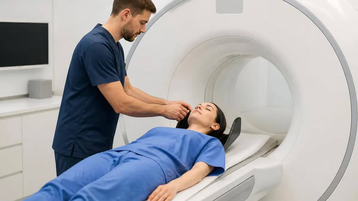
Understanding MRI risks is essential for every patient, technologist, and clinician who works around magnetic resonance imaging. Although MRI uses no ionizing radiation and is considered one of the safest diagnostic tools in modern medicine, the scanner generates an extraordinarily powerful static magnetic field, rapidly switching gradient fields, and radiofrequency energy. Each of these forces can create genuine hazards when proper screening is skipped or when contraindicated implants enter the room. Knowing where the danger truly lies allows you to weigh the benefits against the small but real dangers.
The single most dramatic MRI risk is the projectile, or missile, effect. The static magnet in a typical clinical scanner ranges from 1.5 to 3 tesla, which is tens of thousands of times stronger than the earth's magnetic field. Ferromagnetic objects such as oxygen cylinders, wheelchairs, scissors, and even hairpins can be torn from a hand and flung toward the bore at speeds capable of causing severe injury or death. This force never turns off, even when no scan is running, which surprises many newcomers.
Beyond projectiles, patients face risks tied to implanted medical devices. Pacemakers, certain aneurysm clips, cochlear implants, and some neurostimulators may heat, move, or malfunction inside the magnet. Screening forms exist precisely to capture this history before a person ever approaches the bore. Many devices manufactured in recent years are labeled MR Conditional, meaning they are safe only under specified field strengths and scanning parameters, while older or unlabeled hardware may be absolutely unsafe.
Contrast agents add another layer to MRI risks. Gadolinium-based contrast helps highlight tumors, inflammation, and vascular abnormalities, but it carries a rare association with nephrogenic systemic fibrosis in patients with severe kidney disease. Researchers have also documented gadolinium deposition in brain tissue after repeated dosing, prompting ongoing study and more conservative prescribing. For most patients with normal renal function, the contrast remains well tolerated, yet informed consent should always cover these considerations.
Acoustic noise, peripheral nerve stimulation, and claustrophobia round out the everyday hazards. The gradient coils that build images produce knocking sounds that can exceed 110 decibels, loud enough to damage unprotected hearing. Rapidly changing gradients can trigger tingling muscle twitches, and the enclosed bore provokes anxiety in roughly one in seven patients. None of these are typically dangerous when staff provide hearing protection, monitoring, and reassurance, but they shape the overall patient experience considerably.
This guide walks through every major category of MRI risk in plain language, explains who is most vulnerable, and describes the layered safety systems that keep millions of scans incident free each year. Whether you are preparing for a scan, studying for a registry exam, or supervising an imaging suite, a clear grasp of these hazards turns abstract warnings into practical, life-saving habits. The goal is confidence grounded in knowledge rather than fear of the unknown.
MRI Risks by the Numbers
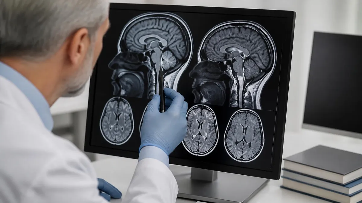
Magnetic Field and Projectile Hazards
The main magnet is always on, even during power loss. It exerts force on ferromagnetic metal continuously, so the scan room must stay controlled around the clock with no exceptions for cleaning or maintenance staff.
Loose ferrous objects accelerate toward the bore with deadly force. Oxygen tanks, mop buckets, IV poles, and keys have all become projectiles, causing crush injuries, broken equipment, and at least one documented patient fatality.
If superconducting magnets warm suddenly, liquid helium boils off rapidly in a quench. This can displace oxygen, freeze skin, or over-pressurize the room if venting fails, so quench buttons are used only in true emergencies.
Suites are divided into four safety zones. Access tightens progressively toward Zone IV, the magnet room, where only screened patients and trained staff may enter after metal detection and verbal verification.
Implanted and embedded devices represent the most common reason a patient is barred from MRI or requires special protocols. The magnetic field can rotate, dislodge, or heat ferromagnetic hardware, while the radiofrequency pulses can induce currents in conductive leads. Because so many devices now exist, manufacturers and the FDA use a three-tier labeling system that every technologist must understand before clearing a patient for the scan room. Misreading these labels has led to serious harm, so verification is never optional or rushed.
MR Safe items pose no known hazard in any MRI environment because they contain no metal and conduct no electricity, such as plastic or silicone components. MR Conditional devices are safe only within precisely defined conditions, including a maximum field strength, a specific gradient slew rate, and a limited specific absorption rate. MR Unsafe items, marked with a red symbol, must never enter the magnet room. When labeling is missing entirely, staff should treat the device as unsafe until documentation proves otherwise.
Cardiac implantable electronic devices once formed an absolute barrier to imaging, but MR Conditional pacemakers and defibrillators are now widespread. Scanning them still demands programming changes, continuous monitoring, and often a cardiologist on standby. Older non-conditional units can experience lead heating, inappropriate pacing inhibition, or reset events that endanger the patient. Each case requires a documented risk-benefit discussion, and many centers maintain dedicated protocols built specifically for these higher-risk encounters.
Cerebral aneurysm clips deserve particular caution. A ferromagnetic clip can torque within the brain and tear the very vessel it was meant to secure, a catastrophic and potentially fatal event. Modern titanium clips are generally safe, but documentation of the exact clip model is mandatory before any scan proceeds. Similarly, cochlear implants, certain neurostimulators, drug infusion pumps, and some older heart valves all require model-specific verification rather than assumptions based on general categories.
Retained metallic foreign bodies create a quieter but serious threat. Metalworkers, welders, and trauma survivors may carry tiny ferrous fragments in the eye or soft tissue without realizing it. A fragment near the orbit can shift in the field and damage the retina or optic nerve. When history suggests this risk, an orbital radiograph or CT screen is ordered first. The principles overlap heavily with broader questions about MRI safety materials and which alloys behave safely.
Finally, everyday accessories cause more denied appointments than patients expect. Insulin pumps, glucose monitors, hearing aids, dentures with magnets, transdermal medication patches with metallic backing, and even certain tattoos with iron-oxide ink can interfere or heat. Thorough screening captures all of these, which is why the intake questionnaire is detailed and repetitive. Honest, complete answers protect the patient far more effectively than any single device on the scanner itself ever could.
MRI Practice Test Questions
Prepare for the MRI - Magnetic Resonance Imaging exam with our free practice test modules. Each quiz covers key topics to help you pass on your first try.
MRI Knowledge
MRI Exam Questions covering Knowledge. Master MRI Test concepts for certification prep.
MRI Physics
Free MRI Practice Test featuring Physics. Improve your MRI Exam score with mock test prep.
MRI Anatomy and Pathology
MRI Test Prep for MRI Anatomy and Pathology. Practice MRI Quiz questions and boost your score.
MRI Anatomy and Positioning
MRI Questions and Answers on MRI Anatomy and Positioning. Free MRI practice for exam readiness.
MRI Contrast Agents
Free MRI Quiz on MRI Contrast Agents. MRI Exam prep questions with detailed explanations.
MRI Patient Care and Positioning
MRI Practice Questions for MRI Patient Care and Positioning. Build confidence for your MRI certification exam.
Gadolinium Contrast and MRI Risks
Gadolinium-based contrast agents shorten the relaxation time of nearby water protons, making blood vessels, tumors, and inflamed tissue glow brightly on T1-weighted images. The free gadolinium ion is toxic, so it is bound inside a chelate that keeps it stable while the body clears it through the kidneys, usually within a day.
This enhancement dramatically improves detection of small lesions, multiple sclerosis plaques, and abnormal vascular structures. For most patients with healthy kidneys, the agent is excreted quickly and safely, and reactions remain uncommon. The clinical value of clearer images frequently outweighs the modest risk involved.
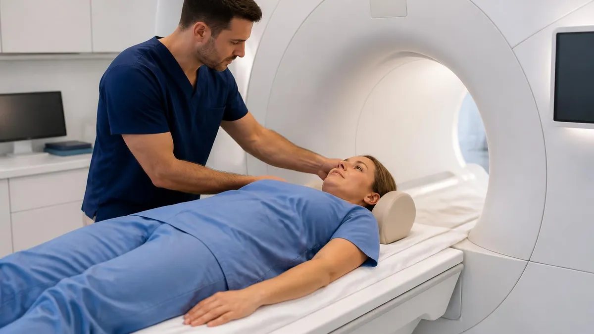
Is MRI Worth the Risk Compared to Other Imaging?
- +Uses no ionizing radiation, unlike CT scans and X-rays
- +Provides superior soft-tissue contrast for brain, spine, and joints
- +Most risks are fully preventable with proper screening
- +Gadolinium reactions are rare in patients with normal kidneys
- +MR Conditional implants allow many more patients to be scanned safely
- +Non-contrast techniques can answer many questions without injection
- −Powerful magnet creates dangerous projectile hazards
- −Certain implants remain absolute contraindications
- −Loud acoustic noise can damage unprotected hearing
- −Claustrophobia affects a meaningful share of patients
- −Gadolinium poses real danger in severe kidney disease
- −Long scan times require patients to hold still
MRI Safety Checklist Before Your Scan
- ✓Complete the full metal and implant screening questionnaire honestly.
- ✓Disclose any pacemaker, defibrillator, or neurostimulator devices.
- ✓Report aneurysm clips, cochlear implants, or surgical hardware.
- ✓Mention prior metalwork, welding, or eye injuries from fragments.
- ✓Remove all jewelry, watches, hairpins, and body piercings.
- ✓Leave phones, credit cards, and keys in a secure locker.
- ✓Tell staff about kidney disease before any gadolinium contrast.
- ✓Inform the technologist if you are or might be pregnant.
- ✓Wear the provided hearing protection throughout the scan.
- ✓Use the emergency squeeze ball if you feel distressed.
The magnet is ALWAYS on
Unlike an X-ray machine, an MRI magnet never powers down between patients. Its field pulls ferromagnetic objects with crushing force around the clock, even during a power outage. This single fact explains why metal screening, controlled zones, and constant vigilance matter every minute the suite is open.
Burns are, perhaps surprisingly, the most frequently reported adverse event during MRI examinations, accounting for the majority of incident reports filed with regulators. They arise from the radiofrequency energy that the scanner deposits into the body to generate signal. When that energy concentrates in a small area, tissue temperature can rise quickly. Most cases are entirely preventable, which makes understanding their causes one of the highest-value safety lessons for both patients and imaging staff alike.
The classic mechanism is a conductive loop. When a patient crosses their hands, lets calves touch, or rests a cable directly against bare skin, the loop concentrates induced current at the contact point. Technologists prevent this by placing foam padding between skin surfaces and between the body and the bore wall. Cables for monitoring equipment are routed carefully and never coiled, because a coiled wire acts like an antenna that focuses heat dangerously at a single spot.
Clothing and cosmetics contribute their own surprises. Athletic fabrics woven with silver or copper threads for odor control can heat sharply, and some compression garments contain metallic fibers. Transdermal medication patches with foil backing have caused skin burns, so they are removed before scanning when medically appropriate. Certain tattoos made with iron-oxide pigment may tingle or warm slightly, though serious tattoo burns remain rare and usually involve very large, dark, older designs.
Acoustic noise is a distinct hazard worth taking seriously. The gradient coils switch on and off thousands of times during a sequence, and the resulting vibrations produce knocking, buzzing, and beeping sounds that can exceed 110 decibels. Without protection, this level threatens permanent hearing damage, especially during long musculoskeletal or neurological studies. Earplugs, padded headphones, or both are mandatory, and many centers pipe in music to make the experience more tolerable for anxious patients.
Peripheral nerve stimulation is a less familiar effect of the rapidly switching gradient fields. As magnetic flux changes quickly, it can induce small currents in the body that cause tingling, twitching, or a tapping sensation, usually across the back, shoulders, or hips. While startling, these sensations are not harmful and stop the moment the sequence ends. Scanner software enforces regulatory limits on gradient slew rate specifically to keep stimulation below the threshold of pain.
Heating of the whole body is governed by the specific absorption rate, a measure of how much RF power tissue absorbs per kilogram. The scanner continuously calculates this value and pauses or slows sequences that would push a patient past safe limits. Vulnerable people, including infants, the elderly, those with fevers, and patients who cannot sweat normally, receive extra caution. These layered electronic safeguards work invisibly to keep core temperature elevation well within a comfortable and physiologically safe range.
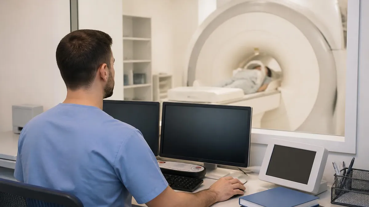
Family members, interpreters, and even hospital staff must complete the same metal screening as patients before crossing into Zone IV. Fatal projectile accidents have involved untrained personnel carrying oxygen tanks or tools. When in doubt, stop and ask the MRI technologist before stepping past the controlled doorway.
Pregnancy raises understandable concern, yet decades of use have produced no proven evidence that MRI harms a developing fetus. Because the technique uses magnetic fields and radio waves rather than ionizing radiation, it is often the preferred advanced imaging choice during pregnancy when ultrasound cannot answer the clinical question. Professional guidelines support scanning at any trimester when the information is needed for the mother's or baby's care, balancing theoretical caution against genuine diagnostic value.
Gadolinium contrast, however, follows stricter rules in pregnancy. The agent crosses the placenta, enters the fetal circulation, and is excreted into amniotic fluid where it may linger. Some studies have associated fetal gadolinium exposure with rare adverse outcomes, so contrast is generally avoided unless it is absolutely essential and no alternative exists. Most prenatal MRI examinations are therefore performed without any injection, relying on the excellent natural soft-tissue contrast the modality provides.
Breastfeeding mothers can usually continue nursing after receiving gadolinium. Only a tiny fraction of the dose enters breast milk, and even less is absorbed by the infant's gut. Major radiology organizations consider continued breastfeeding safe, although anxious parents may choose to pump and discard milk for twenty-four hours for reassurance. Clear counseling helps families make a comfortable, informed decision rather than interrupting feeding out of unnecessary worry.
Children present unique challenges that revolve around movement and comprehension rather than the magnet itself. Young patients struggle to hold still through long sequences, so motion artifact can ruin images. Sedation or general anesthesia is sometimes required, which introduces its own risks that must be weighed carefully. Child-life specialists, mock scanners, and video goggles increasingly reduce the need for sedation by helping children understand and tolerate the loud, enclosed environment more calmly.
Patients with claustrophobia or large body habitus need thoughtful accommodation. Roughly one in seven people experience significant anxiety inside the narrow bore, and a portion cannot complete the scan without help. Strategies include wide-bore and open scanners, mild oral sedation, prism glasses that let patients see outside, and simply entering feet first when anatomy allows. Compassionate communication from the technologist often matters as much as any technical solution for a successful examination.
Critically ill and monitored patients require specialized equipment because standard ventilators, infusion pumps, and monitors are ferromagnetic and unsafe. MR Conditional versions exist but must be positioned correctly and secured. Knowing which materials behave predictably in the field connects directly to questions about MRI safety materials, since the wrong alloy on a single connector can become a projectile. Coordinated planning between nursing, anesthesia, and MRI staff keeps these complex cases safe.
Putting safety knowledge into daily practice is what actually prevents the rare MRI accident, and the habits are surprisingly simple once they become routine. The foundation is rigorous, repeated screening. Every patient should complete the questionnaire, and the technologist should review it verbally before entry rather than trusting paperwork alone. Patients sometimes forget implants from decades earlier, so a calm conversation often surfaces details a hurried checkbox misses. Treat the second verbal screen as essential, never as redundant box-ticking.
Respect the zone system without exception. The four-zone layout exists to create progressively tighter control as you approach the magnet, and shortcuts defeat its purpose entirely. Doors should remain secured, ferromagnetic detection systems should stay active, and no one should prop open the entrance for convenience. If you are a patient or visitor, follow staff instructions precisely, and never assume an object is safe simply because it looks small or harmless to carry inside.
Prepare thoroughly for your appointment to reduce delays and risk. Wear simple clothing free of metal zippers, snaps, and underwire, or expect to change into a gown. Leave valuables, electronics, and jewelry at home or in a locker. Arrive with a list of every surgery, implant, and device you have, including cards from manufacturers, because that documentation lets staff verify MR Conditional status quickly instead of canceling your scan while they hunt for information.
Communicate openly about kidney health and pregnancy before any contrast is considered. If you have diabetes, hypertension, or prior kidney problems, mention them so staff can check your renal function. If pregnancy is possible, say so even if you are unsure. These disclosures never cause judgment; they let the team choose the safest possible protocol, which may simply mean skipping contrast or scheduling a quick blood test before the injection proceeds safely.
During the scan, use the tools provided for comfort and safety. Wear hearing protection the entire time, keep the emergency squeeze ball in hand, and hold as still as you comfortably can to avoid repeat sequences. If you feel burning warmth anywhere, squeeze the ball immediately rather than waiting, because early reporting stops a minor hot spot from becoming a true burn. Staff would always rather pause a scan than allow an injury to develop.
For students and professionals, reinforcement through practice questions cements these principles far better than passive reading. Working through registry-style scenarios about contraindications, contrast policies, and zone control turns memorized facts into fast clinical judgment. Reviewing related topics such as cervical spine protocols or abnormal-finding coding broadens the context in which safety decisions occur. The combination of solid theory, hands-on screening discipline, and regular self-testing is what produces a genuinely safe imaging environment for everyone involved.
Finally, remember that MRI's overall safety record is excellent precisely because the profession takes these hazards seriously. Millions of examinations occur each year with no harm, not because the risks are imaginary, but because layered safeguards neutralize them. Approach the scanner with informed respect rather than fear. When screening is honest, zones are honored, and communication stays open, magnetic resonance imaging delivers extraordinary diagnostic power with a remarkably small and well-managed margin of risk.
MRI Questions and Answers

MRI of Cervical Spine: Complete Guide to Imaging, Anatomy, Pathology, and Patient Preparation
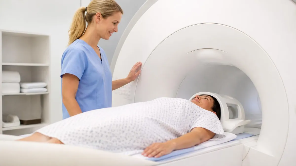
MRI Medical Abbreviation: What MRI Stands For and Why It Matters

Knee MRI Images: A Complete Guide to Reading, Understanding, and Interpreting Knee Scans
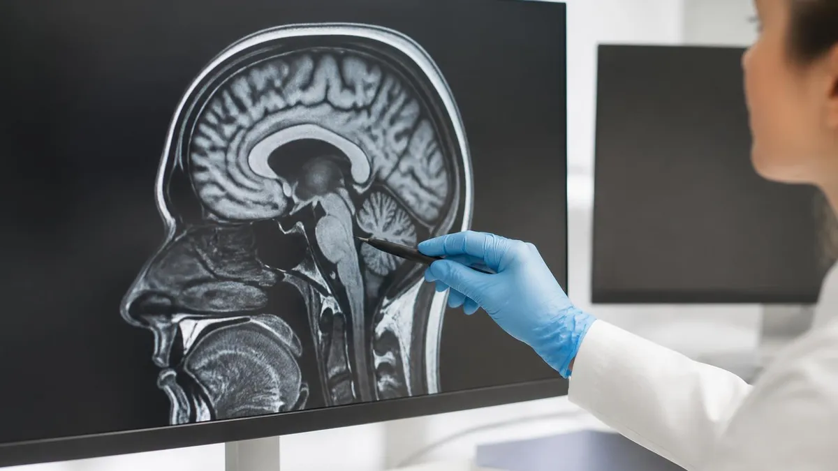
ICD 10 Codes for MRI: Complete Guide to Abnormal MRI Coding, Documentation, and Reimbursement
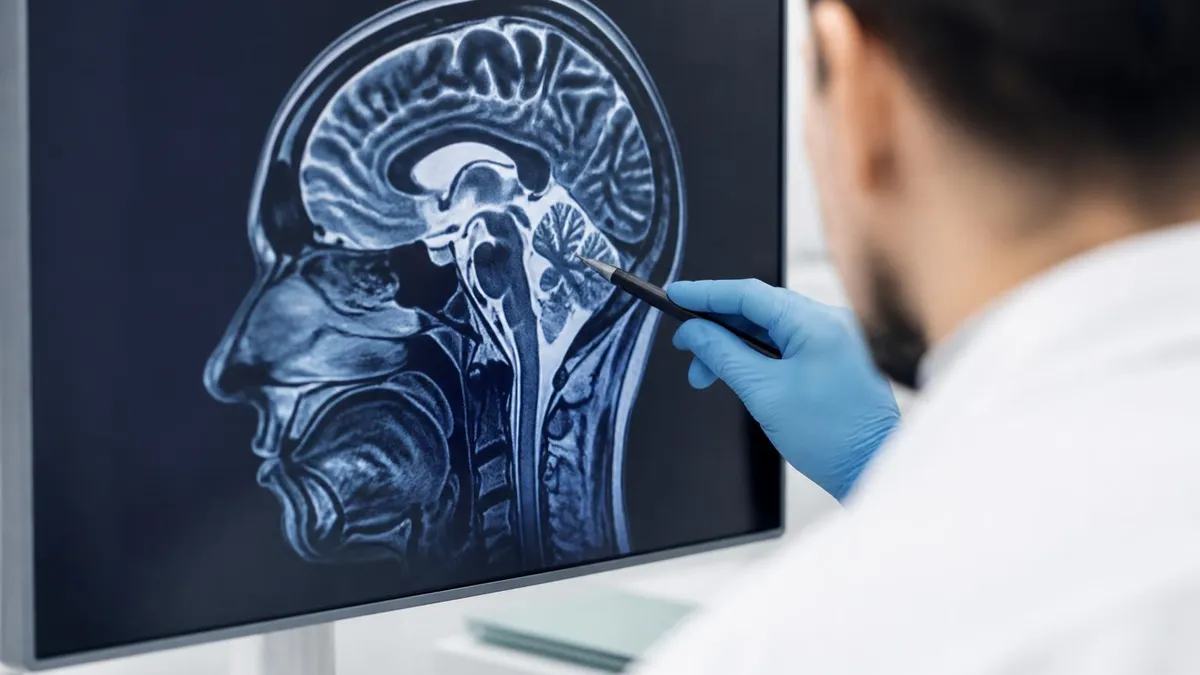
Is Nickel Titanium MRI Compatible? A Complete Guide to MRI Safety Materials
About the Author

Medical Laboratory Scientist & Clinical Certification Expert
Johns Hopkins UniversityDr. Sandra Kim holds a PhD in Clinical Laboratory Science from Johns Hopkins University and is certified as a Medical Technologist (MT) and Medical Laboratory Scientist (MLS) through ASCP. With 16 years of clinical laboratory experience spanning hematology, microbiology, and molecular diagnostics, she prepares candidates for ASCP board exams, MLT, MLS, and specialist certification tests.
Join the Discussion
Connect with other students preparing for this exam. Share tips, ask questions, and get advice from people who have been there.
View discussion (8 replies)