MRI for Cancer: How Breast, Prostate, and Whole-Body MRI Detect and Stage Tumors
MRI for breast cancer and other tumors: how contrast MRI detects, stages, and screens for cancer, who needs it, and how to prepare for the scan. 🔎
MRI for breast cancer has become one of the most powerful tools in modern oncology, offering soft-tissue detail that mammography and ultrasound simply cannot match. When a radiologist needs to know whether a suspicious lump is a benign cyst or an invasive tumor, magnetic resonance imaging often provides the answer without exposing the patient to ionizing radiation. For high-risk women and complex cases, MRI for breast cancer detects lesions earlier and maps their true extent before surgery, shaping the entire treatment plan from biopsy to reconstruction.
But breast imaging is only one chapter in the larger story of mri for cancer. Across oncology, MRI helps physicians find prostate tumors, characterize liver lesions, stage rectal and cervical cancers, and evaluate brain metastases. Each anatomical region uses tailored sequences, coils, and contrast protocols, yet the underlying physics remains the same: hydrogen nuclei aligning in a strong magnetic field, then releasing signal that computers reconstruct into exquisitely detailed cross-sections of living tissue.
What makes MRI especially valuable in cancer care is its sensitivity to changes invisible on other scans. Tumors tend to be more cellular and more vascular than surrounding tissue, and diffusion-weighted imaging captures that crowding while dynamic contrast-enhanced sequences track abnormal blood flow. A 6-millimeter cancer that a CT scan might miss can light up vividly on a properly timed MRI, giving the oncology team a head start measured in months rather than weeks.
For radiologic technologists, MRI safety officers, and radiology students, understanding oncologic MRI is essential. The exams are longer, the contrast timing is unforgiving, and the stakes are high because a missed enhancement can delay a cancer diagnosis. Knowing how to position a patient, when to inject gadolinium, and how to recognize a classic malignant washout curve separates a competent tech from an exceptional one in a busy cancer center.
This guide walks through the full landscape of cancer MRI in plain language. We cover how the scan works, which cancers it serves best, who qualifies for screening, how to prepare, and what the images actually reveal. Whether you are a patient facing your first breast MRI or a student preparing for a registry exam, you will leave with a clear, practical understanding of why this technology has reshaped diagnosis and surveillance across so many tumor types today.
We will also be honest about the limits. MRI is expensive, time-consuming, and prone to false positives that can trigger unnecessary biopsies and anxiety. It is not a replacement for mammography in average-risk women, and gadolinium contrast carries its own considerations for patients with kidney disease. By the end, you will understand both the remarkable strengths and the real-world trade-offs of using MRI to fight cancer effectively.
Cancer MRI by the Numbers
How Cancer MRI Works
A 1.5T or 3T magnet aligns hydrogen protons in the body. Radiofrequency pulses tip them out of alignment, and as they relax they emit signals that build the image with no ionizing radiation involved.
Gadolinium injected into a vein leaks into tumor vessels faster than normal tissue. Dynamic sequences capture this rapid uptake and washout, a hallmark pattern that flags many malignancies during the exam.
DWI measures how freely water moves between cells. Cancers are densely packed, restricting water motion and producing bright signal that distinguishes tumor from benign tissue without any contrast agent at all.
Combining T1, T2, contrast, and diffusion data lets radiologists score lesions for malignancy. This multiparametric approach drives PI-RADS for prostate and BI-RADS for breast cancer reporting.
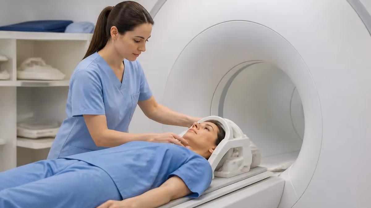
Breast MRI is the most refined oncologic MRI application, and for good reason. The exam uses a dedicated breast coil with the patient lying prone, breasts hanging into openings that reduce motion and improve signal. After a baseline scan, gadolinium is injected and a series of rapid images captures how contrast floods into and drains out of any suspicious area. Malignant tumors typically show fast uptake followed by quick washout, producing a tell-tale curve that radiologists recognize immediately.
Who actually needs breast MRI? The American Cancer Society recommends annual MRI alongside mammography for women with a lifetime breast cancer risk of roughly 20 to 25 percent or higher. This includes BRCA1 and BRCA2 mutation carriers, women with a strong family history, those who received chest radiation between ages 10 and 30, and certain genetic syndromes. For these patients, MRI catches cancers that mammography misses, especially in dense breast tissue where tumors hide behind overlapping glandular shadows.
Beyond screening, breast MRI plays a major staging role. Once cancer is confirmed by biopsy, MRI maps the full extent of disease, hunts for additional tumors in the same breast, and screens the opposite breast for hidden cancer. Surgeons rely on this map to decide between lumpectomy and mastectomy, and to plan margins precisely. Studies show MRI changes surgical management in a meaningful share of cases, though it can also prompt extra biopsies for findings that turn out benign.
The exam also monitors response to neoadjuvant chemotherapy. When a patient receives chemo before surgery to shrink a tumor, MRI tracks whether the cancer is actually melting away. A tumor that fails to respond signals the oncologist to switch drugs, while a complete radiographic response may allow a less aggressive operation. This dynamic feedback loop is one of MRI's quietly transformative contributions to personalized cancer treatment.
Timing matters enormously for premenopausal women. Hormonal fluctuations cause background breast tissue to enhance, mimicking or masking real lesions. For this reason, screening MRIs are ideally scheduled during days 7 through 14 of the menstrual cycle, when hormone-driven enhancement is lowest. Scheduling at the wrong point in the cycle can produce a cluttered, hard-to-read study that may require repeating, wasting time and contrast.
False positives remain the chief drawback of breast MRI. Because it is so sensitive, the scan flags many areas that look suspicious but prove benign on biopsy. This high sensitivity paired with moderate specificity means some women endure additional imaging, biopsies, and anxiety for findings that never threatened their health. Experienced breast radiologists and MRI-guided biopsy capabilities reduce this burden, which is why where you get scanned genuinely matters.
For technologists, breast MRI demands precision. The contrast bolus must be injected and flushed at the right rate, the timing of post-contrast sequences must be exact, and patient positioning must minimize the motion that ruins subtraction images. A single mistimed series can render an entire study non-diagnostic, forcing a costly repeat. Mastering these protocols is a core skill tested heavily on advanced MRI registry and certification exams.
Cancers MRI Detects Best
Breast MRI excels at finding invasive cancers in dense tissue and high-risk patients where mammography falls short. Its sensitivity for invasive disease exceeds ninety percent, making it the gold standard for screening BRCA carriers and staging newly diagnosed tumors before surgery.
It also evaluates breast implants for rupture and assesses how well a tumor responds to chemotherapy. The trade-off is specificity: many benign lesions enhance too, so MRI findings frequently require targeted biopsy to confirm whether an enhancing area is truly cancer or simply normal hormonal activity.
Is MRI Worth It for Cancer Detection?
- +No ionizing radiation, making repeat scans safer over time
- +Superior soft-tissue contrast versus CT and ultrasound
- +Detects small, early tumors hidden in dense tissue
- +Maps the true extent of disease for surgical planning
- +Tracks tumor response to chemotherapy in real time
- +Multiparametric scoring reduces unnecessary biopsies
- +Excellent for screening high-risk patients earlier
- −High cost compared with mammography or ultrasound
- −Long scan times of 30 to 60 minutes per study
- −Frequent false positives that trigger extra biopsies
- −Gadolinium contrast unsuitable for severe kidney disease
- −Claustrophobia and loud noise distress some patients
- −Not safe for many implanted metal devices
- −Limited availability and access in rural areas
How to Prepare for a Cancer MRI
- ✓Tell staff about any pacemaker, implant, or metal in your body.
- ✓Disclose kidney disease so gadolinium dosing can be adjusted.
- ✓Schedule breast MRI for days 7 to 14 of your cycle if premenopausal.
- ✓Remove all jewelry, watches, hairpins, and metal clothing.
- ✓Arrange a current eGFR blood test if contrast is planned.
- ✓Inform the team if you are pregnant or possibly pregnant.
- ✓Mention claustrophobia so sedation can be arranged in advance.
- ✓Wear comfortable clothing without zippers or metal snaps.
- ✓Avoid lotions or deodorants on the day of breast imaging.
- ✓Bring prior imaging and reports for comparison if available.
- ✓Ask whether you should fast for liver or abdominal scans.
- ✓Confirm insurance pre-authorization to avoid surprise bills.
Sensitivity is not the same as accuracy
Breast MRI catches over 90% of invasive cancers, but its high sensitivity means it also flags many benign findings. A positive MRI is a reason for targeted follow-up, not an automatic cancer diagnosis. Always confirm enhancing lesions with biopsy before treatment decisions.
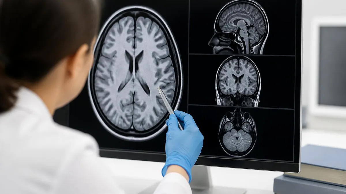
Reading a cancer MRI is a layered process that combines several imaging contrasts into a single judgment. Radiologists start with anatomy, using T1- and T2-weighted images to locate structures and spot obvious masses. T2 images make fluid bright, helping distinguish simple cysts from solid tumors. Then they layer in the functional sequences, diffusion-weighted imaging and dynamic contrast, which reveal the biological behavior of tissue rather than just its shape. A lesion that is bright on diffusion and enhances rapidly draws immediate concern.
The dynamic contrast curve is central to interpretation. After gadolinium injection, the radiologist watches how signal intensity changes over the following minutes. A curve that rises slowly and keeps rising suggests benign tissue. A curve that spikes quickly and then washes out, dropping in intensity, is the classic signature of an aggressive, vascular tumor. Plateau curves fall in between and require careful judgment, often combined with diffusion findings to reach a confident conclusion.
Standardized scoring systems bring consistency to this judgment. For breast imaging, BI-RADS categories range from 1, meaning negative, up to 5, meaning highly suspicious for malignancy, with category 6 reserved for biopsy-proven cancer. Prostate MRI uses PI-RADS, scoring lesions one through five based on combined T2, diffusion, and contrast features. These lexicons let radiologists communicate risk clearly to referring physicians and patients, and they drive whether a biopsy is recommended.
Diffusion-weighted imaging deserves special attention because it works without contrast. Cancer cells are packed tightly, restricting the random motion of water molecules. The apparent diffusion coefficient, or ADC, map quantifies this restriction. Low ADC values, appearing dark on the map and bright on the raw diffusion image, strongly suggest malignancy. For patients who cannot receive gadolinium, diffusion imaging provides a valuable alternative window into tumor biology and cellularity.
Lymph node assessment is another critical task. MRI evaluates the size, shape, and signal of regional nodes to estimate whether cancer has spread. Round, enlarged nodes with irregular borders raise suspicion, though imaging alone cannot replace pathology. In rectal and cervical cancer staging, accurate nodal assessment directly influences whether a patient receives radiation before surgery, underscoring how much treatment hinges on careful image reading.
Artifacts can sabotage interpretation if the technologist and radiologist are not vigilant. Patient motion blurs subtraction images, metal implants create signal voids, and poor fat suppression mimics enhancement. Recognizing these pitfalls prevents both false alarms and missed cancers. A skilled reader always asks whether a suspicious finding could be an artifact before recommending biopsy, and a skilled tech minimizes artifacts at the source through proper positioning and protocol selection.
Finally, comparison with prior studies transforms interpretation. A lesion that is stable over years is far less concerning than a new or growing one. This is why bringing previous imaging matters so much. Cancer MRI is rarely read in isolation; it lives within the patient's history, prior mammograms, ultrasounds, and pathology reports, all woven together to reach the most accurate possible assessment of risk.
Patients with severe kidney impairment face a small but serious risk of nephrogenic systemic fibrosis from older gadolinium agents. Always confirm a recent eGFR before contrast-enhanced cancer MRI, and discuss alternatives if kidney function is reduced. Newer macrocyclic agents have greatly lowered this risk.
Weighing the benefits and limits of cancer MRI helps patients and clinicians use it wisely. The greatest strength is unmatched soft-tissue detail delivered without radiation, which makes MRI ideal for young patients and for repeat scans during long treatment courses. Where mammography sees overlapping shadows and CT sees ambiguous densities, MRI distinguishes tumor from healthy tissue with remarkable clarity, often revealing the precise borders that surgeons and radiation oncologists need to plan curative therapy.
Early detection in high-risk populations is another decisive advantage. For BRCA carriers and women with extremely dense breasts, annual MRI finds cancers years earlier than they would otherwise surface, when tumors are smaller and more curable. The same early-detection logic applies to prostate MRI, which now identifies aggressive cancers while sparing men with indolent disease from unnecessary treatment. Catching cancer early genuinely saves lives and reduces the intensity of treatment required.
Yet the limits are real and must be respected. Cost stands out: a single cancer MRI can run several thousand dollars, far more than mammography or ultrasound, and not every insurer covers screening MRI for borderline-risk patients. Access compounds the problem, since rural and underserved areas may lack the magnets, coils, and subspecialty radiologists needed for high-quality oncologic imaging, creating real disparities in who benefits from this technology.
False positives carry both financial and emotional costs. Because MRI is so sensitive, it flags many benign findings that look suspicious, leading to additional imaging and biopsies. For some patients this cascade brings weeks of anxiety and minor procedures that ultimately reveal nothing dangerous. Centers with high-volume breast and prostate MRI programs and MRI-guided biopsy capability minimize this harm, which is why patients benefit from seeking experienced facilities when possible.
Safety contraindications also restrict who can be scanned. Many older pacemakers, certain aneurysm clips, cochlear implants, and metallic foreign bodies make MRI dangerous or impossible. Even MRI-conditional devices require specific protocols and field-strength limits. Technologists screen every patient meticulously, because a missed implant in a powerful magnetic field can cause serious injury. This rigorous safety culture is fundamental to every oncologic MRI program and is heavily emphasized in registry exams.
The patient experience itself can be a barrier. Scans last 30 to 60 minutes inside a narrow, loud bore, and claustrophobic patients sometimes cannot complete the exam without sedation. Wider bore magnets and open MRI systems help, though open units may sacrifice some image quality. Clear communication, ear protection, and occasional mild sedation usually make even anxious patients comfortable enough to finish a diagnostic study successfully.
For those preparing professionally, mastering these trade-offs is part of the job. Resources like a thorough mri for cancer question bank reinforce how to balance sensitivity against specificity, when contrast is appropriate, and how to screen for safety. Understanding not just how MRI works but when and why to use it is what turns technical knowledge into genuine clinical value for cancer patients.
Practical preparation makes a real difference in how smoothly a cancer MRI goes, both for patients and for the technologists running the scanner. The single most important step is honest, thorough safety screening. Patients should disclose every implant, surgery, and piece of retained metal, even items they assume are irrelevant. A forgotten metallic fragment from an old injury or a non-conditional cardiac device can turn a routine scan into an emergency, so erring on the side of over-disclosure protects everyone involved.
For breast MRI specifically, timing within the menstrual cycle is a detail patients can control. Scheduling during the second week of the cycle minimizes hormonal background enhancement and produces cleaner, more interpretable images. Patients who track their cycles should mention the expected dates when booking, and premenopausal women on hormone therapy should ask whether a brief pause is advisable. These small scheduling choices reduce the chance of an inconclusive study that must be repeated.
Hydration and kidney testing matter whenever contrast is involved. A recent eGFR confirms the kidneys can clear gadolinium safely, and staying well hydrated before and after the exam helps the body flush the agent. Patients with diabetes or known kidney disease should raise this early, because the team may need to choose a specific contrast agent or, occasionally, plan a non-contrast diffusion-based protocol instead to avoid any renal risk entirely.
Comfort planning prevents incomplete scans. Anyone who suspects they may struggle with the enclosed bore should request accommodations in advance rather than discovering panic mid-exam. Options include scheduling on a wide-bore magnet, taking a prescribed mild sedative, bringing music, or arranging a friend to remain nearby. Knowing the scan will be loud and long, and that the technologist is watching and listening the entire time, reassures many anxious patients enormously.
For students and new technologists, the practical lesson is that protocol discipline drives diagnostic quality. Inject contrast at the prescribed rate, run post-contrast sequences on the exact timing the radiologist expects, and double-check fat suppression before letting the patient off the table. A few seconds of verification beats a non-diagnostic study and a recalled patient. Build the habit of reviewing each series as it completes so problems are caught while they can still be fixed.
Studying for certification exams rewards the same systematic mindset. Focus on safety zones, contrast contraindications, sequence purposes, and the scoring systems that translate images into clinical decisions. Practice questions that simulate real oncologic scenarios cement this knowledge far better than passive reading. Pair conceptual study with hands-on protocol practice, and the abstract physics of relaxation times starts to feel like the practical tool it truly is in cancer care.
Above all, remember that cancer MRI is a team effort. The radiologist interprets, but the technologist captures the data, the nurse manages contrast and comfort, and the scheduler sets up the timing. Each role shapes whether a patient receives an accurate, life-changing answer about their disease. Approaching the work with that shared responsibility in mind produces better images, safer scans, and ultimately better outcomes for the people who need them most.
MRI Questions and Answers
MRI Practice Test
MRI of Cervical Spine: Complete Guide to Imaging, Anatomy, Pathology, and Patient Preparation

Knee MRI Images: A Complete Guide to Reading, Understanding, and Interpreting Knee Scans
White Spots on Brain MRI: Causes, What They Mean, and When to Worry
Dental Implants and MRI: What Patients and Techs Need to Know About Safety, Artifacts, and Imaging
About the Author

Medical Laboratory Scientist & Clinical Certification Expert
Johns Hopkins UniversityDr. Sandra Kim holds a PhD in Clinical Laboratory Science from Johns Hopkins University and is certified as a Medical Technologist (MT) and Medical Laboratory Scientist (MLS) through ASCP. With 16 years of clinical laboratory experience spanning hematology, microbiology, and molecular diagnostics, she prepares candidates for ASCP board exams, MLT, MLS, and specialist certification tests.
Join the Discussion
Connect with other students preparing for this exam. Share tips, ask questions, and get advice from people who have been there.
View discussion (6 replies)