Is MRI Contrast Safe? Gadolinium Risks, Side Effects, and What Patients Need to Know
Is MRI contrast safe? Learn about gadolinium risks, side effects, kidney concerns, and what patients should know before a contrast-enhanced MRI scan. ✏️
Is MRI contrast safe? This is one of the most common questions patients ask before undergoing a contrast-enhanced magnetic resonance imaging study, and the honest answer is reassuring but nuanced. For the overwhelming majority of patients, gadolinium-based contrast agents administered during MRI scans are extremely safe, with serious adverse reactions occurring in fewer than 0.04% of injections according to American College of Radiology data. However, safety depends on patient-specific factors including kidney function, allergy history, pregnancy status, and which generation of contrast agent is used.
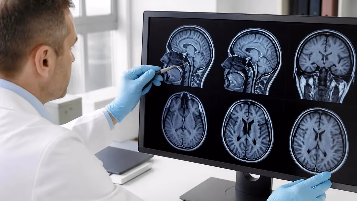
Gadolinium-based contrast agents, commonly abbreviated as GBCAs, have been used clinically since 1988 and have been administered hundreds of millions of times worldwide. They work by altering the magnetic properties of nearby water molecules, which enhances tissue contrast on MRI images and allows radiologists to detect tumors, inflammation, infections, vascular abnormalities, and many other conditions that might otherwise remain invisible. Without contrast, certain diagnoses would be impossible or significantly delayed.
Concerns about MRI contrast safety entered public awareness primarily through two issues: nephrogenic systemic fibrosis (NSF) in patients with severe kidney disease, and gadolinium retention in the brain and other tissues. Both deserve serious attention, but both also have nuance. NSF has become extremely rare with modern macrocyclic agents and proper screening protocols, while the clinical significance of brain gadolinium retention in patients with normal kidney function remains scientifically unresolved.
The FDA, the European Medicines Agency, the American College of Radiology, and the International Society for Magnetic Resonance in Medicine have all reviewed the evidence extensively. Their consensus is that GBCAs remain safe and clinically essential when used appropriately, with the diagnostic benefits substantially outweighing the risks for properly screened patients. This article walks you through what those screening protocols look like, what side effects to expect, and how to advocate for yourself when contrast is recommended.
Patient education matters here because misinformation about gadolinium has caused some people to refuse contrast even when they urgently need it, delaying diagnoses of cancer, multiple sclerosis, stroke, and other time-sensitive conditions. The goal of this guide is balance: presenting the genuine risks honestly while contextualizing them against the very real diagnostic value contrast provides. Understanding the science empowers better conversations with your radiologist and ordering physician.
We will cover the different classes of gadolinium agents, who should avoid contrast entirely, what to expect on injection day, common and rare side effects, the difference between linear and macrocyclic agents, gadolinium retention research, alternatives when contrast is contraindicated, and frequently asked questions. By the end, you should feel equipped to make an informed decision about your scan and to recognize the signs of an adverse reaction if one occurs.
Whether you are preparing for your first contrast MRI or your fiftieth, the same principles apply: disclose your full medical history, hydrate well before and after, and follow up promptly if anything unusual happens within 48 hours. Safety in medicine is rarely absolute, but with modern protocols, gadolinium contrast comes remarkably close.
MRI Contrast Safety by the Numbers
How Gadolinium Contrast Works
Gadolinium is a rare-earth metal with seven unpaired electrons, giving it strong paramagnetic properties. When injected, it shortens the T1 relaxation time of nearby water protons, making tissues appear brighter on T1-weighted images.
Free gadolinium is toxic, so it is bound to a chelating ligand that holds the metal tightly. The chelated complex stays intact in the body and is excreted unchanged through the kidneys within roughly 24 hours.
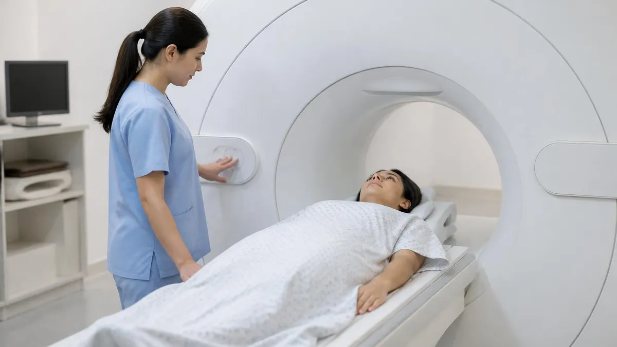
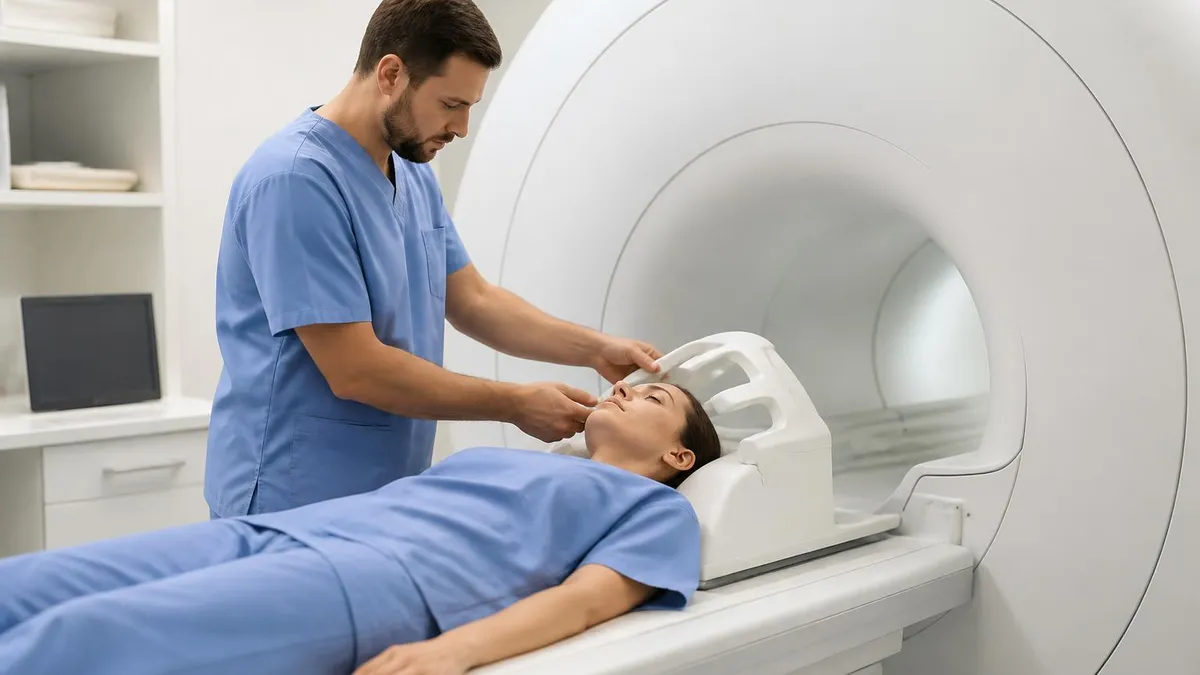
A technologist places an IV line, usually in the arm, and injects the agent at 1-2 mL per second. The dose is weight-based, typically 0.1 mmol per kilogram, followed by a saline flush to clear the line.
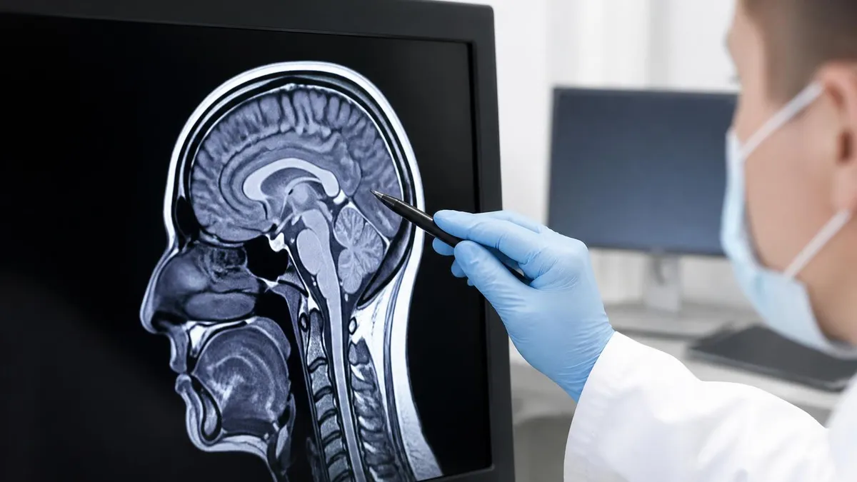
After injection, contrast travels through the bloodstream and distributes into the extracellular space of well-perfused tissues. Areas with increased blood flow or disrupted blood-brain barriers light up dramatically, revealing tumors or inflammation.
Healthy kidneys filter the chelated complex efficiently, with about 98% excreted in urine within 24 hours. Patients with reduced kidney function clear it more slowly, which historically raised NSF concerns with older linear agents.
Understanding the side effects and risk categories of MRI contrast helps patients separate genuine concerns from internet myths. The vast majority of patients experience nothing more than a brief sensation of coolness at the injection site, a metallic taste lasting a few seconds, or mild lightheadedness. These are not adverse reactions in the medical sense; they are normal physiological responses to having a small volume of cold fluid injected into a vein. They resolve on their own without intervention.
Mild adverse reactions occur in roughly 0.07% to 2.4% of injections depending on the specific agent. These include nausea, mild headache, dizziness, warmth or tingling sensations, and small hives at the injection site. Most resolve within minutes without treatment. Moderate reactions, such as significant hives, mild bronchospasm, or vomiting, occur in approximately 0.004% to 0.7% of cases and may require antihistamines or short observation, but rarely escalate.
Severe anaphylactoid reactions are genuinely rare with gadolinium, occurring in roughly 0.001% to 0.01% of injections, which is far lower than the rate seen with iodinated CT contrast. When they do happen, symptoms include difficulty breathing, severe hypotension, throat swelling, and cardiovascular collapse. Every MRI facility maintains emergency equipment and trained staff specifically because, while rare, these reactions can be life-threatening and require immediate epinephrine and resuscitation.
The most discussed serious risk historically is nephrogenic systemic fibrosis, a debilitating condition causing skin thickening, joint contractures, and organ fibrosis that can be fatal. NSF is essentially exclusive to patients with severe kidney impairment, specifically those with an estimated glomerular filtration rate below 30 mL/min/1.73m² or on dialysis. With modern macrocyclic agents and proper screening, NSF has become exceedingly rare, with very few cases reported in the past decade worldwide.
Extravasation, where contrast leaks outside the vein during injection, occurs in roughly 0.05% to 0.1% of administrations. The volume is small and gadolinium chelates are minimally toxic to soft tissue, so most extravasations resolve with elevation and warm or cold compresses. Compartment syndrome from MRI contrast extravasation is exceptionally rare. The team will document the volume extravasated and monitor the site, with surgical consultation only if symptoms suggest pressure-related injury.
For a complete picture of how contrast enhances diagnostic accuracy, review what MRI can detect and its diagnostic capabilities across organ systems. Contrast often makes the difference between confidently diagnosing a small enhancing lesion and missing it entirely, particularly in oncology, neurology, and vascular imaging.
Finally, it is worth noting that pre-existing asthma, atopic conditions, or a prior contrast reaction modestly increase the risk of an allergic-type reaction. Patients with these histories may receive premedication with corticosteroids and antihistamines starting 12-13 hours before the scan, following standard ACR protocols. This dramatically reduces but does not eliminate reaction risk, so vigilance during and after injection remains essential.
MRI Practice Test Questions
Prepare for the MRI - Magnetic Resonance Imaging exam with our free practice test modules. Each quiz covers key topics to help you pass on your first try.
MRI Knowledge
MRI Exam Questions covering Knowledge. Master MRI Test concepts for certification prep.
MRI Physics
Free MRI Practice Test featuring Physics. Improve your MRI Exam score with mock test prep.
MRI Anatomy and Pathology
MRI Test Prep for MRI Anatomy and Pathology. Practice MRI Quiz questions and boost your score.
MRI Anatomy and Positioning
MRI Questions and Answers on MRI Anatomy and Positioning. Free MRI practice for exam readiness.
MRI Contrast Agents
Free MRI Quiz on MRI Contrast Agents. MRI Exam prep questions with detailed explanations.
MRI Patient Care and Positioning
MRI Practice Questions for MRI Patient Care and Positioning. Build confidence for your MRI certification exam.
Linear vs Macrocyclic Gadolinium Agents
Linear gadolinium agents have an open-chain ligand structure that wraps around the gadolinium ion but does not fully encapsulate it. This makes them somewhat less stable, meaning a small fraction of gadolinium can dissociate from the chelate, particularly in patients with prolonged retention. Examples include gadodiamide (Omniscan), gadopentetate dimeglumine (Magnevist), and gadoversetamide (OptiMARK).
Because of stability concerns, the European Medicines Agency restricted several linear agents in 2017, while the FDA added warnings about gadolinium retention to all GBCA labels. Linear agents remain available in the United States for specific clinical applications like liver imaging, but their use has declined significantly. Patients with kidney disease should generally avoid older linear agents whenever a macrocyclic alternative will achieve the diagnostic goal.

Benefits and Drawbacks of Contrast-Enhanced MRI
- +Dramatically improves detection of tumors, infections, and inflammatory lesions
- +Essential for evaluating vascular structures via MR angiography
- +Distinguishes scar tissue from recurrent tumor in post-surgical follow-up
- +Highlights blood-brain barrier breakdown in MS and stroke
- +Allows characterization of liver and kidney masses
- +Improves staging accuracy for cancers across many organ systems
- +Generally lower allergic reaction rate than iodinated CT contrast
- −Requires IV access, which some patients find uncomfortable
- −Small risk of allergic-type reactions in susceptible individuals
- −Contraindicated or restricted in severe kidney disease
- −Trace gadolinium retention occurs in brain and bone tissues
- −Adds time and cost compared to non-contrast studies
- −Not always necessary depending on clinical question
- −Limited use during pregnancy unless benefits clearly outweigh risks
Pre-Scan Safety Checklist for Contrast MRI
- ✓Disclose all known allergies, especially to previous contrast agents or shellfish
- ✓Report any history of asthma, eczema, or other atopic conditions
- ✓Provide a current list of all medications including supplements
- ✓Inform staff about kidney disease, diabetes, or hypertension
- ✓Request a recent eGFR or serum creatinine lab if over age 60
- ✓Tell the technologist if you are or might be pregnant
- ✓Mention if you are breastfeeding so you can plan around feeding times
- ✓Stay well hydrated for 24 hours before and after the injection
- ✓Wear comfortable clothing without metal zippers, buttons, or jewelry
- ✓Arrange a ride home if you received sedation or anti-anxiety medication
- ✓Bring your insurance card and physician order to expedite check-in
- ✓Ask which specific gadolinium agent will be used and why
Hydration matters more than you think
Drinking plenty of water for 24 hours before and after a contrast MRI helps your kidneys clear gadolinium efficiently. Adequate hydration shortens excretion time, reduces the duration of any tissue exposure, and lowers the risk of injection-site discomfort. Aim for clear or pale-yellow urine on scan day.
Gadolinium retention research is one of the most actively debated topics in radiology, and patients deserve a clear-eyed summary rather than reassurance or alarm. Beginning around 2014, multiple studies demonstrated that small amounts of gadolinium remain detectable in the dentate nucleus and globus pallidus of the brain after multiple contrast-enhanced MRI scans, even in patients with completely normal kidney function. This was a significant scientific finding because it contradicted the long-standing assumption that all gadolinium was excreted within 24 hours.
The retention was identified on T1-weighted MRI images as increased signal intensity in specific deep brain structures, and was later confirmed at autopsy and in tissue analysis using mass spectrometry. Gadolinium has been found not only in brain tissue but also in bone, skin, liver, and kidneys at trace concentrations. The amounts are measured in micrograms or nanograms per gram of tissue, vastly smaller than the original injected dose, but they are real and they persist for years.
The critical question is whether this retention causes harm. To date, no peer-reviewed study has definitively linked brain gadolinium retention to neurological symptoms, cognitive decline, or any specific disease in patients with normal renal function. Large registries and longitudinal studies continue to monitor this question, but the current scientific consensus remains that retention has no proven clinical consequence. Some patients have reported a constellation of symptoms they call gadolinium deposition disease, but this entity is not yet medically recognized as causally linked to contrast.
The FDA responded in 2018 by requiring a class-wide warning on all GBCA labels regarding gadolinium retention. The agency also recommended that providers consider the necessity of contrast for each scan, particularly when multiple studies are anticipated, and minimize the number of contrast doses when clinically feasible. This is sometimes called the ALARA principle, meaning as low as reasonably achievable, borrowed from radiation safety practice.
For patients with conditions requiring repeated contrast-enhanced MRI, such as multiple sclerosis monitoring or oncology surveillance, the conversation with your physician should include whether contrast is truly necessary for each follow-up scan or whether some studies could be performed without contrast. Many MS protocols have moved toward less frequent contrast use, reserving it for situations where active inflammation is suspected rather than as routine surveillance.
Patient choice matters here too. Some patients prefer to minimize cumulative gadolinium exposure given the unresolved questions, while others prioritize maximum diagnostic certainty. Both positions are defensible, and a good radiologist will discuss the trade-offs openly. If you are scheduled for repeat contrast studies, ask whether a macrocyclic agent is being used and whether some scans could be performed without contrast.
To understand more about what physicians actually look for on these scans, see common MRI findings across brain, spine and joints. Knowing what is being evaluated helps you have a more informed conversation about whether contrast adds enough value for your specific clinical situation.
Within minutes to hours after a contrast MRI, seek emergency care for difficulty breathing, throat swelling, widespread hives, dizziness, or chest pain. Within days to weeks, contact your physician about new skin thickening, joint stiffness, severe muscle weakness, or persistent severe headache. Save your scan paperwork in case detailed agent information is needed.
Knowing when to skip contrast is just as important as understanding when to use it. There are well-established clinical situations where the risks outweigh the benefits and the radiologist should either use an alternative imaging strategy, perform the MRI without contrast, or choose a completely different modality. The first and most important category is severe kidney impairment, defined as an eGFR below 30 mL/min/1.73m² or active dialysis dependence. In these patients, contrast is avoided unless absolutely essential and no alternative exists.
Pregnancy is another area requiring caution. Gadolinium crosses the placenta and enters fetal circulation, where it is then excreted into amniotic fluid and potentially recirculated through fetal swallowing. Animal studies have shown developmental effects at very high doses, and a large 2016 Canadian study suggested a modest increase in adverse outcomes including stillbirth. Most guidelines recommend avoiding contrast during pregnancy unless the diagnostic information would substantially change maternal management and cannot be obtained any other way.
Patients with a documented prior severe reaction to gadolinium are generally advised to avoid future contrast unless absolutely necessary and the reaction can be characterized to determine whether a different agent class would be tolerated. Some patients with prior moderate reactions can safely receive contrast again with premedication and a switch to a structurally different agent, while those with anaphylaxis are typically labeled allergic and contrast is avoided thereafter.
Children, particularly infants under one year, receive contrast only when truly indicated because their kidneys are still developing and their lifetime cumulative exposure is starting from zero. Pediatric radiologists are typically conservative about contrast use, choosing macrocyclic agents at the lowest effective dose and performing non-contrast protocols when adequate. Discuss alternatives with your pediatric radiologist if your child is scheduled for a contrast study.
For breastfeeding mothers, the news is reassuring. Less than 0.04% of an injected gadolinium dose appears in breast milk, and less than 1% of that tiny amount is absorbed from the infant's gut. The American College of Radiology states that breastfeeding can continue uninterrupted after contrast. Some mothers choose to pump and discard for 24 hours out of personal preference, but this is not medically required.
Finally, there are diagnostic situations where non-contrast MRI is genuinely adequate. Many spine, joint, and orthopedic studies do not require contrast at all. Brain MRI for routine headache evaluation often does not need contrast. For comparison images, see what a normal MRI looks like for brain, spine and knee to better understand what non-contrast studies can show.
If your physician orders a contrast MRI, you can always ask whether non-contrast images would be sufficient. The answer might genuinely be yes, particularly for follow-up imaging or screening exams. Shared decision-making between you and your physician produces better care than blind acceptance of any default protocol.
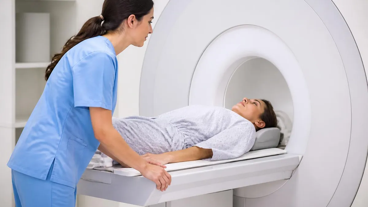
Practical preparation for a contrast-enhanced MRI goes beyond simply showing up at the imaging center. Start hydrating well 24 hours before your scan, aiming for clear or pale-yellow urine. Hydration improves IV placement, accelerates contrast excretion, and reduces the small risk of contrast-related kidney stress in patients with borderline renal function. Avoid heavy meals two hours before the scan, but most facilities do not require complete fasting unless sedation is planned.
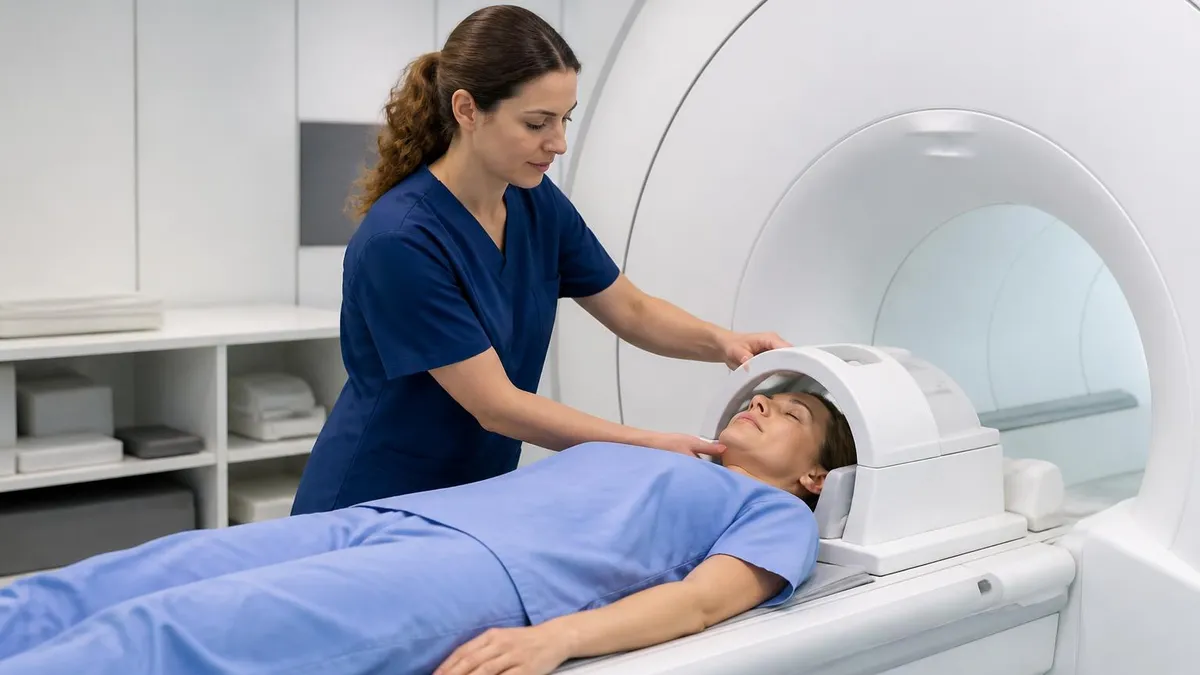
On arrival, expect a thorough screening interview covering allergies, prior contrast reactions, kidney function, pregnancy status, and current medications. Be specific. Vague answers like I think I had a reaction once do not help the radiologist judge risk. If you have lab results showing recent kidney function, bring them. Many centers will check creatinine on arrival for patients over 60 or with diabetes, hypertension, or known kidney disease, since these are the highest-risk groups for contrast complications.
Once on the scanner table, the technologist will place an IV, usually in the antecubital vein. The injection itself happens partway through the exam, often after non-contrast sequences are completed so the radiologist can compare pre and post-contrast images. You may feel a brief coolness traveling up your arm, a metallic taste, or a sensation of warmth. These are normal and last only seconds. Tell staff immediately if you feel pain at the injection site, since this can indicate extravasation.
After the scan, you will be observed for a brief period, typically 15 to 30 minutes, to watch for delayed allergic reactions. Most patients are cleared to leave shortly afterward and can resume normal activities including driving. Continue hydrating well for 24 hours to help flush the contrast from your system. There are no dietary restrictions or activity limitations after a routine contrast MRI in adults with normal kidney function.
Document the specific agent used. Ask the technologist to record the brand name, generic name, dose, and lot number in your medical record. This information becomes invaluable if you ever have a delayed reaction or need to identify which agents you have tolerated for future scans. If you have a smartphone, photographing the empty contrast vial is a reasonable backup record-keeping habit.
Watch for warning signs in the 48 hours following the scan. Most reactions occur within 15 minutes, but delayed reactions including skin rashes, hives, and rarely more serious symptoms can appear hours to days later. Call your physician if you develop a rash, unusual swelling, breathing difficulty, severe headache, or any symptom that feels out of the ordinary. Keep your imaging facility's number accessible.
Finally, advocate for yourself. If anything about the contrast injection process feels rushed or if your concerns are dismissed, speak up. Modern radiology embraces patient-centered care, and reasonable questions about agent choice, dose, alternatives, and cumulative exposure deserve thoughtful answers. The best outcomes come from informed patients working with attentive radiologists, with safety treated as a shared responsibility rather than something that just happens behind the scenes.
MRI Questions and Answers
About the Author
Medical Laboratory Scientist & Clinical Certification Expert
Johns Hopkins UniversityDr. Sandra Kim holds a PhD in Clinical Laboratory Science from Johns Hopkins University and is certified as a Medical Technologist (MT) and Medical Laboratory Scientist (MLS) through ASCP. With 16 years of clinical laboratory experience spanning hematology, microbiology, and molecular diagnostics, she prepares candidates for ASCP board exams, MLT, MLS, and specialist certification tests.
Join the Discussion
Connect with other students preparing for this exam. Share tips, ask questions, and get advice from people who have been there.
View discussion (6 replies)