Metal During MRI: What's Safe, What's Dangerous, and Why It Matters
Metal during MRI can cause burns, projectiles, and image distortion. Learn which metals are safe, screening protocols, and what to remove before scanning.
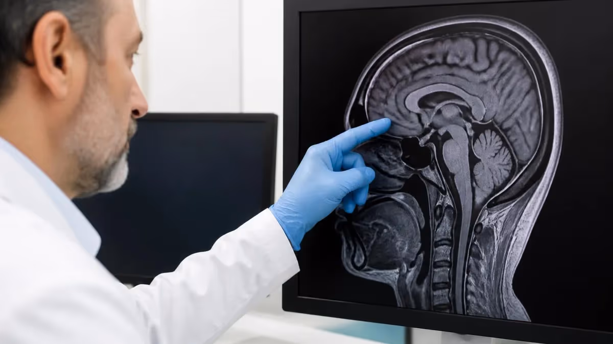
Understanding metal during MRI is one of the most critical safety topics in modern radiology, and it remains the single largest cause of preventable patient injuries inside the scanner suite. Magnetic resonance imaging uses a static magnetic field that is typically 1.5 to 3 Tesla in clinical settings, which is roughly 30,000 to 60,000 times stronger than Earth's natural magnetic field. When ferromagnetic objects enter that field, they can be pulled toward the bore with tremendous force, heated by radiofrequency pulses, or distort the resulting images so badly that the entire scan becomes useless.
The risks associated with metal during MRI fall into three broad categories that every technologist, nurse, and patient should recognize. The first is the projectile effect, where loose metal items become high-velocity missiles attracted to the magnet. The second is heating, where conductive materials absorb radiofrequency energy and can cause first, second, or even third-degree burns on the skin. The third is image artifact, where metal disrupts the local magnetic field and creates dark voids or distorted geometry around implants, sometimes obscuring critical anatomy.
Patients often arrive for an MRI worried about jewelry, dental work, or surgical implants from procedures performed years or even decades ago. Some of these concerns are well-founded, while others reflect outdated information from an era when most implants were not designed with MRI compatibility in mind. Modern orthopedic hardware, vascular stents, and even many pacemakers are now classified as MR Conditional, meaning they can be safely scanned under specific parameters that the manufacturer has tested and documented.
The challenge for healthcare providers lies in identifying which implants are safe, which require special protocols, and which absolutely contraindicate scanning. This article walks through the full spectrum of metal safety in MRI, from the screening process used to catch hidden objects before they enter the room, to the specific manufacturer guidelines that govern how MR Conditional devices must be scanned. We will also explore the science behind why certain alloys behave the way they do in strong magnetic fields.
Whether you are a patient preparing for your first scan, a technologist refining your screening protocol, or a referring clinician trying to determine if your patient can safely undergo imaging, this guide covers the essential information. We will address common items like wedding rings, body piercings, tattoos containing iron oxide pigments, transdermal medication patches with metallic backings, and clothing with metallic threads or hidden snaps that can heat dangerously during a scan.
The good news is that with proper screening, communication, and adherence to established safety protocols, MRI remains one of the safest imaging modalities available. Serious injuries are rare and almost always preventable. The bad news is that complacency kills, and even small lapses in metal screening have resulted in catastrophic incidents that destroyed equipment, injured staff, and in tragic cases caused patient deaths. Respecting the magnet starts with understanding metal.
Metal During MRI by the Numbers

Types of Metal and MRI Safety Classifications
Iron, nickel, cobalt, and most carbon steels are strongly attracted to magnetic fields. These materials pose the greatest projectile and heating risks and are typically classified as MR Unsafe when in their pure or unmodified forms.
Aluminum, platinum, and titanium have weak interactions with magnetic fields. Titanium in particular is widely used in implants because it produces minimal artifact, does not become a projectile, and resists heating under typical clinical RF exposure.
Gold, silver, copper, and bismuth are weakly repelled by magnetic fields and generally pose little projectile risk. However, conductive loops formed by these metals can still heat significantly during longer pulse sequences.
Modern implants tested and labeled as MR Conditional can be safely scanned under specific manufacturer-defined parameters, including maximum field strength, gradient slew rates, and specific absorption rate limits. Always verify the device documentation.
Aneurysm clips made of ferromagnetic alloys, certain older pacemakers, cochlear implants without MRI labeling, and any retained metal fragments from trauma or occupational exposure are absolute contraindications to scanning.
Why is metal during MRI so dangerous, and what exactly happens inside the bore when a ferromagnetic object enters the field? To answer that, we need to look at the physics of magnetism and how the MRI scanner generates its imaging signal. The static magnetic field, often abbreviated as B0, is always on, even when no patient is being scanned. This persistent field is what makes the room hazardous around the clock and why locked doors and zone-based access controls are mandatory at every accredited imaging facility.
The projectile effect occurs because ferromagnetic objects are attracted toward the center of the magnet with a force that increases exponentially as the object gets closer to the bore. A small steel paperclip might be barely noticeable at ten feet but becomes nearly impossible to hold onto at two feet. Larger objects like oxygen tanks, IV poles, wheelchairs, and floor buffers have been pulled into scanners with catastrophic results, including the well-documented death of a six-year-old patient struck by a ferromagnetic oxygen tank in 2001.
Heating presents a more insidious risk because it can occur with metals that are not strongly ferromagnetic. The radiofrequency pulses used to generate images deposit energy into the patient's tissues, and conductive loops or wires can act as antennas that concentrate this energy at specific points. ECG leads, tattoos with metallic pigments, body piercings forming closed loops, and even certain clothing fibers have all caused documented thermal injuries. Burns from MRI are often delayed in presentation and can occur even when the patient feels no warning sensation.
Image artifact is the third major concern and the one most likely to ruin a study without causing physical harm. Metal disrupts the homogeneity of the magnetic field, which the scanner relies on to spatially encode signal. Around an implant, this disruption creates signal voids, geometric distortion, and bright pile-up artifacts that can obscure clinically important anatomy. Specialized sequences like MARS, SEMAC, and VAT have been developed to reduce these artifacts, but they cannot eliminate them entirely.
Beyond these three primary risks, metal in or near the body can also interact with the scanner's gradient coils, which produce rapidly switching magnetic fields used for spatial encoding. These gradient pulses can induce currents in conductive implants, potentially causing peripheral nerve stimulation or interfering with active devices like neurostimulators. Cardiac implantable electronic devices have historically been the most challenging category, though the development of MR Conditional pacemakers has dramatically expanded access to MRI for these patients.
Patients sometimes wonder if their existing imaging history is relevant, and reviewing the history of MRI shows how safety standards have evolved alongside the technology. Early scanners operating at lower field strengths posed different risks than today's 3T systems, and many older implants that were tested decades ago may need re-evaluation under current safety frameworks. This is why a comprehensive screening process must consider not just what the patient has in their body, but also when and where it was placed.
The bottom line is that metal during MRI is never a casual concern. Every object, no matter how small or innocuous it appears, must be evaluated by trained staff before entering Zone IV, the scanner room itself. The cost of vigilance is minor inconvenience. The cost of complacency can be measured in injuries, lawsuits, and lives.
Screening Patients for Metal During MRI
Every MRI screening begins with a structured verbal interview conducted by a trained technologist or nurse. The patient is asked about any history of surgery, trauma involving metal fragments, occupational exposure to grinding or welding, body piercings, tattoos, transdermal patches, and a long list of specific implants. This conversation should never be rushed, and patients should be encouraged to disclose anything they are uncertain about rather than guess.
The verbal screening also covers items the patient might be wearing or carrying that could pose immediate projectile risks. Wallets with magnetic strip cards, hearing aids, dentures with metal frameworks, hairpins, and underwire bras are common items that get missed when patients change quickly. A second verbal verification immediately before entering Zone IV catches items that were forgotten or added after the initial screening was completed.
Should Patients Remove All Metal Before MRI?
- +Eliminates projectile risk from forgotten items in pockets or hair
- +Prevents RF heating injuries from conductive jewelry or piercings
- +Reduces image artifact that could obscure diagnostic findings
- +Simplifies the screening process and reduces decision fatigue for staff
- +Allows the scan to proceed without delays for verification calls
- +Protects valuable items from accidental damage if magnetically attracted
- +Creates a consistent standard that patients can easily understand
- −Some implants cannot be removed and require individual safety assessment
- −Wedding rings and sentimental items create patient anxiety when removed
- −Body piercings may close quickly and require professional re-insertion
- −Dentures and prosthetics may be needed for patient comfort and dignity
- −Transdermal patches contain medication that cannot simply be discontinued
- −Removing all metal is impossible for patients with surgical hardware
- −Some MR Conditional devices are safer left in place than disturbed
Metal During MRI Pre-Scan Safety Checklist
- ✓Remove all jewelry including wedding rings, earrings, body piercings, and watches
- ✓Take off any clothing with metallic threads, snaps, zippers, or hidden hooks
- ✓Empty all pockets including keys, coins, phones, and credit cards
- ✓Remove transdermal medication patches with metallic backings unless physician approved
- ✓Take out hearing aids, dentures, and any removable dental appliances
- ✓Remove cosmetics that may contain metallic pigments, especially eye makeup
- ✓Disclose all surgical history including the year and location of every procedure
- ✓Provide device cards or documentation for any implanted hardware
- ✓Inform staff of any history of metal in the eyes from grinding or welding work
- ✓Confirm pregnancy status and any potential for retained metal from accidents
- ✓Notify staff about tattoos, especially large or older ones with iron-based pigments
- ✓Change into facility-provided scrubs or gown to eliminate clothing concerns
Never Assume an Object Is Safe
The single most important rule in MRI safety is that no object enters Zone IV without verification. A casual assumption that something is non-magnetic has caused fatal accidents involving oxygen tanks, IV poles, and even floor cleaning equipment. When in doubt, test with a handheld ferromagnetic detector or keep it out of the scanner room entirely. Vigilance saves lives.
Implants and MR Conditional devices represent the most nuanced area of metal during MRI safety, and they require a working knowledge of how device labeling has evolved over the past two decades. Before 2005, implants were generally described as either MRI safe or MRI unsafe, with little nuance in between. The current labeling framework, established by ASTM International standard F2503, divides devices into three categories: MR Safe, MR Conditional, and MR Unsafe, each with its own internationally recognized symbol that appears on device packaging and documentation.
MR Safe devices pose no known hazards in any MRI environment and are typically made of non-conducting, non-metallic, non-magnetic materials like plastic or certain ceramics. These items can be brought into the scanner room without restriction. MR Unsafe devices pose unacceptable risks and must never enter the magnet room under any circumstances. The vast majority of modern medical implants, however, fall into the middle category of MR Conditional, which means they can be safely scanned under specific conditions defined by the manufacturer.
The conditions for MR Conditional devices typically include maximum static field strength, often 1.5T or 3T, maximum spatial gradient field, maximum specific absorption rate limits expressed in watts per kilogram, and sometimes restrictions on which anatomical regions can be scanned. For example, a cardiac stent might be MR Conditional at 3T with no SAR restrictions, while a deep brain stimulator might be MR Conditional only at 1.5T with strict SAR limits and specific coil configurations.
Cardiac implantable electronic devices deserve special attention because the field has changed dramatically. The first MR Conditional pacemaker received FDA approval in 2011, and today most major manufacturers offer pacemaker systems specifically designed for MRI compatibility. Even patients with older, legacy non-conditional devices can sometimes be scanned at experienced centers following published protocols, though this requires careful risk-benefit analysis and electrophysiology involvement. For detailed device-specific guidance, the St Jude pacemaker MRI compatibility list illustrates how manufacturer documentation guides scanning decisions.
Orthopedic implants, including joint replacements, plates, screws, and spinal hardware, are generally MR Conditional and pose minimal projectile risk because they are firmly anchored to bone. The primary concerns with orthopedic hardware are image artifact in the immediate vicinity of the implant and modest heating during long sequences. Patients with hip or knee replacements can almost always be scanned safely, though the imaging quality near the implant will be limited regardless of optimization efforts.
Cochlear implants and other active hearing devices remain among the more restrictive categories. Some newer cochlear implant models are MR Conditional at 1.5T with specific head positioning requirements, while others require surgical removal of an internal magnet before scanning. Patients with these devices should always have their implant card available and should consult with their audiologist before scheduling any MRI. The risk of demagnetizing the implant or causing painful displacement of internal components is real and well-documented.
Any patient with a history of metal grinding, welding, machining, or penetrating trauma to the eye should receive orbital radiographs or CT before MRI. Even tiny intraocular metal fragments can shift in the magnetic field and cause permanent blindness. This screening question is not optional and should never be skipped, even for cooperative adult patients who feel certain they have no retained metal.
Beyond the well-known categories of implants and obvious jewelry, several less obvious sources of metal during MRI deserve dedicated attention because they are commonly overlooked. Tattoos, particularly large or older ones, sometimes contain iron oxide pigments that can heat during scanning. The risk is generally low for most modern tattoos, but cases of first and second-degree burns have been documented, especially with tattoos placed before the mid-1990s when pigment formulations were less standardized. Patients with tattoos should be advised to report any burning, tingling, or warming sensation immediately during the scan.
Transdermal medication patches present another frequently missed hazard. Many of these patches contain a thin metallic foil backing that improves drug stability and prevents medication from leaching through the outer surface. When exposed to RF energy, this foil can heat dramatically and cause burns at the patch site. Common offenders include nicotine patches, clonidine patches, fentanyl patches, and certain hormone replacement patches. Always remove these before scanning and ensure that a replacement is available afterward if the medication is essential.
Cosmetics can contain surprising amounts of metallic pigment, especially mascara, eyeliner, eyeshadow with shimmer, and permanent makeup applied as cosmetic tattooing around the eyes, lips, and eyebrows. Permanent eyeliner has caused localized burns and significant artifact on orbital imaging. Patients scheduled for head or neck MRI should be specifically asked to remove all eye makeup before the scan and to disclose any permanent cosmetic procedures during screening.
Clothing is a category where seemingly innocuous items can create significant hazards. Athletic wear often contains metallic threads for moisture-wicking or antimicrobial properties. Some bras have underwire that is missed if the patient is in a hurry. Compression garments, sports tape, and even some socks contain conductive fibers. The safest approach is to require all patients to change into facility-provided gowns or scrubs, eliminating the possibility of overlooked metallic clothing components entirely.
Hair accessories and styling products deserve mention because they cause frequent problems. Bobby pins, hair clips with metal components, hair extensions secured with metal beads, and even some hair sprays containing aluminum particles can create artifacts on head and cervical spine imaging. Patients with elaborate hairstyles should be screened carefully and may need to undo braids or remove extensions before scanning, which can add significant time to the appointment.
Finally, dental work is a frequently asked-about category that creates more anxiety than necessary. Most dental fillings, crowns, and bridges are made of materials that are safe in MRI, though they may cause local artifact on facial and skull base imaging. Orthodontic braces are typically MR Conditional and produce significant artifact but do not pose safety risks at standard clinical field strengths. The MRI with and without contrast protocols may need adjustment when extensive dental hardware is present, and your radiologist should be informed in advance.
The pattern across all these categories is consistent: thorough screening, patient education, and conservative removal of optional items produces the safest possible scanning environment. The few seconds spent confirming that a patient has no metallic eye makeup or hair accessories prevents the kind of incidents that result in burns, ruined scans, and frightened patients.
Practical tips for patients preparing for an MRI begin well before arriving at the imaging center. Read any pre-appointment instructions carefully, paying special attention to the screening form that most facilities mail or email in advance. Fill it out at home where you have time to look up your surgical history, find old discharge papers, and locate device cards for any implants. If you have records from procedures performed at other hospitals, gather them and bring copies to your appointment. Information that seems trivial may turn out to be crucial.
On the day of your scan, wear simple clothing without metal closures, decorative beading, or metallic prints. Sweatpants, t-shirts, and elastic-waist garments work well, though most facilities will ask you to change into a gown regardless. Leave valuable jewelry at home rather than bringing it and risking loss in the changing room. If you must wear a wedding ring or other jewelry, plan to remove it and store it safely in a lockable cubby provided by the facility.
For staff working in MRI departments, the practical reality is that safety culture must be reinforced constantly through training, drills, and accountability. Every employee with access to Zone IV should complete annual MRI safety training, and contractors, cleaning staff, and visiting consultants need orientation before entering controlled areas. Posted signage, locked doors, and access control systems are essential but never sufficient on their own. The human element is always the last and most important safeguard.
Communication during the scan is another practical consideration that affects safety outcomes. Patients should know how to operate the squeeze ball or call button and should be encouraged to report any warming, tingling, or unusual sensation immediately. Technologists should establish a baseline expectation that interrupting the scan is always acceptable and never inconveniences the staff. Burns and other adverse events are far easier to prevent than to treat, and patient feedback during scanning is often the earliest warning sign.
For patients with complex medical histories, scheduling extra time for the appointment can dramatically reduce stress. If you have multiple implants, recent surgeries, or any uncertainty about your screening answers, arriving thirty minutes early gives the staff time to verify device documentation, contact other providers if needed, and adjust protocols appropriately. Rushed screening is dangerous screening, and most facilities appreciate patients who acknowledge the complexity of their case in advance.
Documentation is critically important for patients with implants. Carry your device card or a printed list of your implants whenever you travel or seek medical care. Include the manufacturer name, model number, and serial number when available. This information allows any facility to look up MR Conditional parameters quickly and make appropriate decisions about scanning. Many patients find it helpful to store this information digitally in their phone's medical ID feature for emergency access.
Finally, remember that the goal of all this caution is not to prevent MRI scans but to make them as safe as possible. MRI is an extraordinarily valuable diagnostic tool that has transformed medicine, and most patients with implants can be safely imaged with appropriate planning.
The screening process exists to identify the small minority of cases that need special handling, not to disqualify patients from the benefits of advanced imaging. When in doubt, ask questions, advocate for yourself, and trust that your healthcare team wants the same outcome you do: a safe, successful, diagnostic scan that helps guide your care.
MRI Questions and Answers
About the Author
Medical Laboratory Scientist & Clinical Certification Expert
Johns Hopkins UniversityDr. Sandra Kim holds a PhD in Clinical Laboratory Science from Johns Hopkins University and is certified as a Medical Technologist (MT) and Medical Laboratory Scientist (MLS) through ASCP. With 16 years of clinical laboratory experience spanning hematology, microbiology, and molecular diagnostics, she prepares candidates for ASCP board exams, MLT, MLS, and specialist certification tests.