What Does an EEG Test Measure? Brain Waves, Signals, and What to Expect
What does an EEG test measure? Learn how brain waves are recorded, what conditions it diagnoses, costs, side effects & how long it takes. 🧠

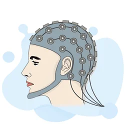
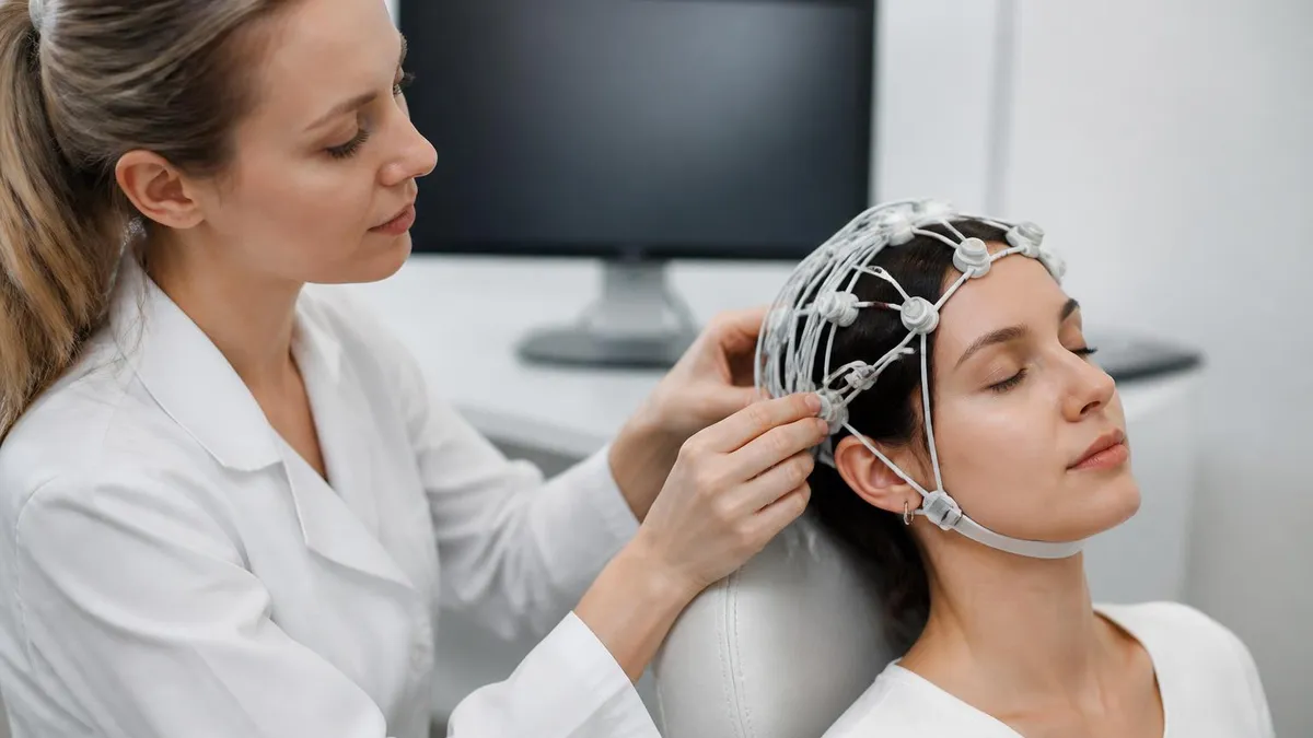
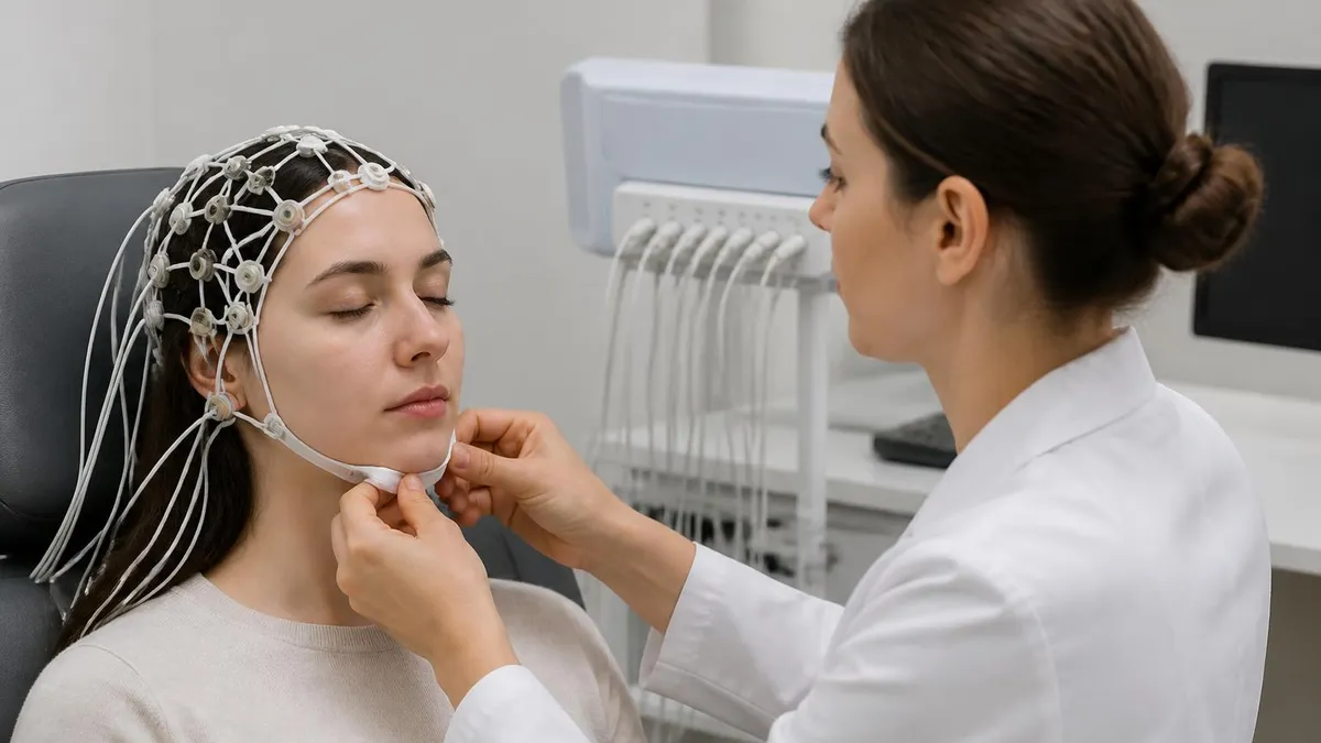
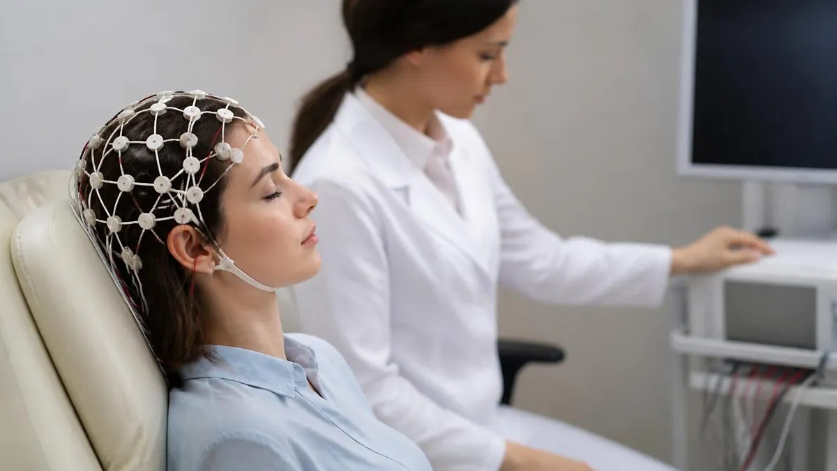
An EEG test — short for electroencephalogram — is one of the most powerful, non-invasive diagnostic tools in modern neurology. At its core, what does an EEG measure? It measures the electrical activity generated by millions of neurons firing simultaneously in your brain. Thin metal electrodes placed on the scalp detect these tiny voltage fluctuations and translate them into wave patterns on a graph, giving clinicians a real-time window into how your brain is functioning at any given moment.
The human brain is never truly silent. Even during deep sleep, neurons constantly communicate through electrochemical signals, and these signals produce distinctive rhythmic patterns known as brain waves. An EEG test captures these patterns across a range of frequencies — from slow delta waves during deep sleep to fast gamma waves during intense cognitive processing. The relative proportion, location, and synchrony of these waves tell a trained electroencephalographer an enormous amount about neurological health.
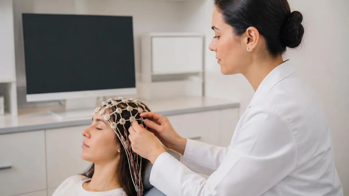
If you're wondering what is eeg test in a clinical context, the answer is that it is an EEG medical test that records electrical brain activity over a defined period, typically 20 to 40 minutes for a routine outpatient study. The test is painless, involves no radiation, and requires no injections. Electrodes are applied with a conductive gel and simply rest against the scalp, passively picking up signals rather than sending any electricity into the body.
Neurologists use the EEG test to diagnose and monitor a wide range of conditions. Epilepsy is the most well-known application — certain seizure types produce unmistakable spike-and-wave discharges that are diagnostic on their own. Beyond seizures, an EEG medical test can help evaluate encephalopathies, sleep disorders, brain tumors affecting cortical function, metabolic disturbances affecting the brain, and disorders of consciousness such as coma or brain death assessment.
Understanding the cost and accessibility of an EEG test is also important for patients. In the United States, an EEG test price can range from roughly $200 for a basic outpatient study at a community clinic to over $2,000 at a hospital-based epilepsy monitoring unit, depending on the type of study, duration, geographic location, and whether insurance covers the procedure. Many insurers do cover EEGs when there is a documented medical indication such as unexplained seizures or fainting episodes.
The EEG test side effects are minimal compared to many other diagnostic procedures. Some patients report mild scalp irritation from the electrode gel, or temporary skin redness at electrode sites. There are no known lasting side effects from standard EEG recording. Certain activation procedures — such as hyperventilation or strobe light stimulation — may temporarily provoke symptoms in susceptible individuals, but these are conducted in controlled settings with trained personnel present at all times to manage any reactions safely.
For anyone preparing to take an EEG technician certification exam, or for patients who simply want to understand what happens during their upcoming study, this article provides a thorough breakdown of everything an EEG captures, how different brain wave frequencies are interpreted, what conditions the test can and cannot diagnose, and practical guidance on what to expect before, during, and after the procedure.
EEG Test by the Numbers

The Five Main Brain Wave Types an EEG Records
The slowest brain waves, dominant during deep, dreamless sleep in healthy adults. Abnormal delta activity during wakefulness can indicate serious cortical dysfunction, structural brain lesions, or encephalopathy. In infants, delta activity is normal even while awake.
Commonly seen during drowsiness, light sleep, and meditative states. Focal theta activity in a fully awake adult can indicate a localized area of cortical dysfunction such as a tumor, stroke, or traumatic injury affecting underlying brain tissue.
The hallmark rhythm of a relaxed, awake adult with eyes closed. Alpha activity is typically maximal over the occipital (visual) regions. It attenuates or disappears when the eyes open or when the patient engages in active mental processing — a normal and expected response.
Associated with active thinking, concentration, and alertness. Beta rhythms are dominant during normal waking consciousness. Excessive beta activity, particularly generalized, is a common finding in patients taking benzodiazepines or barbiturates and is considered a drug effect rather than pathology.
The fastest rhythms recorded on a standard EEG, linked to higher cognitive processing, memory binding, and sensory integration. Gamma analysis requires careful filtering to distinguish true brain-generated activity from muscle artifact, which occurs in a similar frequency range.
One of the primary reasons physicians order an EEG medical test is to evaluate for epilepsy and classify seizure type. Epilepsy affects approximately 3.4 million Americans, and a single routine EEG captures epileptiform abnormalities — spikes, sharp waves, or spike-and-wave complexes — in roughly 50% of people with confirmed epilepsy. Serial EEGs or prolonged monitoring significantly increase the diagnostic yield. The specific pattern, location, and morphology of epileptiform activity guide treatment decisions and help determine whether a patient is a surgical candidate.
Beyond epilepsy, an EEG test is invaluable for evaluating altered mental status. When a patient presents with unexplained confusion, agitation, or impaired consciousness, EEG can quickly identify whether nonconvulsive status epilepticus (NCSE) is the cause — a condition that can mimic psychiatric illness or medication toxicity but requires urgent anticonvulsant treatment. Studies estimate that NCSE accounts for up to 37% of cases of unexplained altered consciousness in the ICU setting, making continuous EEG monitoring a standard of care in many neurocritical care units.
Sleep disorders are another important domain for EEG. A what is a eeg test in the context of sleep medicine involves recording brain waves overnight alongside other physiological signals — respiratory effort, oxygen saturation, eye movements, and muscle tone — in what is called a polysomnogram. The EEG portion identifies sleep stages (N1, N2, N3, and REM) and detects abnormal events such as sleep spindle irregularities, K-complex abnormalities, or seizures that only occur during sleep, which are easily missed by daytime studies.
Encephalopathies — whether metabolic, toxic, infectious, or autoimmune in origin — produce characteristic EEG changes. Triphasic waves, once considered pathognomonic of hepatic encephalopathy, are now recognized as a nonspecific marker of diffuse cortical dysfunction seen in various metabolic disturbances. Diffuse slowing, with theta and delta rhythms replacing normal alpha background activity, indicates impaired cortical processing and its severity often correlates with the degree of clinical impairment, providing a useful objective measure of a patient's neurological status over time.
An EEG test also plays a critical role in brain death determination. In the United States, electrocerebral silence (ECS) — defined as no EEG activity above 2 microvolts across all electrode pairs — is one confirmatory test used alongside clinical examination criteria to establish brain death in appropriate clinical contexts. Strict technical standards must be followed, including minimum recording time, electrode impedance requirements, and the exclusion of confounding factors like hypothermia or sedating medications, to ensure the EEG recording is interpretable.
Researchers have increasingly used EEG to study cognitive function, attention, and processing speed. Event-related potentials (ERPs) — tiny voltage changes time-locked to specific stimuli — allow investigators to probe discrete cognitive operations with millisecond precision. The P300 component, for instance, reflects attentional allocation and working memory updating. These research applications have translated into clinical use for assessing disorders of consciousness in patients who are unable to communicate, potentially detecting covert awareness in patients who appear unresponsive at bedside.
For patients undergoing brain surgery, intraoperative EEG monitoring tracks cortical function in real time, alerting surgeons to ischemia or seizure activity during anesthesia. Electrocorticography (ECoG) — EEG recorded directly from the brain surface via electrodes placed on the cortex — provides the highest spatial resolution possible, enabling precise mapping of epileptogenic zones before surgical resection. Understanding these diverse applications underscores why EEG remains one of the most clinically versatile neurophysiological tests available despite being invented nearly a century ago.
How Long Is an EEG Test? Types, Durations, and What Each Involves
A standard outpatient EEG test typically lasts between 20 and 40 minutes of actual recording time, though the entire appointment — including electrode application and removal — runs closer to 60 to 90 minutes. During the recording, patients rest quietly with eyes closed, then open, while the technician captures baseline brain activity across all standard electrode positions using the International 10-20 System of electrode placement.
Activation procedures are typically included in a routine EEG to increase the likelihood of capturing abnormalities. These include three to five minutes of hyperventilation, which can provoke absence seizures in susceptible individuals, and photic stimulation — a strobe light flashed at varying frequencies between 1 and 30 Hz — to detect photoparoxysmal responses. Brief drowsiness or sleep may also be encouraged because many epileptiform abnormalities are sleep-activated and may only appear when the patient transitions out of full wakefulness.
EEG Test: Benefits and Limitations Compared to Other Brain Tests
- +Non-invasive and painless — no needles, injections, or radiation exposure required
- +Excellent temporal resolution — detects brain activity changes in milliseconds, far better than MRI
- +Can be performed at the bedside for critically ill or immobile patients who cannot access MRI
- +Relatively low EEG test cost compared to MRI or CT scan, especially for routine outpatient studies
- +Can capture dynamic, moment-to-moment changes in brain state during sleep, seizures, or activation
- +Ambulatory versions allow 24-72 hour monitoring in a patient's natural home environment
- −Poor spatial resolution — cannot precisely localize sources deep within the brain like MRI can
- −A normal EEG does not rule out epilepsy — about 50% of epilepsy patients have a normal first EEG
- −Highly susceptible to artifact from muscle movement, eye blinks, electrode pop, and electrical interference
- −Requires expert interpretation — misreading normal variants as pathology is a significant risk
- −Standard EEG captures only 20-40 minutes of activity, potentially missing infrequent abnormalities
- −EEG test side effects from activation procedures (hyperventilation, photic stimulation) may provoke symptoms in some patients
How to Prepare for an EEG Test: 10-Step Pre-Test Checklist
- ✓Wash your hair the night before or the morning of the test — do NOT apply conditioner, oils, sprays, or styling products afterward.
- ✓Ask your doctor whether to take or hold your regular medications; most are continued, but clarify any anticonvulsants or sedatives specifically.
- ✓Avoid caffeine for at least 8 hours before the appointment — coffee, energy drinks, and caffeinated sodas can alter brain wave patterns.
- ✓Sleep deprivation is sometimes requested before an EEG to increase the chance of capturing sleep-activated abnormalities — confirm with your ordering physician.
- ✓Eat a light meal before the test; low blood sugar from fasting can cause symptoms that confound the EEG recording.
- ✓Arrive with clean, dry hair and no hair extensions, wigs, or hairpieces that would prevent electrode placement on the scalp.
- ✓Wear comfortable, loose clothing — you will be lying or sitting still for 60 to 90 minutes total during the appointment.
- ✓Bring a list of all current medications, including over-the-counter drugs and supplements, to give the technician before recording begins.
- ✓Inform the technician of any recent head injuries, surgeries, skull defects, or implanted devices before the electrodes are placed.
- ✓Plan for a driver if your doctor requested a sleep-deprived EEG — drowsiness after the test makes it unsafe to operate a vehicle alone.
A single normal EEG result does not mean you don't have epilepsy.
Studies consistently show that approximately 40–50% of people with confirmed epilepsy will have a normal first routine EEG. Epileptiform discharges are intermittent and may simply not occur during the brief recording window. If your EEG is normal but your physician still suspects seizures, repeat testing, sleep-deprived studies, or prolonged ambulatory monitoring significantly increase the diagnostic yield — sometimes up to 80–90% after three serial recordings.
The EEG test cost in the United States varies considerably based on the type of study, the facility performing it, your geographic location, and your insurance status. A routine outpatient EEG at a private neurology clinic typically ranges from $200 to $700 before insurance adjustments.
Hospital-based outpatient departments often charge more — sometimes $800 to $1,500 — due to higher facility fees. If your test is ordered by a physician and there is a documented medical indication, most major insurance plans, including Medicare and Medicaid, cover the procedure at standard in-network rates, leaving patients responsible only for their deductible and copay.
Ambulatory EEG studies, which involve renting portable recording equipment for 24 to 72 hours, typically cost $300 to $1,000 depending on the duration and the provider's billing practices. Video-EEG monitoring during an inpatient epilepsy monitoring unit admission is substantially more expensive, with total costs frequently ranging from $5,000 to $20,000 or more depending on the length of stay, which can span several days to two weeks.
For uninsured patients, some academic medical centers and community health programs offer reduced fees on a sliding scale. To understand what is eeg test used to diagnose and whether the cost is justified for your specific situation, you can review detailed pricing and insurance guidance at what is eeg test used to diagnose on our dedicated cost page.
Geographic variation is significant. EEG test prices in rural areas or smaller markets tend to be lower than in major metropolitan areas like New York City, Los Angeles, or Boston, where facility costs and specialist fees are higher.
Comparing prices across facilities using your insurer's cost estimation tool or requesting a good faith estimate — which providers are now required to provide under the No Surprises Act — can help you avoid unexpected bills. Some patients also use Health Savings Account (HSA) or Flexible Spending Account (FSA) funds to pay for EEG costs, as these studies qualify as medical expenses under IRS guidelines.
For patients who are uninsured or underinsured, telehealth neurology consultations followed by a referral to a federally qualified health center (FQHC) or community hospital can significantly reduce out-of-pocket costs. University hospital training programs may also offer EEGs at reduced rates because the studies support resident and fellow education under attending supervision. Prices in these settings can be 30 to 50 percent lower than a comparable private practice or commercial imaging center.
Understanding the full scope of EEG test cost also means accounting for interpretation fees, which are billed separately from the technical component in most US healthcare settings. The technical component covers electrode application, recording, and basic report preparation by the EEG technician. The professional interpretation fee is charged by the reviewing neurologist or epileptologist who analyzes the recording and prepares the diagnostic report. Both components are typically billed to insurance individually, and patients may receive two separate Explanation of Benefits (EOB) statements for a single EEG study.
EEG test side effects are another consideration worth addressing in the context of cost-benefit analysis. Because the test is essentially risk-free under normal circumstances — no radiation, no contrast agents, no sedation in most cases — the benefit-to-risk ratio is extremely favorable.
The most common EEG test side effects are minor scalp irritation or redness at electrode sites due to the abrasive gel used to improve conductivity, and these resolve within a few hours. Rarely, a patient with a known photosensitive epilepsy syndrome may experience a seizure provoked by photic stimulation, but this occurs in a monitored setting with trained clinicians who can respond immediately.
For patients who experience claustrophobia or anxiety about medical procedures, an EEG is typically much more comfortable than MRI scanning, which requires lying still in a loud, enclosed tube for 30 to 60 minutes. EEG is performed in an open room, usually while the patient reclines in a comfortable chair or on a padded table. The presence of electrodes may feel unusual at first, but most patients adapt within a few minutes and find the experience far less stressful than anticipated before the appointment.

Many common medications significantly alter EEG patterns and can lead to misinterpretation if not disclosed. Benzodiazepines and barbiturates increase fast beta activity; lithium can cause diffuse slowing or even spike-like activity at toxic levels; stimulants increase arousal rhythms; and some antidepressants lower the seizure threshold. Always provide a complete medication list — including over-the-counter drugs and herbal supplements — to both the ordering physician and the EEG technician before recording begins.
Interpreting EEG results requires specialized training because the brain produces an enormous variety of patterns, and many normal variants closely resemble pathological findings. The 14-and-6-Hz positive spikes, wicket spikes, and small sharp spikes of sleep (SSSS) are classic benign normal variants that have historically been misread as epileptiform abnormalities, leading to inappropriate diagnoses of epilepsy and unnecessary medication trials. This is one reason EEG interpretation is considered a subspecialty within neurology and neurophysiology, with formal fellowship training required for competency.
When your neurologist receives your EEG report, it will typically describe the background activity (the dominant rhythm and its frequency, amplitude, and symmetry), the presence or absence of normal features (such as sleep spindles, vertex sharp waves, or the posterior dominant rhythm), and any abnormalities detected. Abnormalities are classified as either epileptiform — suggesting a predisposition to seizures — or non-epileptiform, such as focal or diffuse slowing that may indicate structural or metabolic dysfunction but does not by itself indicate epilepsy.
A focal abnormality is one that is limited to a specific region of the brain. Focal slowing over the left temporal region, for example, might indicate a structural lesion such as a prior stroke, a low-grade tumor, or mesial temporal sclerosis — one of the most common pathological substrates of drug-resistant focal epilepsy. Focal epileptiform discharges in the same region would strongly support the diagnosis of temporal lobe epilepsy and guide decisions about which antiseizure medication to select or whether to pursue surgical evaluation.
Generalized abnormalities, by contrast, involve the entire brain simultaneously. Generalized 3-Hz spike-and-wave complexes are the classic EEG signature of childhood absence epilepsy. Generalized polyspike-and-wave discharges point toward juvenile myoclonic epilepsy (JME), a common syndrome that typically responds well to certain anticonvulsants but has a high relapse rate if medication is discontinued. Understanding the EEG pattern not only confirms the diagnosis but directly determines the treatment approach, since medications effective for one epilepsy syndrome may actually worsen another.
For anyone studying to become an EEG technologist or preparing for the ABRET (American Board of Registration of Electroencephalographic and Evoked Potential Technologists) certification exam, mastering the classification and recognition of these patterns is essential. The ABRET exam tests candidates on normal variants, artifacts, epileptiform abnormalities, encephalopathy patterns, and technical standards for recording quality. Understanding how to differentiate true pathology from benign variants or artifact is the cornerstone of competent EEG practice. To understand how EEG differs from cardiac monitoring, check out what is the eeg test compared to ECG and EKG in our dedicated comparison article.
EEG technology has advanced considerably since Hans Berger recorded the first human EEG in 1924. Digital EEG systems now allow post-hoc filtering, montage reformatting, and quantitative analysis that were impossible with analog paper recordings. High-density EEG systems using 128 or 256 electrodes — compared to the standard 19-21 — offer significantly improved spatial resolution and enable source localization algorithms that estimate where in the brain a given electrical signal originates. While these systems are primarily used in research settings, they are increasingly appearing in clinical epilepsy centers for pre-surgical evaluations.
Functional connectivity analysis and machine learning algorithms applied to EEG data represent the frontier of the field. Researchers are developing automated seizure detection systems capable of identifying ictal patterns in long-term recordings faster and more reliably than human review alone — a critical need given that a 72-hour ambulatory EEG can contain over a million pages of data if printed on paper. These computational advances are expected to reduce interpretation time, improve diagnostic consistency, and eventually enable real-time seizure forecasting systems that could alert patients to an impending seizure before it occurs.
For EEG technologists and students preparing for board certification, developing a systematic approach to reading EEG tracings is far more important than memorizing individual patterns in isolation. Every EEG should be evaluated in a consistent sequence: first assess the overall background activity for organization and symmetry, then identify the dominant posterior rhythm and confirm it is appropriate for the patient's age and level of arousal, then evaluate the response to eye opening, then systematically review each electrode pair for focal abnormalities, and finally assess for any paroxysmal events or epileptiform discharges.
Artifact recognition is one of the most practically important skills an EEG technologist must develop. Common sources of artifact include muscle activity (high-frequency irregular signals from scalp and facial muscles), electrode pop (brief, high-amplitude transient from poor electrode contact), EKG artifact (rhythmic QRS complexes seen through scalp electrodes in thin patients), movement artifact, and 60-Hz electrical interference from power lines. Distinguishing these from true brain activity requires experience, and misidentifying artifact as pathology — or vice versa — are equally serious errors with clinical consequences.
Practical preparation for the ABRET Registered EEG Technologist (R.EEG T.) examination requires both conceptual understanding and hands-on experience. The examination covers neuroanatomy, neurophysiology, electrode placement, instrumentation and calibration, digital EEG principles, artifact identification, normal EEG patterns across the lifespan, abnormal EEG patterns, and special procedures including ambulatory EEG and intraoperative monitoring. Most candidates complete a formal EEG technology training program accredited by CAAHEP, which typically includes one to two years of didactic coursework combined with supervised clinical hours in an accredited EEG laboratory.
Time management during the ABRET exam is a common challenge. The exam consists of 225 questions administered over a timed session, and candidates often report that the EEG pattern recognition sections — which involve interpreting actual EEG samples — require the most careful and deliberate thinking. Developing speed and confidence with waveform identification during training is critical, as rushed reading under examination conditions increases the likelihood of misidentifying normal variants as abnormal findings or overlooking subtle epileptiform discharges buried within complex background activity.
Practice questions are one of the most effective study tools available to ABRET candidates, and targeted quiz practice specifically on abnormal epileptiform patterns and activation procedures — two heavily tested domains — can significantly improve examination scores. Reviewing case-based questions that present actual EEG scenarios forces active recall rather than passive reading, reinforcing both pattern recognition and the clinical reasoning needed to select the correct interpretation from similar-appearing options. Spaced repetition with timed practice sessions mirrors the real examination environment.
Beyond the initial certification, continuing education in EEG is an ongoing professional requirement. The field evolves continuously as new syndromes are classified, new ACNS (American Clinical Neurophysiology Society) guidelines are published, and new technology expands what is measurable. Staying current with the literature, attending annual meetings of the American Epilepsy Society or ACNS, and maintaining certification through the ABRET recertification process every five years are all components of professional excellence in clinical neurophysiology.
Whether you are a patient curious about an upcoming procedure, a student preparing for board certification, or a clinician expanding your neurophysiology knowledge, understanding what an EEG test measures — from the biophysics of neuronal voltage generation to the clinical interpretation of complex waveform patterns — provides an essential foundation. The EEG remains irreplaceable in the neurological toolkit precisely because it measures brain function in real time, offering insights into neurological health that no structural imaging study can replicate.
EEG Questions and Answers
EEG Electroencephalography Practice Test PDF (Free Printable 2026)
Sleep EEG: How Overnight Brain Wave Testing Works and What Your Results Mean
EEG Test Cost in 2026: Prices, Insurance Coverage, and How to Save
EEG vs ECG vs EKG: Brain and Heart Test Differences
Abnormal EEG Results: What They Mean, Common Patterns, and Next Steps
About the Author
Educational Psychologist & Academic Test Preparation Expert
Columbia University Teachers CollegeDr. Lisa Patel holds a Doctorate in Education from Columbia University Teachers College and has spent 17 years researching standardized test design and academic assessment. She has developed preparation programs for SAT, ACT, GRE, LSAT, UCAT, and numerous professional licensing exams, helping students of all backgrounds achieve their target scores.