
What Does an EEG Test Measure? Purpose, Procedure, Cost, Safety, and Results
🧠 What does an EEG test measure? Learn how it works, how long it takes, typical cost, side effects, and what your results mean before your appointment.

So, what does an EEG test actually measure? An electroencephalogram, or EEG, records the tiny electrical signals produced when neurons in your brain communicate. Small metal discs called electrodes sit on the scalp, pick up those signals, and send them to an amplifier that draws them as wavy lines on a screen. The result is a real-time picture of brain function, not brain structure, which is why doctors order it when they suspect seizures, sleep disorders, or unexplained confusion. Practice with the free EEG - Electroencephalography practice test on PracticeTestGeeks (11 questions with answer explanations).
The EEG test is one of the oldest tools in neurology. German psychiatrist Hans Berger recorded the first human EEG in 1929, and the basic idea has not changed since: neurons fire, ions move, and the combined electrical field is measurable at the scalp. Modern digital systems sample the signal 256 to 512 times per second and store it for review. That speed lets a specialist see changes lasting only a fraction of a second.
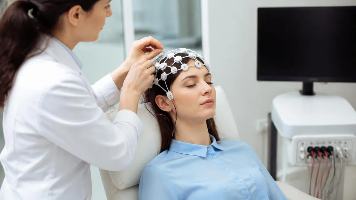
People often search for "what is an EEG test" because a doctor mentioned it in passing, and the term sounds intimidating. In reality it is a painless, noninvasive outpatient procedure. Nothing is sent into your head. The electrodes only listen. You lie on a bed or recline in a chair, a technologist places the sensors, and the recording runs while you rest, breathe deeply, or watch a flashing light. Most people describe it as boring rather than uncomfortable.
Doctors order an EEG medical test for several reasons. The most common is suspected epilepsy, since abnormal bursts of electrical activity called spikes and sharp waves can appear between seizures. Others include unexplained fainting, episodes of staring or memory gaps, head injury, encephalopathy, sleep disorders, and monitoring depth of sedation or coma. An EEG test for brain activity also helps separate true seizures from look-alike events such as psychogenic nonepileptic spells.
It helps to know what an EEG test does not do. It cannot read your thoughts, diagnose a tumor, or measure intelligence. It will not show a stroke the way an MRI does, and it cannot tell by itself whether you have a mental health condition. Instead, it answers a narrow but valuable question: is the brain's electrical rhythm normal for your age and state of alertness? That answer guides treatment decisions, medication choices, and whether further imaging is needed.
This guide walks through everything patients and aspiring technologists want to know. You will learn the brain wave bands the test records, how long an EEG takes from check-in to checkout, what it typically costs in the United States, whether it has side effects, how results are interpreted, and what duties an EEG technologist performs. Practice questions at the end let you test your knowledge of the patterns discussed here.
Free EEG - Electroencephalography Study Tools
Interactive EEG - Electroencephalography resources on PracticeTestGeeks, free to start:
- Free EEG Routine Questions and Answers 1 — 11 questions with instant scoring and answer explanations
- EEG - Electroencephalography flashcards — review key terms and concepts card by card
- EEG - Electroencephalography study guide — topic-by-topic overview of what the exam covers
- EEG - Electroencephalography cheat sheet — one-page printable summary for last-minute review
The EEG Test by the Numbers
What Does an EEG Test Record? The Five Brain Wave Bands
Slow, high-amplitude waves normal in deep sleep and in infants. In an awake adult, excess delta activity can point to a focal lesion, encephalopathy, or sedation, so technologists note when and where it appears.
Theta appears in drowsiness, light sleep, and normally in children. Persistent theta over one brain region in an alert adult may signal localized dysfunction, and bursts of it can accompany certain seizure types.
The classic relaxed-wakefulness rhythm, strongest over the back of the head when your eyes are closed. It fades when you open your eyes, which is why technologists ask you to do exactly that during recording.
Fast, low-amplitude activity linked to alertness and concentration. Sedatives such as benzodiazepines increase beta, so reviewers always check your medication list before calling frontal beta abnormal.
Very fast rhythms associated with higher cognitive processing. They are small and easily masked by muscle artifact, so they play a smaller role in routine clinical reading than the lower bands.
Cost is one of the first questions patients ask, and the honest answer is a wide range. A routine outpatient EEG test price in the United States commonly runs from about $200 to $3,000 before insurance, depending on the facility, region, and whether a physician reading fee is bundled. Hospital outpatient departments usually bill more than freestanding neurodiagnostic clinics. The same recording can carry very different charges across town, so asking for an itemized estimate is reasonable.
Insurance changes the picture dramatically. Medicare and most commercial plans cover an EEG when it is medically necessary, such as evaluating seizures or altered mental status. Many plans still apply a deductible or coinsurance, so a patient with a $1,500 charge might pay anywhere from $0 to several hundred dollars. Prior authorization is more common for long-term recordings than for routine ones, so confirm requirements before you schedule.
Different EEG formats carry different costs. A routine 20 to 40 minute recording sits at the low end. A sleep-deprived EEG costs about the same, though it may add a separate technologist fee. Ambulatory EEG, which records at home for one to three days, often runs $1,000 to $3,500. Inpatient video EEG monitoring in an epilepsy unit is the most expensive, because the bill includes the room, nursing, and continuous review across several days.
Several practical steps can lower your EEG test cost. Ask whether a freestanding outpatient lab is available instead of the hospital, since facility fees are often smaller. Request the cash or self-pay price, which can be lower than the billed rate. Check that both the facility and the interpreting neurologist are in network. Finally, ask the billing office about financial assistance, because nonprofit hospitals are required to offer charity-care policies. Finding an EEG test near me that is in network is the single biggest saver.
People also ask what makes this an EEG medical test rather than a research or consumer product. The difference is clinical purpose and oversight. A medical EEG is ordered by a licensed clinician, recorded by a trained technologist using a standardized electrode layout, and interpreted by a physician, usually a neurologist with epilepsy or clinical neurophysiology training. Consumer headbands and wellness apps record far fewer channels and are not diagnostic, so insurers do not reimburse them.
One more cost factor is the report itself. Billing typically splits into a technical component, covering equipment and technologist time, and a professional component, covering the physician's interpretation. You may receive two separate statements and think you were charged twice. Reviewing both lines against your explanation of benefits prevents confusion. If a repeat study is ordered because the first recording was inconclusive or full of artifact, ask whether the second test counts toward the same deductible and authorization.
How Long Is an EEG Test? What Happens Before, During, and After
Plan on about 60 to 90 minutes for the full appointment, even though the recording itself is often only 20 to 40 minutes. Most of the extra time is setup. The technologist measures your head, marks electrode positions with a washable pencil, gently scrubs each spot, and attaches the sensors with conductive paste or gel. Applying 19 to 21 electrodes carefully takes 20 to 30 minutes.
Your instructions beforehand matter. You will usually be asked to wash your hair the night before and skip conditioner, gels, and sprays, since residue weakens contact. Eat normally and take prescribed medicines unless told otherwise, but avoid caffeine on the day. If your doctor wants a sleep-deprived study, you may be asked to stay up late or wake very early so you fall asleep during recording.
Is an EEG Test Worth It? Benefits and Limits
- +Completely noninvasive and painless, with nothing entering the body
- +No radiation exposure, unlike CT scans
- +Millisecond-level timing shows brain activity as it changes
- +Can detect epileptiform discharges between seizures
- +Safe for children, pregnant patients, and older adults
- +Widely available in hospitals and outpatient clinics
- −A single routine EEG can miss abnormalities, with roughly 50 percent sensitivity for epilepsy on the first study
- −Poor spatial detail compared with MRI
- −Muscle movement and eye blinks create artifact that complicates reading
- −Setup takes time and leaves paste in the hair
- −Results require expert interpretation and may take days
- −Costs can be high without insurance coverage
Pros and Cons at a Glance
| Pros | Cons |
|---|---|
| Completely noninvasive and painless, with nothing entering the body | Poor spatial detail compared with MRI |
| No radiation exposure, unlike CT scans | Muscle movement and eye blinks create artifact that complicates reading |
| Millisecond-level timing shows brain activity as it changes | Setup takes time and leaves paste in the hair |
| Can detect epileptiform discharges between seizures | Results require expert interpretation and may take days |
| Safe for children, pregnant patients, and older adults | Costs can be high without insurance coverage |
How to Prepare for Your EEG Test: A Practical Checklist
- ✓Wash your hair the night before and skip oils, gels, and conditioner.
- ✓Remove braids, extensions, and hairpieces if you can.
- ✓Avoid caffeine, energy drinks, and cola on the day of the test.
- ✓Eat a normal meal so low blood sugar does not alter the tracing.
- ✓Take your regular medications unless your doctor says to hold them.
- ✓Bring a written list of every medicine and supplement you take.
- ✓Follow sleep-deprivation instructions exactly if they were given.
- ✓Wear a button-front shirt so nothing pulls over the electrodes.
- ✓Arrange a ride home if you were told to stay up late.
- ✓Confirm insurance authorization and your estimated cost before the visit.
A Normal EEG Does Not Rule Out Epilepsy
Epileptic discharges come and go, so a short recording can easily miss them. Repeating the study, adding sleep, or extending monitoring can raise detection from about 50 percent on the first routine EEG to roughly 90 percent after several recordings. Ask your doctor about next steps if symptoms continue.
After the recording, the real work begins: interpretation. A neurologist reads the tracing page by page, usually in 10 to 20 second windows, looking at background rhythm, symmetry between hemispheres, reactivity to eye opening, and the presence of sleep features. Reviewers compare what they see with age-appropriate norms, because a pattern normal in a toddler would be abnormal in a 60-year-old. They also consider medications, since many drugs alter the EEG.
Abnormal findings fall into a few broad categories. Epileptiform abnormalities include spikes, sharp waves, and spike-and-wave complexes, and they strongly support a tendency toward seizures. Non-epileptiform abnormalities include focal slowing, which may reflect a structural problem, and diffuse slowing, which suggests encephalopathy from infection, metabolic disease, or medications. Some patterns, such as triphasic waves or periodic discharges, point toward particular underlying causes and prompt further workup.
Reports are written in standardized terminology so every clinician reads them the same way. A typical report lists the patient's state during recording, a description of the background activity, any epileptiform or slow patterns with their location, and a clinical correlation sentence. The conclusion is usually graded as normal, borderline, or abnormal. It is important to remember the report is one input. Your doctor combines it with symptoms, exam, imaging, and sometimes blood tests.
The EEG technologist, also called a neurodiagnostic technologist, is the person who makes good interpretation possible. Daily duties include verifying the physician's order, taking a brief history, measuring the head using the 10-20 system, applying electrodes, checking impedances, and monitoring signal quality throughout. During recording they annotate events such as blinking, coughing, or movements, and they document any clinical signs that appear during a seizure. Their notes often decide whether a questionable wave is real or artifact.
Technologists also carry safety responsibilities. They must recognize a seizure, protect the patient from injury, note timing and behavior, and call a nurse or physician when needed. They clean and maintain equipment, follow infection-control rules, and keep records accurate. In an EEG test hospital setting, they may also set up portable bedside studies in intensive care units, where sedation, ventilators, and lines create heavy electrical interference and demand extra problem-solving skill.
Many technologists pursue credentials to advance. The ABRET registered EEG technologist credential, R. EEG T., is the best-known certification, and further credentials exist for long-term monitoring and intraoperative monitoring. Employers in the United States often prefer or require registration, and it generally supports better pay and more independence. Anyone studying for this career should understand electrode placement, artifacts, montages, and recognition of common patterns, which is exactly what the practice quizzes in this section help reinforce.
If you have ever had a seizure, a head injury, or photosensitivity, tell the technologist before the strobe light or hyperventilation begins. They can adjust or skip activation procedures and will have an emergency plan ready, which keeps the study both safe and useful.
The next most common question after cost is whether there are EEG test side effects. For the vast majority of people the answer is no. The electrodes record only; they deliver no current to the brain. You will not feel shocks, tingling, or heat. Because there is no radiation, no contrast dye, and no anesthesia in a routine study, you can resume driving, working, and eating immediately afterward unless you were sleep-deprived or given a mild sedative for a child.
Minor, temporary effects do occur. Some people notice a bit of redness or irritation where paste or adhesive touched the skin, particularly those with sensitive skin or latex or adhesive allergies. Hair may feel sticky until you wash it, and the scalp may be slightly tender where electrodes were scrubbed in. Let the technologist know about allergies beforehand so they can choose hypoallergenic materials. These symptoms usually fade within hours and rarely require treatment.
The more important safety consideration involves activation procedures. Hyperventilation can cause lightheadedness, tingling in the lips or fingers, and in rare cases triggers a seizure in someone with epilepsy. Photic stimulation can likewise provoke a seizure in people with photosensitive epilepsy, which affects only a small percentage of those with the condition. Because monitored settings are prepared for this, a seizure during an EEG is considered clinically useful and handled quickly.
Certain groups need extra caution. People with severe asthma, sickle cell disease, significant heart or lung disease, or recent stroke may be excused from hyperventilation. Pregnant patients can have an EEG safely, and hyperventilation is often modified. Infants and young children usually receive a shorter setup with careful distraction, and parents may stay in the room. Sleep deprivation, if requested, is the main reason a seizure might occur, so follow your doctor's instructions about how much sleep to skip.
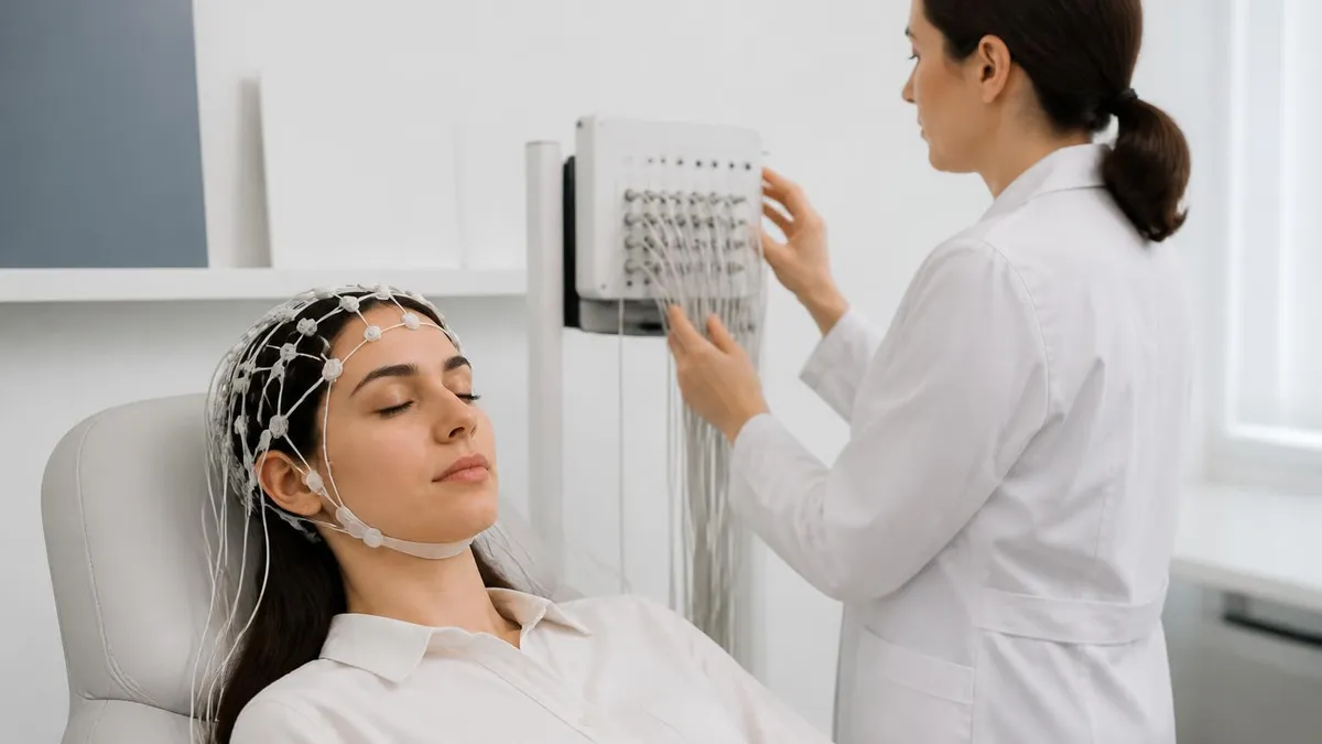
Different test formats carry slightly different experiences. Routine and sleep-deprived EEGs are short and low-burden. Video EEG adds a camera so staff can match behavior to brain waves, and it usually requires a hospital stay of several days. Ambulatory EEG lets you wear a recorder at home for 24 to 72 hours, which means managing skin comfort, keeping the cap dry, and logging events in a diary, but no one needs to hospitalize you.
If you feel unwell afterward, contact your clinician. Persistent severe headache, prolonged confusion, or a rash spreading beyond the electrode sites deserve a call. Otherwise, wash your hair gently with warm water and mild shampoo, and use a little conditioner to loosen leftover paste. Understanding what an EEG brain test involves, including its electrodes and paste, usually removes most of the anxiety people feel before their first appointment.
Whether you are a patient or a student technologist, a few practical habits make the EEG experience smoother. Start by asking your doctor exactly why the test was ordered. Knowing the clinical question, such as ruling out seizures versus evaluating confusion, helps you understand which procedures will be used and whether sleep, hyperventilation, or flashing lights will be required. It also helps you describe your symptoms accurately to the technologist, who will ask about spells, triggers, and recent events.
Keep a short symptom diary for a week before the appointment. Record the date, time, duration, and what you were doing during each episode, along with how you felt before and afterward. A witness description is especially valuable because many seizures impair awareness. Bring this log, plus your medication list and any prior imaging reports. The more context the interpreting neurologist has, the more accurately they can match tracing patterns to your story.
On test day, arrive about fifteen minutes early with clean, dry hair and comfortable clothes. Use the restroom beforehand, since you will be connected to wires for a while. If you are anxious, tell the technologist; most are experienced at calming nervous patients and will explain each step as it happens. Try to stay relaxed and still, because the cleanest recording is the one where tension, swallowing, and head movement are minimal.
Parents of children should prepare them in simple language. Explain that the stickers or paste sit on the head to take pictures of brain waves, and that nothing hurts. Bring a favorite blanket, book, or tablet for the setup stage, and ask whether a parent may lie beside the child. For toddlers, scheduling around naptime can help, since many young patients are asked to sleep during recording and a rested child is easier to settle.
Students who plan to become technologists can use the same experience as a study tool. Learn the 10-20 system landmarks: nasion, inion, and the preauricular points. Practice recognizing common artifacts such as eye blink, EMG, electrode pop, and 60 Hz interference. Review the activation procedures and their expected effects, then drill spike versus sharp wave morphology and normal variants that mimic them. Shadowing in an actual lab, even for a few hours, builds recognition faster than reading alone.
Finally, follow up on results. Ask when the report will be ready, who will call you, and what happens if the study is normal but symptoms continue. Request a copy for your records. If the recording was limited by artifact or you did not fall asleep, ask whether repeating it or switching to ambulatory or video monitoring makes sense. Being an active participant helps ensure that your EEG leads to a clear plan rather than more uncertainty.
Sample EEG - Electroencephalography Practice Questions
Try these questions from our free EEG - Electroencephalography practice tests. The correct answer and an explanation follow each question.
An alert and oriented adult patient provides consent for an EEG but adamantly refuses the hyperventilation (HV) activation procedure due to a history of panic attacks. Which of the following is the most appropriate course of action for the technologist?
- A. Insist that the patient attempt HV, as it is a required part of the standard procedure.
- B. Cancel the entire EEG study because it will be incomplete without the activation.
- C. Respect the patient's refusal, document it on the record, and proceed with the remainder of the EEG.
- D. Ask a family member to convince the patient to perform the procedure.
Answer: C. Respect the patient's refusal, document it on the record, and proceed with the remainder of the EEG.
Competent adult patients have the right to refuse any part of a medical procedure. The technologist must respect the patient's autonomy. The proper action is to document the refusal clearly on the technologist's report and continue with the other parts of the study, such as photic stimulation and obtaining a sleep recording.
Which activation procedure is particularly valuable for diagnosing juvenile myoclonic epilepsy (JME)?
- A. Sleep deprivation followed by drowsiness recording
- B. Hyperventilation alone
- C. Intermittent photic stimulation
- D. Cognitive task activation
Answer: C. Intermittent photic stimulation
Intermittent photic stimulation is especially effective for provoking generalized spike-wave discharges in juvenile myoclonic epilepsy due to its high photosensitivity prevalence.
To minimize the risk of electrical shock to the patient, what is the single most important electrical safety practice in the EEG laboratory?
- A. Using a notch filter to remove 60 Hz interference.
- B. Ensuring all electrodes have an impedance below 1 kOhm.
- C. Plugging all patient-connected equipment into the same properly grounded, hospital-grade outlet cluster.
- D. Applying a separate ground electrode for each piece of monitoring equipment.
Answer: C. Plugging all patient-connected equipment into the same properly grounded, hospital-grade outlet cluster.
Using a single, common ground point for all equipment connected to the patient prevents the formation of ground loops and minimizes the risk of leakage currents passing through the patient. Plugging the EEG machine and any other connected devices into the same outlet block ensures they share a common ground reference. Applying multiple ground electrodes from different pieces of equipment can be dangerous and is known as 'double grounding'.
Which of the following is NOT a standard activation procedure in a routine adult EEG?
- A. Hyperventilation
- B. Photic stimulation
- C. Sleep deprivation prior to recording
- D. Cold caloric stimulation
Answer: D. Cold caloric stimulation
Cold caloric stimulation is a vestibular bedside test, not a standard EEG activation procedure; HV, photic stimulation, and sleep deprivation are standard.
EEG Questions and Answers

EEG Artifacts: How to Identify, Eliminate, and Document Common Recording Interference

ERP vs EEG: What Each Test Measures, How They Differ, and What to Expect

EEG Cap Electrodes: Everything You Need to Know About the EEG Test
EEG Monitoring: What Is an EEG Test, How It Works, Cost, and What to Expect
Ambulatory EEG: 24-72 Hour Home Recording for Suspected Seizures
How this page was made: written with AI tools and published by Nathan Beja, founder of Practice Test Geeks, who is responsible for its content. Exam facts are checked against the official sources listed below. Not reviewed by a subject-matter expert. Editorial policy
Sources & references
Exam facts on this page (fees, format, dates, passing scores) are checked against these official sources; the date is our last check. Editorial policy
- abret.org — Late registration, score validity, test delivery, fees, exam format, passing score, retake policy, score release, test dates · verified Oct 2026