
EEG Test: Does EEG Measure Action Potential? How the Test Works, Cost, Duration, and Side Effects
🧠 Does EEG measure action potential? Learn what an EEG test records, how long it takes, typical cost, side effects, and how brain waves are read.

Does EEG measure action potential activity? Not directly. An EEG test records the summed electrical fields produced by thousands of neurons working together, and those fields come mostly from postsynaptic potentials rather than the brief spikes known as action potentials. This distinction matters whether you are studying for a technologist exam, preparing for a medical appointment, or simply curious about what the wires on the scalp actually pick up. Here is a clear, practical explanation. Practice with the free EEG - Electroencephalography practice test on PracticeTestGeeks (11 questions with answer explanations).
An action potential is a rapid, all-or-nothing voltage change that travels down an axon, lasting roughly one to two milliseconds and swinging about 100 millivolts across the cell membrane. It is the signal neurons use to communicate over distance. A postsynaptic potential, by contrast, is a slower, graded change in voltage that occurs on the dendrites and cell body when neurotransmitters arrive. These graded changes last tens of milliseconds, and they are the real source of the waves a technologist sees on screen.
Why do spikes mostly disappear from scalp recordings? Action potentials are brief and travel in different directions along many axons, so their tiny electrical fields cancel each other out almost completely. Postsynaptic potentials last longer, overlap in time, and occur in parallel rows of cortical pyramidal cells that all point toward the brain surface. Their fields add together instead of cancelling. That is why the signal reaching the scalp electrodes reflects synaptic activity and not individual spiking.
The numbers show how faint the signal is. A single neuron produces far too little voltage to detect from outside the head. Researchers estimate that roughly 10,000 or more neurons across several square centimeters of cortex must be active at nearly the same moment to produce a measurable scalp signal. By the time it crosses cerebrospinal fluid, bone, and skin, the voltage has shrunk to about 10 to 100 microvolts, which is why sensitive amplifiers are essential.
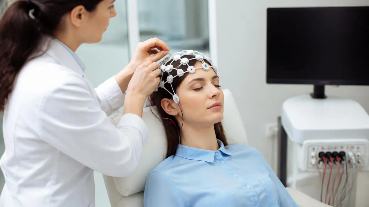
So what does a patient actually experience during an EEG test? Small metal discs, usually 19 of them in a standard layout, are attached to the scalp with paste or a cap. They do not send any electricity into the head. They only listen. A clinician then reviews the rhythms for signs of epilepsy, sleep disorders, or encephalopathy. To see how eeg works in practice, including what can distort the trace, read our artifact guide.
Although the question is often framed as a trick, the honest answer has nuance. Invasive recordings, such as depth electrodes or microelectrode arrays placed in the brain during surgery, can capture action potentials from nearby neurons. Scalp EEG cannot. Some high-frequency activity above 80 hertz may carry traces of spiking synchrony, but it remains an indirect, blurred reflection. Understanding that boundary prevents common misunderstandings on exams and in conversations with your neurologist.
This article walks through the full picture: the biology behind the signal, the step-by-step procedure, the brain waves clinicians interpret, and the practical questions patients ask most, including how long an EEG test takes, what an EEG test costs, and whether side effects exist. Use the practice quizzes along the way to check your understanding, especially if you are preparing for an EEG technologist credential or a neurodiagnostics course.
Free EEG - Electroencephalography Study Tools
Interactive EEG - Electroencephalography resources on PracticeTestGeeks, free to start:
- Free EEG Routine Questions and Answers 1 — 11 questions with instant scoring and answer explanations
- EEG - Electroencephalography flashcards — review key terms and concepts card by card
- EEG - Electroencephalography study guide — topic-by-topic overview of what the exam covers
- EEG - Electroencephalography cheat sheet — one-page printable summary for last-minute review
EEG by the Numbers
How an EEG Test Works Step by Step
Check-In and History
Measuring and Marking the Scalp
Attaching the Electrodes
Baseline and Activation Recording
Removal and Interpretation
How an EEG Test Detects Brain Activity
The main generators of scalp EEG are pyramidal neurons in the cerebral cortex. These cells have long apical dendrites aligned perpendicular to the surface, so when excitatory synapses activate near the top, current flows along the cell like a tiny battery. The result is a dipole, a pair of positive and negative poles. Thousands of aligned dipoles firing together create an electrical field strong enough to be measured outside the skull. Randomly arranged neurons cannot do this.
Each electrode records the voltage at one location relative to a reference. EEG machines use differential amplifiers that compare two inputs and boost only the difference between them. Anything common to both inputs, such as electrical noise from the room, is subtracted away. This is called common-mode rejection, and it allows a clean trace to appear even though the brain signal is a million times weaker than household current. A ground electrode keeps the patient at a stable baseline.
The signal is then filtered and digitized. Typical clinical settings use a low-frequency filter near 1 hertz, a high-frequency filter near 70 hertz, and a notch filter at 60 hertz to remove US power line interference. Modern systems sample at 256 to 512 times per second, and research systems may exceed 1,000. Higher sampling rates preserve fast activity but produce larger files. Over-filtering can hide real spikes or even create misleading waveforms.
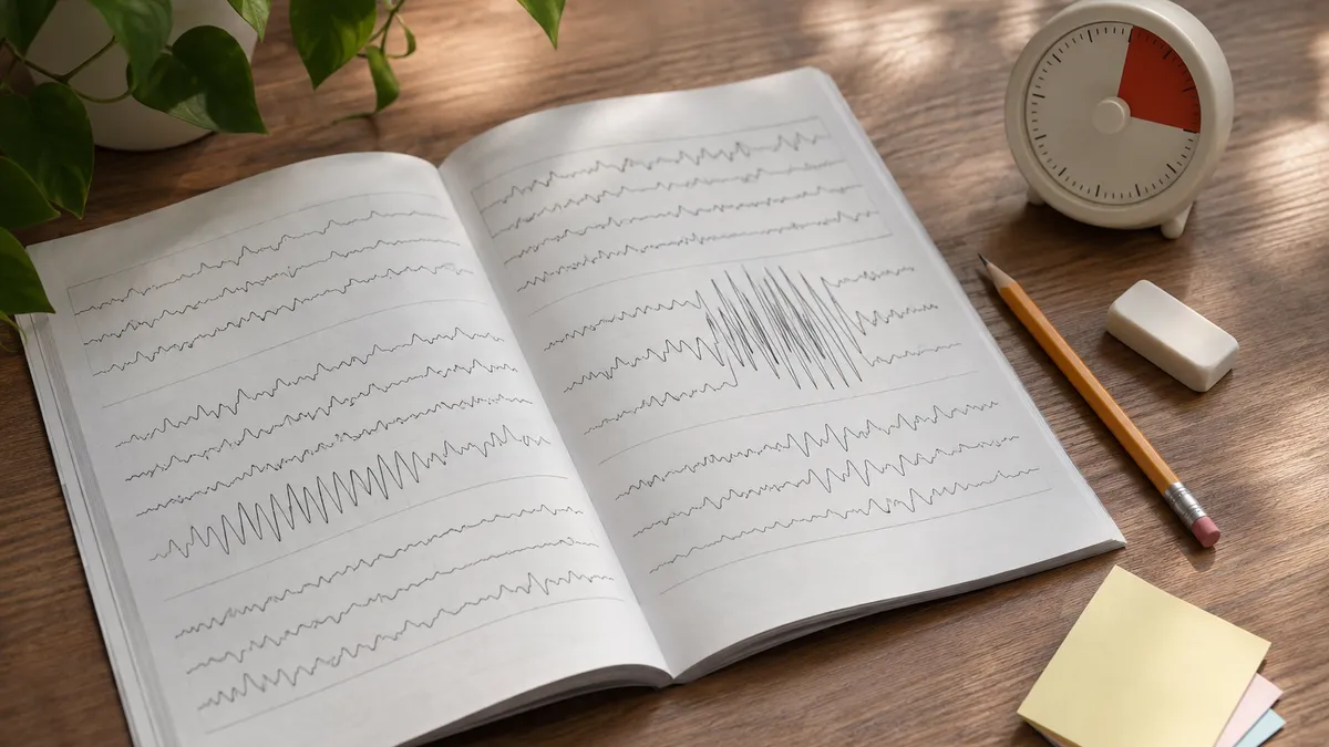
Brain rhythms are grouped by frequency. Delta waves, from 0.5 to 4 hertz, dominate deep sleep. Theta, 4 to 8 hertz, appears in drowsiness and some childhood states. Alpha, 8 to 13 hertz, is the classic posterior rhythm seen when a relaxed adult closes their eyes. Beta, 13 to 30 hertz, appears with alertness and sedative medications. Gamma, above 30 hertz, relates to focused processing. Each band has normal and abnormal contexts, which is why age and state matter.
EEG excels at timing. It resolves changes within a millisecond or two, far faster than MRI or PET scans, which track blood flow over seconds. That makes it ideal for catching a seizure as it begins or measuring how quickly the brain responds to a sound. The tradeoff is spatial precision. Because the skull smears the electrical field, localization is limited to roughly a few centimeters, and deep structures such as the hippocampus contribute only weakly.
This blurring is called volume conduction. Electrical current spreads through cerebrospinal fluid, bone, and scalp, so a source in one area can appear at several electrodes. Skull thickness, hair, and tissue conductivity all change the picture. Clinicians compensate by using multiple montages, which display the same data using different electrode pairings. A bipolar montage highlights local differences, while a referential montage shows each electrode against a common point. Comparing both helps pinpoint where an abnormal discharge truly begins.
Because EEG is dominated by postsynaptic activity, a spike on the screen is not literally an action potential. Epileptiform spikes represent large, synchronous bursts of depolarization across many neurons, a phenomenon called a paroxysmal depolarization shift, which summates into a sharp wave. The name spike describes its shape on the page, not the cellular event. Keeping this vocabulary straight is a frequent exam trap, and our practice questions on epileptiform patterns test it directly.
What Is an EEG Medical Test Used to Diagnose?
The most common reason doctors order an EEG medical test is suspected epilepsy. A routine recording can reveal interictal epileptiform discharges, brief spikes or sharp waves that occur between seizures. Finding them supports a diagnosis and helps classify the seizure type as focal or generalized. However, a single routine study captures abnormalities in only about 30 to 50 percent of people with epilepsy, so a normal result never fully rules the condition out.
When the first test is inconclusive, neurologists often repeat it, add sleep deprivation, or move to longer monitoring. Sleep increases the chance of capturing discharges because drowsiness activates epileptic networks. Video EEG in a hospital unit, or an ambulatory device worn at home for 24 to 72 hours, records actual events so doctors can match behavior to brain activity. This guides medication choices and, in some cases, referral for epilepsy surgery evaluation.

Is an EEG Test Worth It? Strengths and Limits
- +Painless, noninvasive, and free of radiation
- +Millisecond timing captures seizures as they happen
- +Widely available in hospitals and outpatient labs
- +Far less expensive than MRI, PET, or MEG scanning
- +Can run for hours or days, including at home
- +Helps classify seizures and guide medication decisions
- −Poor spatial resolution of only a few centimeters
- −Does not directly record individual action potentials
- −A normal routine result does not rule out epilepsy
- −Sensitive to artifacts from muscle, eye, and electrical noise
- −Deep brain structures are hard to detect from the scalp
- −Interpretation requires an experienced reader
Pros and Cons at a Glance
| Pros | Cons |
|---|---|
| Painless, noninvasive, and free of radiation | Poor spatial resolution of only a few centimeters |
| Millisecond timing captures seizures as they happen | Does not directly record individual action potentials |
| Widely available in hospitals and outpatient labs | A normal routine result does not rule out epilepsy |
| Far less expensive than MRI, PET, or MEG scanning | Sensitive to artifacts from muscle, eye, and electrical noise |
| Can run for hours or days, including at home | Deep brain structures are hard to detect from the scalp |
| Helps classify seizures and guide medication decisions | Interpretation requires an experienced reader |
EEG Test Preparation Checklist Before Your Appointment
- ✓Wash your hair the night before with plain shampoo.
- ✓Skip conditioner, gel, hairspray, and oils on test day.
- ✓Remove braids, extensions, and tight hair accessories if possible.
- ✓Follow your lab's caffeine instructions exactly.
- ✓Eat a normal meal so blood sugar stays steady.
- ✓Bring a current list of all medications and doses.
- ✓Ask your doctor whether to take or hold antiseizure drugs.
- ✓Follow sleep deprivation instructions if your study requires it.
- ✓Wear a comfortable button-front shirt that avoids pulling over the head.
- ✓Arrange a driver if you will be very tired afterward.
Scalp EEG Records Synaptic Fields, Not Single Spikes
Scalp EEG measures summed postsynaptic potentials from aligned cortical pyramidal neurons, not individual action potentials. Only invasive electrodes placed in or on the brain can record spiking from nearby cells. Remember this distinction for exams and for conversations with your care team.
EEG Test Price, Cost, and How Long an EEG Test Takes
The EEG test price varies widely across the United States. A routine outpatient EEG often bills between $200 and $1,000 at an independent clinic, while hospital-based facilities may charge $1,000 to $3,000 or more once technical and professional fees are combined. The EEG test cost depends on region, facility type, recording length, and whether a neurologist's interpretation is billed separately. Because list prices rarely match what insurers pay, asking for an estimate before scheduling is the smartest first step.
Longer or more complex studies cost more. An ambulatory EEG worn for 24 to 72 hours commonly runs from about $1,000 to $3,500 or higher, and inpatient video EEG monitoring is billed per day alongside room charges, often totaling many thousands of dollars. Sleep-deprived studies and tests that include activation procedures may add modest fees. Pediatric studies sometimes require extra staff time, which can raise the technical charge, so confirm the exact billing codes with the facility.
Most commercial insurers, Medicare, and Medicaid cover EEG when it is medically necessary, such as evaluating seizures, unexplained loss of consciousness, or altered mental status. Coverage still depends on your plan's deductible, copay, and prior authorization rules. Patients with high-deductible plans may owe the full allowed amount until the deductible is met. Ask your insurer about the CPT code, commonly 95816 for awake and drowsy recordings or 95819 for awake and asleep studies.
If you are uninsured, ask about self-pay discounts, which many hospitals offer, and request an itemized bill. Nonprofit and academic centers may have financial assistance programs, and some independent neurodiagnostic labs publish flat cash rates. Comparing two or three providers can reveal large differences for the same study. Do not skip a recommended test because of the sticker price before asking, since delayed diagnosis of epilepsy can carry greater medical and financial costs.
Now the question of time: how long is an EEG test? A routine recording usually lasts 20 to 40 minutes of actual data collection. Including check-in, measuring your head, applying electrodes, and removing them afterward, expect the appointment to take about 60 to 90 minutes. Studies that include sleep may take up to two or three hours. Plan accordingly, especially if you need to arrange transportation, childcare, or time off work for the visit.
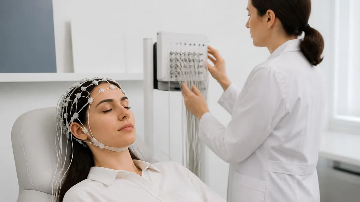
Extended studies take far longer by design. An ambulatory EEG is worn at home for one to three days, and a hospital video EEG may continue for several days until enough events are captured. Sleep-deprived EEGs usually require staying up late the night before, which adds preparation but not necessarily recording time. Technologists often note events with a push button or diary, so keep a written log of symptoms, medication times, and any unusual sensations.
Results do not appear instantly. After recording, a technologist saves the data, and a board-certified neurologist or epileptologist reviews the hours of tracing, often scanning pages of ten seconds each. A written report generally reaches your ordering physician within a few days, though urgent inpatient studies may be read the same day. Ask your doctor how and when you will receive results, and whether a follow-up visit is needed to discuss next steps.

Never stop or skip antiseizure medication before an EEG unless your neurologist specifically tells you to. Abruptly changing doses can provoke seizures. Always confirm medication instructions with the ordering physician or the EEG lab before test day.
EEG Test Side Effects, Safety, and Limitations
People often worry about EEG test side effects, but the recording itself is painless and safe. The electrodes only detect activity; they never deliver current to the brain. There is no radiation, no needle, no contrast dye, and no sedation in a routine study. Most patients return to normal activities immediately, including driving, unless they were sleep deprived or a child received a sedative. Infants and adults alike tolerate the procedure well.
The most common complaints are minor. Conductive paste or adhesive can cause temporary skin redness or itching, and removing the paste may require a damp cloth and a good shampoo. Some people feel mild soreness where electrodes were pressed or taped, especially with collodion glue, which has a strong odor. Anyone with a known allergy to adhesives, latex, or tape should tell the technologist so an alternative can be used.
Activation procedures carry a small additional risk. Hyperventilation, breathing deeply and quickly for about three minutes, can cause lightheadedness, tingling in the fingers, or dizziness; it is generally avoided in people with certain heart, lung, or vascular conditions. Photic stimulation uses flashing lights to look for sensitivity, and in rare cases it can trigger a seizure in someone who is photosensitive. Trained staff are present and prepared to respond immediately.
Because capturing a seizure is sometimes the goal of longer monitoring, patients may have seizures during inpatient video EEG, especially if medications are reduced under supervision. This is a planned, controlled setting with nurses and padded safety rails on beds. Ambulatory EEG poses little added risk, though skin irritation can build after several days. Follow instructions about keeping the electrodes dry, since showering during monitoring can damage equipment and ruin the recording.
EEG has genuine limitations. It records mainly cortical surface activity, so deep sources are hard to see, and a normal result does not exclude epilepsy. Artifacts from eye blinks, muscle tension, sweat, and electrical equipment can mimic or hide real findings. Learning to separate them is a core technologist skill, and clear documentation of every problem during the recording helps the interpreting physician avoid mistaking noise for pathology.
Another limitation is interpretation. Normal variants, such as wicket spikes or benign small sharp spikes, can look like epileptiform activity and lead to overdiagnosis when read by inexperienced clinicians. Medications, age, drowsiness, and even caffeine change the background. For that reason, results must always be matched to the clinical story. A reader who sees a questionable discharge should weigh its shape, field, and behavior before calling it abnormal.
Finally, return to the opening question. Scalp EEG does not directly measure action potentials, and only invasive electrodes such as electrocorticography grids, stereo-EEG depth leads, or single-unit microelectrodes can record spikes from individual cells. Even then, researchers distinguish local field potentials, which reflect synaptic activity, from multi-unit activity, which reflects firing. Knowing which signal a technique captures helps you choose the right tool and interpret every result correctly.
Practical Tips for Your EEG Test and Exam Prep
If you are a patient, preparation begins the day before. Wash your hair with plain shampoo, skip conditioner, gels, sprays, and oils, and avoid braids, extensions, or tight styles when possible. Product residue raises electrode impedance and weakens the signal. Eat normally unless instructed otherwise, since low blood sugar changes brain rhythms. Bring a list of current medications, and ask your physician whether any antiseizure drugs should be taken or held before the appointment.
Caffeine is a common question. Many labs ask you to avoid coffee, tea, cola, and energy drinks for several hours beforehand because stimulants can alter background activity and make drowsiness harder to achieve. If your test is sleep deprived, follow the exact schedule your lab gives you, such as sleeping only four or five hours. Arrange a driver, because tired patients should not travel alone, especially after being awake most of the night.
During the test, relax and follow instructions. You will lie or recline on a bed with eyes mostly closed. Try to avoid clenching your jaw, frowning, or moving your head, since muscle and movement artifacts hide brain waves. The technologist may ask you to open and close your eyes, breathe deeply, or watch flashing lights. These steps are routine. If you feel dizzy or uncomfortable at any moment, say so right away.
After the test, expect some paste in your hair. Most is removed at the clinic with gauze and a gentle solvent, and the rest washes out at home with warm water and shampoo. Parents helping a child can bring a hat or change of clothes. Write down any questions that come up and schedule a follow-up. If symptoms continue after a normal EEG, tell your doctor, because further testing may still be appropriate.
If you are studying for a technologist credential, build your preparation around the 10-20 system, montages, filters, sensitivity settings, and artifact recognition. Draw the electrode map from memory until you can label Fp1, F3, C3, P3, O1, and the matching right-side sites without hesitation. Practice predicting how a change in sensitivity alters the apparent height of a wave. Hands-on repetition builds speed far better than rereading notes.
Use question banks regularly. Short daily quizzes on epileptiform patterns, activation procedures, and instrumentation expose weak areas quickly, and reviewing the explanation for each wrong answer is where most learning occurs. Keep a running list of missed topics and revisit them every few days. Mix timed sets with untimed review, and simulate test conditions before your exam so that pacing feels natural and nerves do not drive your decisions.
Finally, connect concepts rather than memorizing isolated facts. Link postsynaptic potentials to dipoles, dipoles to scalp fields, and scalp fields to what you see on a montage. When you can explain why EEG does not capture individual action potentials, you understand the foundation for everything else. Review the related guides below on electrodes, artifacts, ambulatory monitoring, and ERP, then return to the practice quizzes to confirm that the knowledge has stuck.
Sample EEG - Electroencephalography Practice Questions
Try these questions from our free EEG - Electroencephalography practice tests. The correct answer and an explanation follow each question.
A K-complex is defined as a well-delineated negative sharp wave followed by a positive component with a total duration of at least:
- A. 0.5 seconds
- B. 1.0 second
- C. 1.5 seconds
- D. 0.25 seconds
Answer: A. 0.5 seconds
AASM criteria define a K-complex as a biphasic wave lasting ≥0.5 seconds, maximal over frontal regions.
Informed consent for EEG should include information about which potential discomfort or risk?
- A. Risk of permanent brain damage from the recording
- B. Skin irritation from electrode adhesive and small possibility of seizure provocation during activation procedures
- C. Risk of electric shock from the EEG machine
- D. Radiation exposure during the recording
Answer: B. Skin irritation from electrode adhesive and small possibility of seizure provocation during activation procedures
Informed consent should cover possible skin irritation from electrode paste or adhesive (collodion), mild discomfort from skin preparation, and the small risk that activation procedures could provoke a seizure in susceptible individuals.
When performing intermittent photic stimulation (IPS) according to standard laboratory protocols, what is the most appropriate action for the technologist?
- A. Instruct the patient to keep their eyes open for the entire duration of the procedure.
- B. Continue flashing for at least 30 seconds after a generalized discharge appears to confirm its persistence.
- C. Begin with the highest frequency (30 Hz) and decrease in 5 Hz increments.
- D. Present flashes at varying frequencies, typically starting low and increasing, testing with both eyes open and eyes closed.
Answer: D. Present flashes at varying frequencies, typically starting low and increasing, testing with both eyes open and eyes closed.
Standard IPS protocol involves presenting flash trains of several seconds at various frequencies (e.g., 1, 3, 5, 10, 15, 20, 25, 30 Hz). It is critical to test under both eyes-open and eyes-closed conditions, as photosensitivity can vary. The stimulus should be stopped immediately if a clear, self-sustaining epileptiform discharge is triggered to prevent inducing a seizure.
What is the recommended duration for hyperventilation during a routine EEG recording?
- A. 30 seconds
- B. 1 minute
- C. 3 minutes
- D. 10 minutes
Answer: C. 3 minutes
Standard hyperventilation is performed for 3 minutes with continued recording for at least 1-2 minutes afterward to capture the recovery phase and any provoked abnormalities.
Take the full EEG - Electroencephalography practice test
EEG Questions and Answers

EEG Artifacts: How to Identify, Eliminate, and Document Common Recording Interference

ERP vs EEG: What Each Test Measures, How They Differ, and What to Expect

EEG Cap Electrodes: Everything You Need to Know About the EEG Test
EEG Monitoring: What Is an EEG Test, How It Works, Cost, and What to Expect
Ambulatory EEG: 24-72 Hour Home Recording for Suspected Seizures
How this page was made: written with AI tools and published by Nathan Beja, founder of Practice Test Geeks, who is responsible for its content. Exam facts are checked against the official sources listed below. Not reviewed by a subject-matter expert. Editorial policy
Sources & references
Exam facts on this page (fees, format, dates, passing scores) are checked against these official sources; the date is our last check. Editorial policy
- abret.org — Late registration, score validity, test delivery, fees, exam format, passing score, retake policy, score release, test dates · verified Oct 2026