
EEG Test Explained: Does EEG Measure Brain Activity, What It Costs, and What to Expect
🧠 Does EEG measure brain activity? Learn what an EEG test is, how long it takes, what it costs, possible side effects, and what to expect on test day.

Does EEG measure brain activity? Yes, and that is exactly what an EEG test is designed to do. Electroencephalography records the tiny electrical signals produced when networks of neurons communicate. Small metal sensors on the scalp pick up those signals, an amplifier magnifies them thousands of times, and a computer draws them as wavy lines a neurologist can read. The test is painless, noninvasive, and one of the most widely used diagnostic tools in neurology.
If you have been told to schedule an EEG test, you probably have practical questions. Why was it ordered? Will it hurt? How should you prepare? Doctors most often order it for suspected seizures, unexplained fainting or blackouts, sleep disorders, confusion, or changes in alertness. The test gives a real-time picture of how the brain is functioning, which an MRI or CT scan cannot do because those scans show structure rather than electrical behavior.
The brain runs on electricity. Each neuron generates small voltage changes when it receives signals from neighbors. One neuron is far too weak to detect from outside the skull, but when thousands of neighboring cortical neurons fire together, their combined signal reaches the scalp at roughly 10 to 100 microvolts. That is about a thousand times weaker than a AA battery, which is why EEG equipment needs sensitive amplifiers and careful skin preparation to get a clean recording.
The history is short but impressive. German psychiatrist Hans Berger recorded the first human EEG in 1924 and noticed a steady rhythm of about ten cycles per second when a person relaxed with closed eyes. That rhythm, now called the alpha rhythm, is still one of the first things a technologist looks for. A century later, the basic principle is unchanged, although digital systems, dense electrode caps, and video monitoring have made recordings far more detailed.
It helps to be clear about what EEG can and cannot do. It measures the timing and pattern of electrical activity with millisecond precision, so it is excellent at capturing seizures, sleep stages, and sudden changes in brain state. It does not read thoughts, measure intelligence, or diagnose mental illness by itself. It also records mostly from the outer cortex, so very deep structures contribute only faintly. Knowing those limits keeps your expectations realistic and the results easier to understand.
Many people searching for what is an eeg test also want to know about variations. A routine EEG lasts about 20 to 40 minutes. A sleep-deprived EEG asks you to stay up late beforehand to make abnormal activity more likely to appear. An ambulatory EEG records for one to three days at home. Each version answers the same basic question, whether brain electrical activity is normal, but with different chances of catching infrequent events.
This guide walks through the full picture: how the test works, what happens step by step, what it costs in the United States, how long it takes, possible side effects, and how to prepare. You will also find practice resources for anyone studying EEG as a technologist or student. Whether you are a patient, a caregiver, or a learner, the goal is plain-language answers so that the day of your test feels familiar instead of intimidating.
The EEG Test by the Numbers
What Is an EEG Test? The Process Step by Step
Check-In and Health History
Measuring and Marking the Scalp
Attaching the Electrodes
Resting Recording
Activation Procedures
Removal and Reading
To understand how an EEG test works, start with the cells behind the signal. The EEG mostly reflects postsynaptic potentials in pyramidal neurons of the cerebral cortex, which are lined up in parallel like a forest of trees. When thousands of these neurons receive input at the same moment, their small voltage changes add together into a signal strong enough to travel through cerebrospinal fluid, bone, and skin to the electrodes.
Each electrode does not work alone. The machine records the voltage difference between pairs of electrodes, or between each electrode and a shared reference, and displays the result as a channel. A standard recording has 16 to 32 channels running at the same time. By comparing channels across the head, a neurologist can see whether a pattern is local, such as the left temporal lobe, or widespread across both hemispheres, which changes the diagnosis considerably.
Brain waves are described by their speed, measured in cycles per second, or hertz. Delta waves run below 4 Hz and dominate deep sleep. Theta waves run 4 to 8 Hz and appear in drowsiness. Alpha waves, 8 to 13 Hz, appear over the back of the head when you relax with eyes closed. Beta waves, 13 to 30 Hz, accompany active thinking, and gamma waves above 30 Hz relate to fast processing.
Normal does not mean one single pattern. A healthy adult who is awake shows posterior alpha that disappears when the eyes open, mild beta over the front of the head, and symmetrical activity between the two sides. Children show slower rhythms that speed up as the brain matures, and older adults may show slight slowing. Interpreting a recording therefore depends on age and state of alertness, which is why a trained specialist, not software alone, makes the final call.
Abnormal patterns usually fall into two families. Epileptiform discharges, such as spikes and sharp waves, are brief electrical events that point toward a tendency for seizures. Slowing, meaning too much delta or theta while awake, suggests general or focal brain dysfunction from causes like injury, infection, medication effects, or encephalopathy. Neither finding is a diagnosis on its own, and doctors combine the EEG with your symptoms, exam, and imaging.
EEG has a major strength that other brain tests lack: speed. It resolves changes within about a millisecond, far faster than functional MRI, which tracks blood flow over several seconds. That makes EEG the best everyday tool for catching the start of a seizure or watching how the brain responds to a sound or flash. Its weakness is spatial precision, since it cannot pinpoint exactly where deep activity begins.
So when someone asks whether EEG measures brain activity, the accurate answer is yes, directly and in real time, but with some nuance. It measures electrical activity of the cortex, not chemical activity, blood flow, or structure. That is why neurologists often pair it with MRI, which shows anatomy, and with clinical history. Together these tools give a fuller picture than any single test could, and EEG supplies the timing piece that the others cannot.
EEG Medical Test Types: Routine, Sleep-Deprived, and Ambulatory
A routine EEG is the standard version of the eeg medical test. It takes place in a clinic or hospital lab, lasts about 20 to 40 minutes of actual recording, and includes resting periods plus hyperventilation and photic stimulation. It is the usual first step when a doctor suspects seizures, unexplained spells, or altered mental status, and it requires almost no preparation beyond clean, dry hair.
The limitation of a routine EEG is its short window. If abnormal discharges are infrequent, a 30 minute recording can miss them entirely, and a normal result does not rule out epilepsy. Roughly half of people with epilepsy show abnormal activity on a first routine study, and repeating the test raises the yield considerably. Your neurologist may therefore suggest a longer or sleep-based recording if suspicion stays high.
Is an EEG Test Worth It? Benefits and Drawbacks
- +Completely noninvasive, with no needles, radiation, or contrast dye
- +Records brain electrical activity in real time with millisecond precision
- +Helps confirm epilepsy and classify seizure types
- +Useful for evaluating sleep disorders, confusion, and altered consciousness
- +Widely available and typically covered by insurance when medically necessary
- +Safe for children, older adults, and pregnant patients
- −A normal routine EEG does not rule out epilepsy
- −Mostly records the cortex and misses very deep brain activity
- −Muscle movement and blinking can create artifacts that complicate reading
- −Setup with paste and electrodes can be messy and time consuming
- −Cannot show brain structure, so imaging is often needed too
- −Cost varies widely between hospitals and outpatient labs
EEG Test Preparation Checklist
- ✓Wash your hair the night before and skip conditioners, oils, and gels.
- ✓Remove braids, extensions, clips, and wigs if the lab asks you to.
- ✓Eat normally and do not skip meals, since low blood sugar can alter the tracing.
- ✓Avoid caffeine for 8 to 12 hours if your lab instructs you to.
- ✓Follow sleep instructions exactly, including staying up late for a sleep-deprived study.
- ✓Take your regular medications unless your doctor says otherwise.
- ✓Bring a list of all medications and supplements with doses.
- ✓Wear a comfortable shirt that buttons or zips, not one pulled over the head.
- ✓Arrange a ride home if you were asked to sleep-deprive or may feel drowsy.
- ✓Call your insurer beforehand to confirm coverage, copay, and any prior authorization.
A Normal EEG Does Not Always Mean Nothing Is Wrong
Epileptiform discharges come and go, so a single 30 minute recording can look completely normal in someone who has epilepsy. Repeating the study, adding sleep, or choosing a longer recording meaningfully raises the chance of catching abnormal activity. Ask your neurologist how your result fits your symptoms rather than treating one tracing as final.
One of the most common questions is how long an EEG test takes. Plan for about 60 to 90 minutes at the facility for a routine study. Roughly 30 to 45 minutes goes to measuring and attaching electrodes, 20 to 40 minutes to recording, and a few minutes to removal. Sleep-deprived studies often run 90 minutes or longer, and ambulatory recordings run 24 to 72 hours, though you spend only about an hour in the clinic for hookup.
Cost is the other big question, and the honest answer is that it varies. In the United States, the billed charge for a routine outpatient EEG commonly ranges from about $200 at a small independent lab to $3,000 or more at a hospital, because hospitals add facility fees. The amount you actually pay depends on your insurance, deductible, and whether the facility is in network, so the sticker price is rarely your final bill.
Insurance usually covers an EEG when it is medically necessary, such as evaluation of seizures, syncope, or suspected encephalopathy. Medicare and most commercial plans cover it, though you may owe a copay or coinsurance of 10 to 20 percent after the deductible. Some plans require prior authorization, especially for ambulatory or video EEG. Calling your insurer before the appointment and asking for the CPT code from the clinic can prevent surprises.
Extended studies cost more. An ambulatory EEG, which runs 24 to 72 hours, often bills between $1,000 and $3,500 or more depending on length and whether video is included. Inpatient video EEG monitoring in an epilepsy unit, which can last several days, is usually billed as part of a hospital stay and can reach tens of thousands of dollars before insurance. These are the studies where authorization and cost estimates matter most.
If you are uninsured or have a high deductible, there are ways to lower the price. Ask the scheduler for the self-pay or cash discount, which can be significantly lower than the billed charge. Compare a hospital outpatient department with an independent neurology office, since the same test often costs less in the latter. Nonprofit hospitals must offer financial assistance programs, and you can request an itemized estimate in writing before committing.
Also remember that the recording fee is not the only line item. The neurologist who interprets the tracing usually bills separately for the professional reading, so you may receive two statements. Some labs also charge for a technologist’s supplies or a follow-up visit. Asking whether the quote is global, meaning technical plus professional components together, helps you compare prices between providers more accurately and avoid bills you did not anticipate.
Results timing is the last piece. The technologist cannot interpret the tracing during the study, and the neurologist reviews it afterward, often within one to five business days. Your ordering doctor then explains the findings and next steps. If you want to understand how to how long is an eeg test reports and tracings are reviewed, our guide on reading EEGs explains the patterns doctors look for in each stage.
Because EEG test cost can differ by thousands of dollars between facilities, ask for a written estimate that includes both the technical and professional fees before you book. Confirm your insurer has approved the study if authorization is required. A short phone call can save a surprise bill.
Safety is usually the first concern for people facing their first test, and the reassuring news is that an EEG is among the safest procedures in medicine. The machine only listens. It sends no electricity into your head, so you cannot be shocked, and there is no radiation, injection, or sedation involved in a routine study. Children, older adults, and pregnant patients can all have the test. Most people describe it as boring rather than uncomfortable.
That said, people ask about eeg test side effects, and a few mild ones do exist. The most common is skin irritation: redness, itching, or small marks where electrodes or adhesive sat, which usually fades within hours or a day. Some people find the paste sticky and hard to wash out, and sensitive skin may react to the adhesive or tape. Tell the technologist about allergies before hookup so they can choose gentler materials.
The more notable risk involves the activation procedures. Hyperventilation can cause lightheadedness, tingling in the hands and lips, or dizziness, which resolves within a minute or two of normal breathing. Flashing lights, called photic stimulation, can very rarely trigger a seizure in people who are photosensitive. This risk is small, and the lab is prepared to respond right away. Always mention a seizure history so the team can take precautions.
Sleep deprivation, when requested, brings its own temporary effects. You may feel tired, irritable, or foggy the next day, and because sleep loss itself can lower the seizure threshold, doctors only request it when the benefit is worth it. Do not drive if you are drowsy afterward. If you take seizure medication, never stop it on your own for a test. Your neurologist will tell you if any adjustment is appropriate.
Long recordings carry practical, not medical, burdens. During ambulatory EEG you wear a head wrap and a recorder for up to three days. You cannot shower normally, sleeping may be awkward, and the adhesive can irritate skin. Inpatient video monitoring may involve a planned reduction of seizure medicine under supervision to capture events, which carries real risk and is only done in a hospital with trained staff watching continuously.
Know when to call for help. Contact the lab or your doctor if you develop a spreading rash, blistering, or significant swelling at electrode sites, or if you feel unwell in a way that worries you. After a seizure during a test, the team will evaluate you before you leave. For ordinary side effects such as mild redness or a headache from missing coffee or sleep, rest, hydration, and gentle washing are usually enough.
Finally, remember the emotional side. Waiting for results can feel stressful, particularly if the test was ordered after a frightening episode. Bring a family member, write your questions down in advance, and ask when and how results will be shared. Understanding that the procedure itself is low risk, and that any abnormal finding is a starting point for a plan rather than a verdict, makes the whole experience far easier to manage.
Good preparation makes the recording cleaner and the results more useful, and most of it is simple. Arrive with freshly washed, completely dry hair and no styling products, because oils and gel keep electrodes from making good contact. Wear comfortable clothing that does not need to be pulled over your head. Bring your insurance card, photo ID, and any referral paperwork. Arriving ten minutes early lets you finish forms without feeling rushed.
Follow the sleep and caffeine instructions on your appointment sheet to the letter, since labs differ. For a routine study, most want you to sleep normally and eat beforehand. For a sleep-deprived test, they may ask you to sleep only four or five hours or stay up past a certain time. Caffeine can mask drowsiness and change the tracing, so many labs ask you to skip coffee, tea, soda, and energy drinks that morning.
Medication questions deserve a call ahead. Do not stop anti-seizure medicine, antidepressants, or anything else on your own, because sudden changes can trigger seizures and may not help the test anyway. Instead, bring a complete list with doses and tell the technologist what you took and when. Sedatives and some sleep aids can alter brain waves, and the neurologist reading your study needs that context to interpret the pattern correctly.
During the test, your main job is to cooperate calmly. Stay as still as you comfortably can, since blinking hard, clenching your jaw, and moving your head create artifacts that look like brain activity. Follow instructions to open and close your eyes, breathe deeply, or watch the light. If you feel dizzy or uncomfortable, say so immediately. Technologists are trained to adjust, pause, or stop the recording whenever you need it.
It is fine, even helpful, to fall asleep. Many abnormal patterns show up in light sleep, so a short nap during the recording can increase the diagnostic value. If you tend to feel anxious in medical settings, bring headphones for the waiting room, take slow breaths, and let the technologist know. A relaxed patient produces cleaner data, which means fewer repeat studies and a more confident report for your doctor.
Afterward, plan a simple routine. Wash the paste out with warm water and shampoo, and use a little conditioner if it feels stubborn, since the gel is water soluble. Eat, rehydrate, and rest if you were sleep-deprived. Ask the front desk when results will be sent and to whom. Then schedule or confirm the follow-up appointment so findings are reviewed with you rather than sitting unread in a portal.
If you are studying EEG as a future technologist or nursing student, turn your experience into learning. Review the 10-20 electrode system, memorize frequency bands, and practice spotting artifacts versus true epileptiform discharges. Our free practice questions let you test pattern recognition at your own pace, with explanations for every answer. Pairing real-world understanding with repeated question practice is the fastest way to build confidence before a credentialing exam or clinical rotation.
EEG Questions and Answers

Sleep Deprivation EEG: What It Is, How It Works, and What to Expect

How to Read an EEG Test: A Complete Guide to Brain Wave Patterns and Results
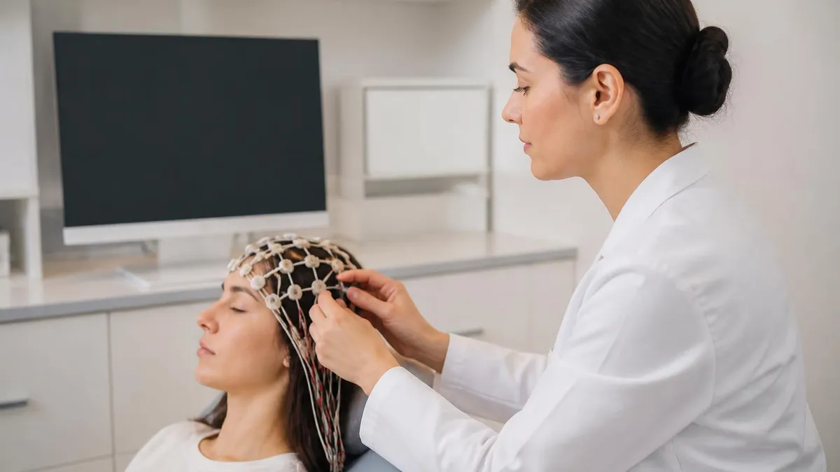
Abnormal EEG Results: What They Mean, Common Patterns, and Next Steps
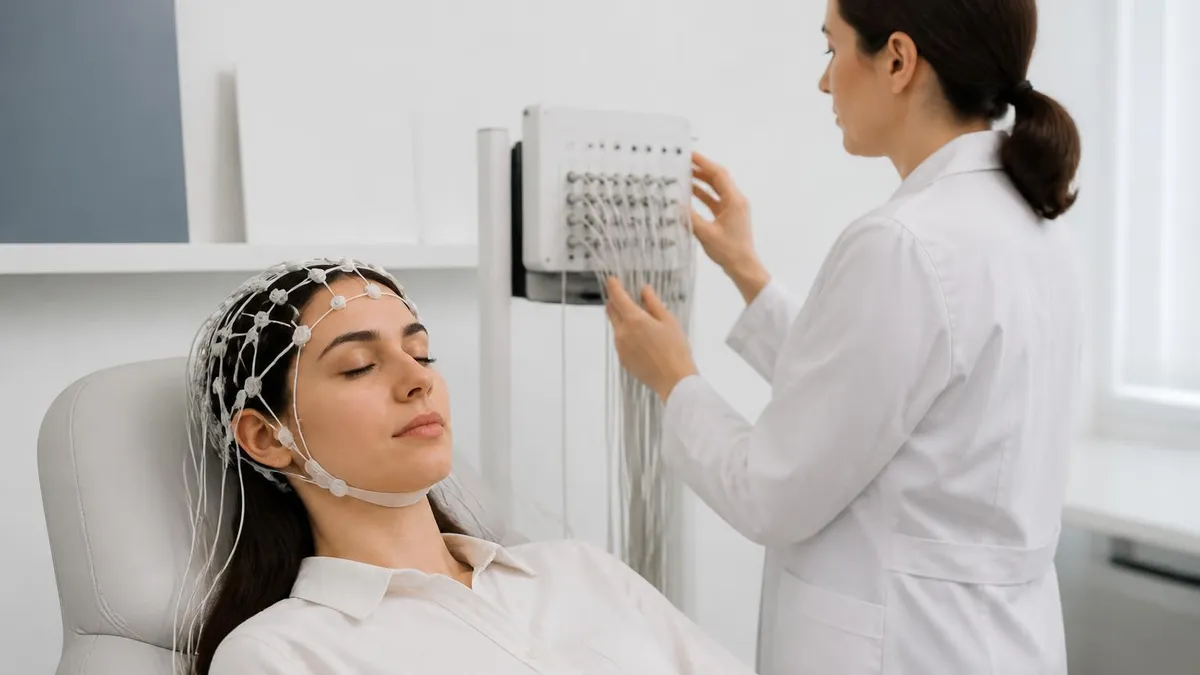
Ambulatory EEG: 24-72 Hour Home Recording for Suspected Seizures
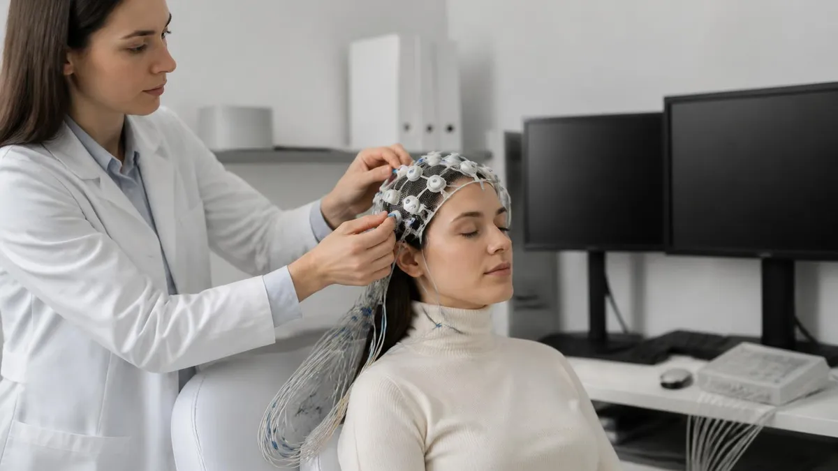
EEG for Seizures: How the Test Diagnoses Epilepsy and What to Expect
How this page was made: written with AI tools. Exam facts are checked against the official sources listed below. Not reviewed by a subject-matter expert. Editorial policy
Sources & references
Exam facts on this page (fees, format, dates, passing scores) are checked against these official sources; the date is our last check. Editorial policy
- abret.org — Test delivery, eligibility, fees, exam format, passing score, retake policy, score release, test dates, late registration, score validity · verified Oct 2026