
EEG Test Technical Specifications: How a 32 EEG Recording Works, What It Costs, and What to Expect
🧠 Learn what an EEG test is, how long it takes, what it costs, its side effects, and the technical specs behind a 32 EEG channel recording in this US guide.
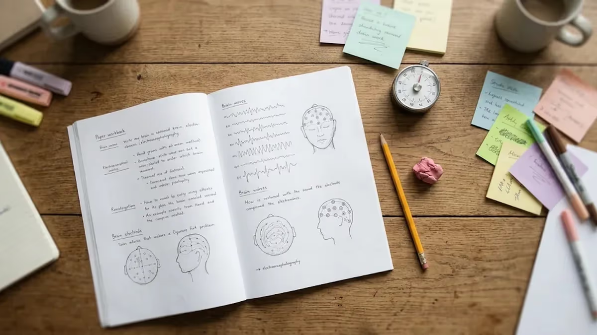
An EEG test records the brain's electrical activity through small metal discs placed on the scalp, and the number of discs matters more than most patients realize. A 32 EEG setup uses 32 recording electrodes, which is more than the 21 used in a standard clinic study and fewer than the 64 or 128 used in many research labs. This guide explains the technical specifications behind that number in plain language for patients, students, and new technologists.
If you are asking what is an EEG test, the short answer is a painless medical test that amplifies tiny voltage fluctuations produced by cortical neurons and displays them as wavy lines. Physicians use it to evaluate seizures, sleep disorders, encephalopathy, and unexplained loss of consciousness. The signals captured are roughly 10 to 100 microvolts, thousands of times weaker than a AA battery, so hardware specifications decide whether the result is readable.
Understanding the specifications helps in practical ways. A patient comparing providers can ask whether a quote covers a 21-channel or a 32 EEG montage, and how many minutes of recording are included. A student preparing for a registry exam needs to know sampling rates, filter settings, and impedance limits. A caregiver booking an ambulatory study needs to know battery life and storage capacity. Each of those questions has a concrete numerical answer that this article provides.
Technical choices also affect how artifacts appear on the tracing. Eye blinks, muscle tension, and electrical line noise can mimic or hide real brain activity when settings are poorly chosen. For a deeper look at how these problems are recognized and corrected, read our companion guide on eeg technical specifications and recording interference, which pairs well with the hardware details you will read below, and keep it handy while studying.
We will cover four areas. First, what the equipment physically consists of: electrodes, amplifiers, converters, and software. Second, the settings a technologist chooses, such as sampling rate, filters, and sensitivity. Third, real-world logistics, including how long is an EEG test, plus EEG test cost and EEG test price ranges in the United States. Fourth, safety, including EEG test side effects, which are minimal but worth understanding before your appointment.
Throughout the article, numbers reflect typical practice in US hospitals and outpatient neurology clinics that follow American Clinical Neurophysiology Society guidelines. Individual labs may differ slightly, and your neurologist always decides which protocol fits your clinical question. Treat this page as educational background rather than medical advice, and bring your own questions to the ordering provider, who knows your history and can tailor the study to your situation.
Finally, you will find practice resources at the end. Quiz questions on abnormal epileptiform patterns and activation procedures let you test recall while the technical details are fresh. Whether you are a patient preparing for a first recording or a trainee building foundations for an exam, the goal is the same: to turn a confusing list of specifications into knowledge you can actually use on test day.
EEG Technical Specifications by the Numbers
What Is an EEG Test? The Recording Process Step by Step
Preparation and History
Scalp Measurement and Cap Placement
Impedance Check
Baseline Recording and Activation
Review and Reporting
The electrodes are the first link in the chain. Most clinical systems use tin, silver, or silver-chloride discs about 10 millimeters wide, chosen because they hold a stable electrical potential. In a 32 EEG cap, sensors are embedded in fabric or silicone at fixed positions, which speeds setup. Gold cup electrodes held with paste remain common for longer studies because they stay attached for hours and usually give excellent signal quality.
After the electrodes, the signal enters an amplifier. Clinical amplifiers typically provide input impedance above 100 megohms and common-mode rejection of at least 100 decibels, meaning they cancel interference that appears equally on every electrode. Gain converts microvolts into volts the converter can digitize. These numbers explain why a quiet, grounded room matters: the amplifier is designed to ignore noise, but only up to certain limits of strength.
The analog-to-digital converter then samples the voltage. The ACNS minimum is generally 200 samples per second per channel, and most US labs record at 256, 500, or 512 Hz. Resolution is commonly 16 to 24 bits, which determines how finely amplitude differences are measured. Higher sampling rates capture fast activity such as spikes and high-frequency oscillations, but they produce larger files, a trade-off especially important during multi-day monitoring.
Filters shape what the reader sees. A typical low-frequency filter is set near 0.5 to 1 Hz to remove slow drift from sweat and movement, while the high-frequency filter sits around 70 Hz to cut muscle noise. In the United States, a 60 Hz notch filter reduces power-line interference, though technologists avoid relying on it because it can distort sharp waves. Good electrode contact is always the preferred fix over aggressive filtering.
Brain rhythms are classified by frequency. Delta waves run 0.5 to 4 Hz and dominate deep sleep. Theta runs 4 to 8 Hz, alpha 8 to 13 Hz, and beta 13 to 30 Hz. Gamma exceeds 30 Hz. A healthy awake adult with closed eyes shows a posterior alpha rhythm of roughly 9 to 11 Hz, and its presence, symmetry, and reactivity to eye opening are among the first things a reader checks.
Display settings are equally standardized. A common sensitivity is 7 microvolts per millimeter, with a paper-speed equivalent of 30 millimeters per second, so each screen page shows about 10 seconds of data. Readers switch between montages, such as longitudinal bipolar, transverse bipolar, and referential, because a given abnormality may be obvious in one arrangement and subtle in another. Those same settings are covered on registry exams.
Impedance deserves its own mention. Clinical guidelines generally call for electrode impedances between 100 ohms and 5 kilo-ohms, with matched values across sites. Mismatched impedance turns environmental noise into a visible difference between channels. That is why technologists recheck contact during the recording, and why a 32 EEG cap takes longer to prepare than a smaller montage: every additional channel is another connection to verify and maintain.
EEG Test Specifications by Type: Routine, 32 EEG, and Ambulatory
A routine EEG test is the most common EEG medical test. It uses the standard 21 electrodes of the 10-20 system and records for 20 to 40 minutes, often capturing both wakefulness and drowsiness. Sampling is typically 256 to 500 Hz. The patient lies in a hospital bed or recliner while the technologist watches the screen and annotates events in real time.
Activation procedures are built in. Hyperventilation lasts about three minutes, and photic stimulation uses flashing lights at frequencies from roughly 1 to 30 flashes per second. Because the window is short, a routine study can miss abnormalities that occur only occasionally. A normal result therefore does not rule out epilepsy, and repeat or longer testing is common when suspicion remains high.
32 EEG Channels vs Standard 21 Channels: Which Is Better?
- +Higher spatial resolution helps localize where abnormal activity begins
- +Better coverage of temporal and inferior regions often missed by 21 electrodes
- +Supports source localization and advanced research analysis
- +Useful in epilepsy surgery planning and complex diagnostic questions
- +Provides redundancy if a single electrode loses contact
- +Allows flexible montage choices when reviewing the recording
- −Setup takes longer and needs more technologist time
- −More electrodes can mean more discomfort for sensitive scalps
- −Larger data files increase storage and review time
- −Often costs more than a routine study
- −Rarely needed for a straightforward first-seizure evaluation
- −Extra channels also bring extra opportunities for artifacts
How to Prepare for an EEG Test: Pre-Appointment Checklist
- ✓Wash your hair the night before and skip conditioner, gel, or oil.
- ✓Avoid braids, weaves, extensions, and hairspray on test day.
- ✓Ask your doctor whether to continue or hold your medications.
- ✓Eat a normal meal so low blood sugar does not alter the tracing.
- ✓Skip caffeine for the period your lab recommends, usually 8 to 12 hours.
- ✓Follow any sleep-deprivation instructions exactly if they are given.
- ✓Bring a list of all medications and supplements.
- ✓Wear a button-front shirt so nothing pulls over the electrodes.
- ✓Arrive 15 minutes early to complete forms and insurance checks.
- ✓Ask the lab for the quoted EEG test cost and the number of channels.
More channels do not automatically mean a better diagnosis
A clean 21-channel recording with low impedance often beats a noisy 32 EEG study. Electrode quality, patient state, and recording length usually matter more than channel count alone. Ask what clinical question the study answers before assuming that more electrodes is always the better choice.
How long is an EEG test? A routine recording lasts 20 to 40 minutes, but the full appointment usually takes 60 to 90 minutes because of cap placement, impedance checks, and cleanup. A sleep-deprived or sleep-recorded study can last 1 to 2 hours. Ambulatory EEG runs 24 to 72 hours, while inpatient video monitoring can last several days. Plan your day with the longer appointment window in mind, not just the recording time.
Cost is the next question patients ask most. The EEG test cost in the United States ranges widely, from roughly $200 for a basic outpatient study to $3,000 or more for hospital-based or extended recordings. The EEG test price depends on the facility fee, the technical component, and the physician's interpretation fee, which are often billed separately. Freestanding clinics generally charge less than hospital outpatient departments for the same procedure.
Billing uses CPT codes. A routine awake and drowsy study is commonly billed as 95816, while a recording that includes sleep is billed as 95819. Longer monitoring uses separate codes, and ambulatory studies have their own set. Knowing the code lets you request a precise quote and compare providers. Ask whether the quote includes the physician reading fee, because a surprise second bill is one of the most frequent complaints.
Insurance coverage is usually strong when the test is ordered for a clear medical reason, such as suspected seizures, but it often requires prior authorization. Medicare and most commercial plans cover an EEG medical test with a deductible or coinsurance. If you have a high-deductible plan, you may pay a large share. Call your insurer with the CPT code and the facility's name before scheduling to learn your expected out-of-pocket amount.
Uninsured patients have options. Many hospitals offer self-pay discounts of 30 to 60 percent, and some publish price lists as required by federal transparency rules. Ask about financial assistance programs and payment plans, and compare outpatient neurology offices with hospital departments. A 32 EEG study or any extended recording will usually cost more, so confirm in writing which test is being ordered and how many hours of recording are included.
Staffing and equipment also influence price. A registered EEG technologist prepares the patient, and a board-certified neurologist or clinical neurophysiologist interprets the recording. Facilities that maintain 24-hour video monitoring or ICU continuous EEG carry higher overhead, which shows up in billing. That is why the same technical procedure can cost three or four times as much at one location compared with another nearby.
Finally, remember hidden items. Some clinics charge separately for sleep-deprivation setup, video, or after-hours scheduling, and a follow-up appointment to discuss results may also be billed. Request an itemized estimate, keep records of what was quoted, and ask about the date results will be sent. Transparent answers to these questions are a good sign of a well-run lab and help you avoid surprises later.
Never stop anti-seizure or other prescribed medications before an EEG test unless your neurologist explicitly tells you to. Stopping suddenly can trigger seizures. If your doctor wants you to adjust medication for the recording, follow their written instructions exactly and ask about safety precautions.
EEG test side effects are minimal because the test only records activity; it does not send electricity into the body. The electrodes are passive sensors, and the amplifier is battery-isolated or medically isolated from the wall outlet. Most people feel nothing beyond the light pressure of the cap and a cool, sticky sensation from the gel. You can usually drive yourself home and return to normal activities immediately afterward.
The most common complaint is skin irritation. Abrasive prep, adhesive paste, or collodion can cause temporary redness, itching, or small marks where electrodes sat. These typically fade within a day or two. People with sensitive skin or known adhesive allergies should tell the technologist beforehand, because alternatives exist, including different gels and hypoallergenic tape. Washing the scalp gently at home removes residue, though some pastes need a warm, soapy soak.
Activation procedures carry the only meaningful risk. Hyperventilation can cause lightheadedness, tingling in the fingers, or lip numbness for a minute or two. Photic stimulation, the flashing lights, may provoke a seizure in a small number of people with photosensitive epilepsy. Labs are prepared for this, with trained staff and emergency equipment present. If a seizure occurs during testing, it is also diagnostically valuable, because it is recorded live.
Hyperventilation is generally avoided or modified for people with certain conditions, including recent stroke, significant cardiopulmonary disease, sickle cell disease, or moyamoya. Tell your provider about any such history. Children may have the procedure adapted, since breathing exercises are often done by blowing a pinwheel. Pregnancy is not a contraindication for an EEG test, and the study uses no radiation, which is a major advantage over many imaging approaches.
Sleep deprivation instructions can leave you tired, irritable, or headachy, so arrange a ride home if you slept very little. Dehydration or caffeine withdrawal can also cause headache. Neither is a result of the machine itself. Bring water, a snack, and a book or phone to pass the time, and tell the technologist if you feel unwell so they can pause the recording and keep you comfortable.
Data privacy and equipment safety are rarely discussed but important. Recordings are protected health information under HIPAA, and clinics must store them securely. Reusable electrodes are cleaned and disinfected between patients according to infection-control guidelines, while many labs now use single-use sensors. If you have concerns about cleaning practices, ask the technologist directly; they should be able to explain exactly how equipment is disinfected and handled.
Artifacts can also affect safety indirectly, because a misread tracing may lead to the wrong conclusion. Movement, sweat, and poor contact are common culprits. Reviewing our guide to EEG cap electrodes is not required, but it can help you understand why technologists ask you to stay still and keep your eyes closed. Cooperation produces a cleaner recording and a more accurate report.
Start practical preparation a week ahead by calling the lab. Confirm the type of study, the number of electrodes, the expected length, and the quoted price. Ask whether sleep deprivation is required and whether you need to bring anyone with you. Writing down answers prevents misunderstandings on the day, and it also gives you a record if a billing question arises later with the facility or your insurance company.
Make a symptom diary before the appointment. Record when events happen, how long they last, what you felt beforehand, and what a witness observed. Neurologists use these details to decide how to interpret the tracing. If you can, bring a phone video of an event, since a clear video often adds more diagnostic value than a verbal description. Share the diary with the technologist so they can watch for similar behavior.
On the day, dress comfortably, avoid hair products, and eat a normal meal unless told otherwise. Use the restroom before cap placement because you will be lying still for a while. Tell the technologist about any scalp sensitivity, claustrophobia, or anxiety. Technologists handle these concerns every day and can adjust their approach, whether that means explaining each step or giving you time to relax before the recording starts.
During the recording, follow instructions closely. Keep your eyes closed when asked, avoid clenching your jaw, and try not to talk or move your head. Muscle activity creates fast, spiky artifact that can resemble pathological activity on the screen. If you need to cough, swallow, or scratch, tell the technologist so they can mark the event. These small notes save the reader from misinterpreting a harmless movement.
After the test, expect some leftover gel. A warm shower and gentle shampooing remove most of it, and a comb or washcloth helps with stubborn paste. Do not scrub hard if your scalp is irritated. If you wore a long-term ambulatory setup, follow the removal instructions closely and return the equipment on time. Ask when your neurologist will receive the report and who will contact you with results.
Students and trainees should connect specifications to patterns. Memorize sampling rates, filter ranges, impedance targets, and frequency bands, then practice recognizing how they change what appears on screen. Use flashcards for 10-20 electrode names and write out each montage. Our EEG monitoring guide covers what an EEG test is, how it works, and what to expect in more clinical detail, and it is a useful second read.
Finally, take the practice quizzes below to measure retention. Spend extra time on questions you miss and revisit the matching section of this article. If you are a patient, bring your remaining questions to your provider; if you are studying for a credential, repeat the quizzes weekly. Whether you are comparing EEG test price options or learning about a 32 EEG layout, repetition turns technical detail into confident understanding.
EEG Questions and Answers

EEG Artifacts: How to Identify, Eliminate, and Document Common Recording Interference

ERP vs EEG: What Each Test Measures, How They Differ, and What to Expect

EEG Cap Electrodes: Everything You Need to Know About the EEG Test
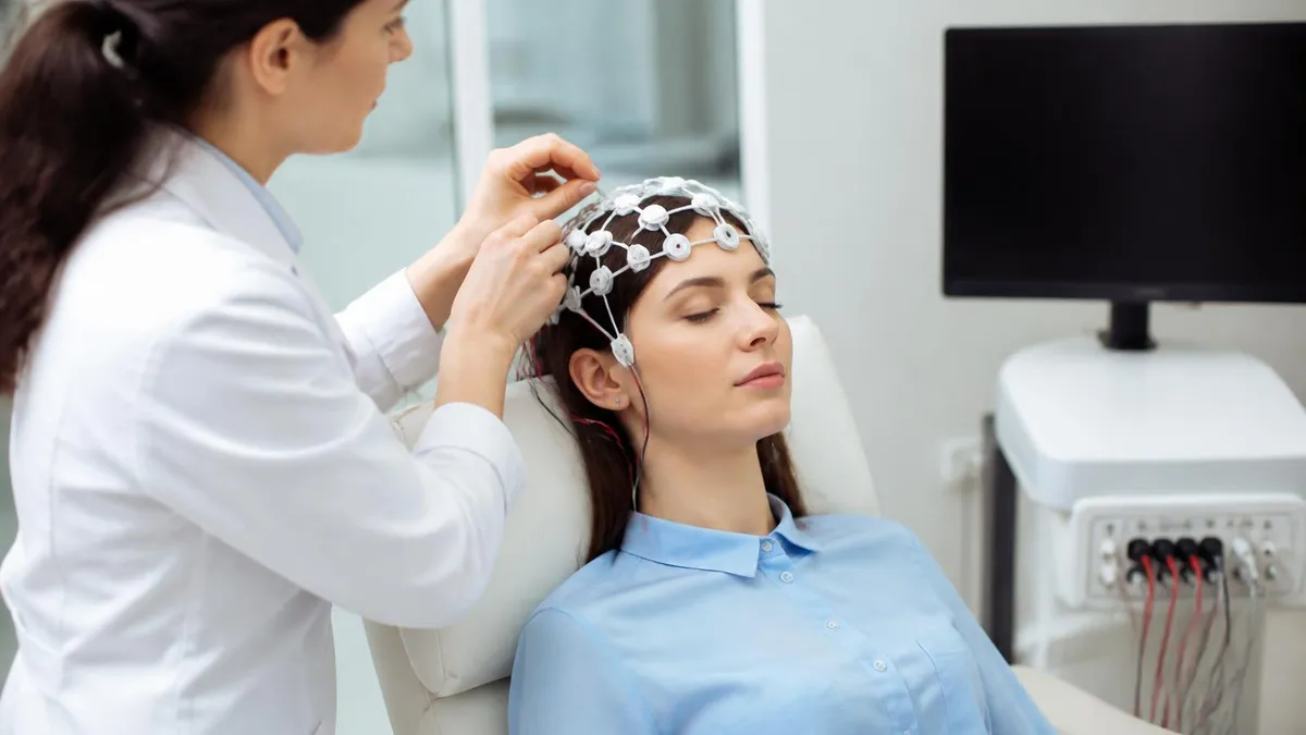
EEG Monitoring: What Is an EEG Test, How It Works, Cost, and What to Expect
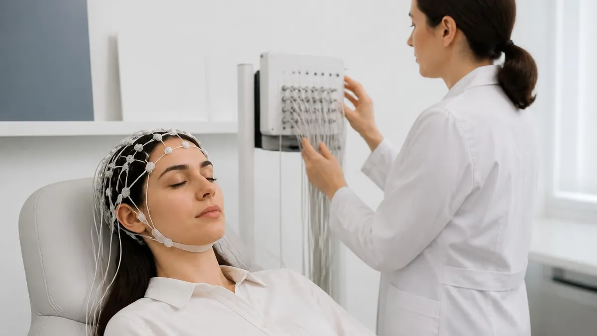
Ambulatory EEG: 24-72 Hour Home Recording for Suspected Seizures
How this page was made: written with AI tools and published by Nathan Beja, founder of Practice Test Geeks, who is responsible for its content. Exam facts are checked against the official sources listed below. Not reviewed by a subject-matter expert. Editorial policy
Sources & references
Exam facts on this page (fees, format, dates, passing scores) are checked against these official sources; the date is our last check. Editorial policy
- abret.org — Late registration, score validity, test delivery, fees, exam format, passing score, retake policy, score release, test dates · verified Oct 2026