
EEG Monitoring for Patients: What the EEG Test Is, How It Works, and What to Expect
Everything about the EEG test — what it is, how long it takes, cost, side effects & cardiac arrest monitoring. 🧠 Real patient guide.

An EEG test — short for electroencephalogram — is one of medicine's most powerful tools for recording the brain's electrical activity. If your doctor has ordered an EEG medical test, you may have questions about what to expect, how it works, and why it matters. EEG monitoring for cardiac arrest patients has become especially critical in modern intensive care units, where neurologists use continuous brain-wave recordings to detect silent seizures and assess the likelihood of neurological recovery in the hours and days after a cardiac event.
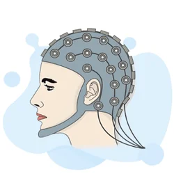
The test itself is painless and non-invasive. Small metal discs called electrodes are placed on the scalp using a conductive paste or cap. These electrodes detect tiny voltage fluctuations generated by the firing of neurons and transmit those signals to a computer that graphs them as wavy lines — the classic EEG readout. Neurologists, epileptologists, and critical care specialists are trained to read these patterns and identify abnormalities ranging from epileptiform spikes to the slow delta waves associated with metabolic encephalopathy.
One of the most common reasons patients and families encounter an EEG is after a seizure. When someone has an unexplained loss of consciousness, convulsions, or episodes that might be seizure-related, an EEG provides objective electrical evidence of what is happening in the brain. A routine outpatient EEG typically lasts 20 to 40 minutes, but extended or ambulatory recordings can run for 24 to 72 hours, capturing events that occur infrequently and would be missed on a short study.
Beyond seizure detection, EEG is used to evaluate sleep disorders, diagnose encephalitis or metabolic disturbances, confirm brain death in certain clinical protocols, and monitor anesthesia depth during surgery. In academic medical centers, EEG data is also analyzed for research purposes, helping scientists understand consciousness, memory consolidation, and the neural correlates of various psychiatric conditions. The technology has been in clinical use since Hans Berger recorded the first human EEG in 1924.
Cost is a practical concern for many patients. The EEG test price and EEG test cost vary widely depending on the type of study, the facility, and insurance coverage. A routine outpatient EEG at a hospital or neurology clinic in the United States typically ranges from $200 to $700 before insurance adjustments, while a prolonged video-EEG monitoring stay in an epilepsy monitoring unit can cost several thousand dollars. We cover cost in detail in a dedicated section below.
For healthcare professionals preparing for EEG certification exams, or for students learning the fundamentals of neurodiagnostic technology, understanding how eeg monitoring for patients works in clinical practice is foundational knowledge. Real-world patient scenarios — including cardiac arrest recovery, pediatric epilepsy, and post-operative monitoring — appear frequently on registry examinations administered by ABRET and similar credentialing bodies.
This guide covers everything patients, caregivers, and EEG students need to know: the types of EEG tests available, what the procedure feels like, how to interpret common findings, what side effects (if any) to expect, how much the test costs, and how EEG monitoring is used in some of the most critical medical situations a person can face. Read through each section or use the table of contents to jump directly to what matters most to you.
EEG Test by the Numbers

Types of EEG Tests Available to Patients
A standard 20–40 minute recording performed in a neurology clinic or hospital. Electrodes are placed on the scalp and the patient rests while brain activity is recorded. Commonly ordered after a first seizure or to evaluate headaches, blackouts, or confusion.
A portable recorder worn by the patient at home captures brain activity over one to three days. Ideal for detecting infrequent seizures or events that do not occur during a brief in-office study. Patients keep a diary of symptoms for correlation.
Combines simultaneous video recording with EEG in a hospital epilepsy monitoring unit. Clinicians can match observable behavior with electrical brain patterns, which is essential for classifying seizure type, localizing seizure onset, and evaluating candidates for epilepsy surgery.
Used in intensive care units for critically ill patients, including those recovering from cardiac arrest, stroke, or traumatic brain injury. Electrodes remain in place for days, allowing neurologists to detect nonconvulsive status epilepticus and guide treatment in real time.
Performed during neurosurgery to monitor brain function and protect eloquent cortex. Can be combined with electrocorticography (ECoG), in which electrodes are placed directly on the brain surface for the highest possible spatial resolution of electrical activity.
Understanding what actually happens during an EEG medical test removes much of the anxiety patients feel before their appointment. The procedure begins with scalp preparation. A technologist measures the patient's head and marks electrode positions according to the international 10-20 system, a standardized grid that ensures reproducible placement across different sessions and facilities. Positions are named using letter-number combinations: Fp1 and Fp2 for the frontal poles, C3 and C4 for the central regions, O1 and O2 for the occipital lobes, and so on.
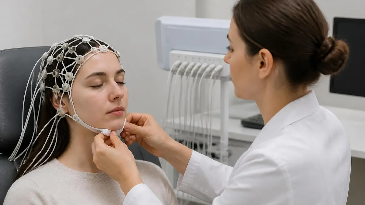
Next, each electrode site is lightly abraded with a mild scrub to reduce skin impedance, which improves the quality of the electrical signal. Electrodes are then secured with conductive gel or paste — a substance that feels cold and slightly tacky. Some facilities use electrode caps, which look like swim caps embedded with small metal contacts. The cap method is faster and increasingly preferred in research and pediatric settings. For a standard 21-electrode recording, setup takes approximately 20 to 30 minutes.
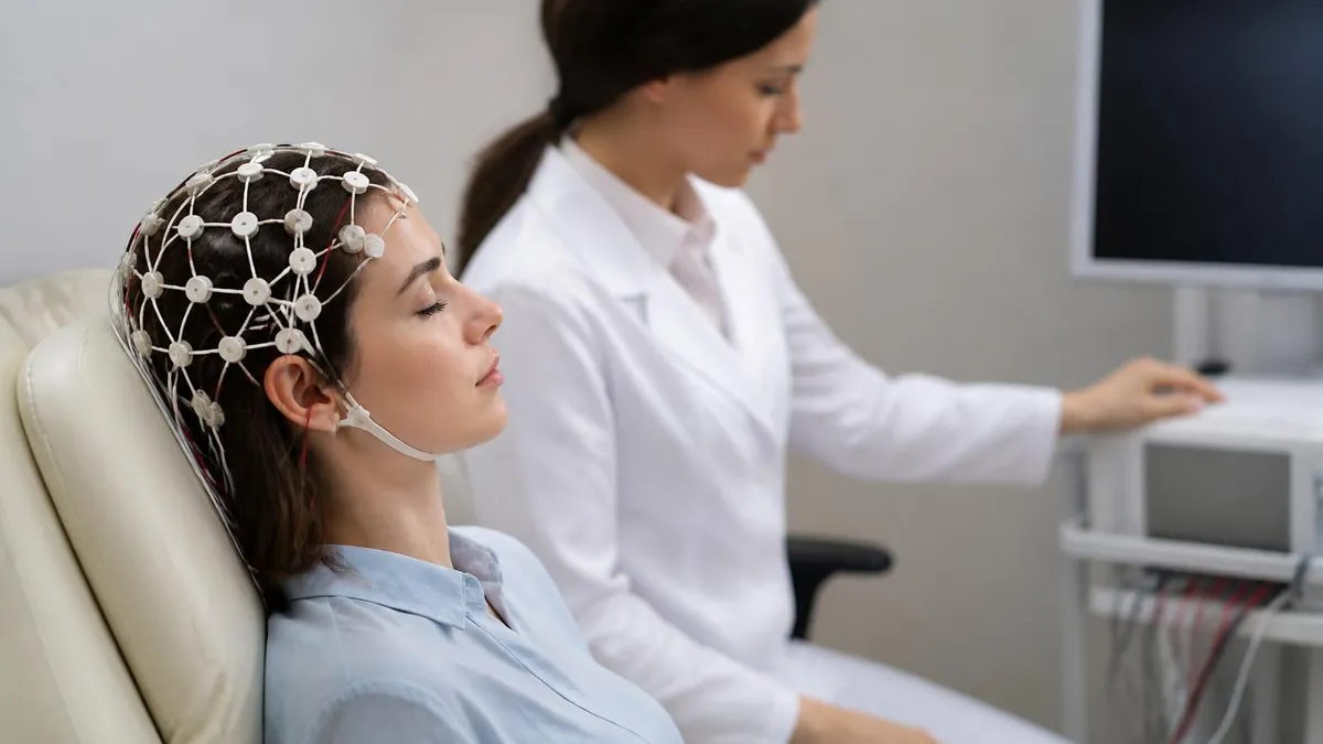
Once electrodes are in place, the technologist asks the patient to lie still or sit comfortably in a reclining chair. Relaxation is important because muscle artifact — the electrical noise generated by jaw clenching or scalp movement — can obscure brain signals. The recording begins with a baseline of eyes-open and eyes-closed activity, during which the technologist watches for normal alpha wave suppression (the alpha waves that appear when your eyes are closed should diminish when you open them).
Activation procedures are a key part of most routine EEGs. Hyperventilation asks the patient to breathe rapidly and deeply for three minutes. This temporarily lowers carbon dioxide in the blood, causing cerebral vasoconstriction and sometimes activating abnormal electrical discharges that would not otherwise appear. Photic stimulation exposes the patient to a strobe light flashing at frequencies between 1 and 30 Hz. Some patients with photosensitive epilepsy will show spike-and-wave discharges — a finding that has direct implications for seizure management and driving restrictions.
Sleep recording is often included because seizure discharges are more likely to appear during drowsiness and sleep. Patients scheduled for a routine EEG are sometimes asked to sleep-deprive themselves the night before, making it easier to fall asleep naturally during the recording. Alternatively, a sleep-deprived EEG may be specifically ordered when a routine study has been non-diagnostic but seizures are still clinically suspected.
At the conclusion of the recording, electrodes are removed and the conductive paste is washed out. Most patients return to normal activities immediately. The raw EEG data is reviewed and interpreted by a neurologist or clinical neurophysiologist, who produces a written report describing the dominant background rhythm, any asymmetries, sleep architecture if present, findings from activation procedures, and any abnormal patterns.
Turnaround time for reports varies from same-day in emergencies to several days for routine outpatient studies. For those studying to become EEG technologists or preparing for registry exams, the eeg monitoring for patients resource provides practice scenarios drawn from real clinical situations.
It is worth emphasizing that a normal EEG does not rule out epilepsy. Seizure activity is episodic, and the electrodes can only detect what is happening electrically at the cortical surface during the recording period. Studies show that a single routine EEG captures epileptiform discharges in only about 50 percent of people with confirmed epilepsy. Repeat recordings, sleep-deprived studies, and prolonged ambulatory monitoring all increase diagnostic yield significantly.
What Is an EEG Test Used to Diagnose?
EEG is the gold-standard test for evaluating seizures and diagnosing epilepsy. Neurologists look for interictal epileptiform discharges — spikes, sharp waves, and spike-and-wave complexes — that occur between seizures and indicate a brain region with increased excitability. The location of these discharges helps classify the epilepsy syndrome (generalized versus focal) and guides medication selection. Generalized spike-and-wave at 3 Hz is classic for absence epilepsy, while focal temporal sharp waves suggest mesial temporal lobe epilepsy.
When a seizure is captured during the recording — an ictal event — the EEG provides direct evidence of seizure onset zone and spread pattern. This information is critical for patients considering epilepsy surgery. A negative routine EEG does not exclude epilepsy, and many patients require sleep-deprived studies, 24-hour ambulatory recordings, or inpatient video-EEG monitoring before a definitive electrical abnormality is confirmed. Approximately 2.3 million Americans live with active epilepsy, and EEG plays a central diagnostic role for virtually all of them.

Pros and Cons of EEG Monitoring for Patients
- +Non-invasive and painless — no needles, radiation, or sedation required for standard recordings
- +High temporal resolution captures brain events millisecond by millisecond, unmatched by MRI or CT
- +Ambulatory versions allow patients to be monitored at home during their normal daily routine
- +Relatively low cost compared to MRI or PET scanning, making it accessible in most hospital settings
- +Continuous ICU monitoring detects silent seizures that would otherwise go unrecognized and untreated
- +Provides real-time feedback during epilepsy surgery and intraoperative procedures to protect brain function
- −Limited spatial resolution — scalp electrodes cannot precisely localize deep brain generators
- −A normal EEG does not rule out epilepsy, creating diagnostic uncertainty for some patients
- −Electrode setup is time-consuming and the conductive paste can be difficult to wash out of hair
- −Motion artifact and muscle noise can obscure recordings, particularly in agitated or uncooperative patients
- −Interpretation requires significant specialist training; findings can be subtle and require expert review
- −Prolonged monitoring requires the patient to remain connected to equipment, limiting mobility and comfort
How to Prepare for Your EEG Test: 10-Step Patient Checklist
- ✓Wash your hair the night before or morning of the test — avoid conditioner, oils, or styling products that increase scalp impedance.
- ✓Ask your neurologist whether to continue, reduce, or temporarily stop antiseizure medications before the study.
- ✓Sleep-deprive yourself if instructed — this means staying awake until 2 a.m. and waking no later than 6 a.m.
- ✓Eat a normal meal before the appointment — low blood sugar can affect brain wave patterns and cause discomfort.
- ✓Avoid caffeine on the day of the test unless your doctor specifically advises otherwise.
- ✓Arrive at least 15 minutes early to complete paperwork and allow the technologist adequate setup time.
- ✓Inform the technologist of all medications, supplements, and any recent illnesses that might affect results.
- ✓Bring a list of your symptoms, their frequency, and any triggers you have identified for the neurologist's review.
- ✓Plan for the paste removal — bring a comb or fine-tooth pick; some patients shower at the facility after the study.
- ✓Arrange transportation if sleep deprivation is required, as drowsy driving is unsafe after an overnight restriction.
A Normal EEG Does Not Mean No Epilepsy
Studies consistently show that a single routine EEG detects epileptiform discharges in only about 50 percent of people with confirmed epilepsy. If your EEG comes back normal but your doctor still suspects seizures based on your history, ask about a sleep-deprived study, 24-hour ambulatory recording, or video-EEG monitoring — each significantly increases the chance of capturing diagnostic findings.
The EEG test cost in the United States depends on several variables: the type of EEG ordered, whether it is performed in a hospital outpatient department or an independent neurology clinic, your geographic region, and your insurance coverage. For patients paying out of pocket, a routine 20–40 minute EEG typically costs between $200 and $700 at community hospitals and independent neurology practices. Academic medical centers and large hospital systems often charge higher facility fees, pushing prices toward $1,000 or more before insurance adjustments are applied.
Prolonged monitoring significantly increases the Eeg test price. A 24-hour ambulatory EEG, which includes equipment rental, technologist time for hookup and download, and physician interpretation, commonly runs $800 to $2,000. A hospital-based video-EEG stay in an epilepsy monitoring unit — which can last three to seven days and involves continuous nursing care, telemetry equipment, and daily neurologist review — can total $5,000 to $30,000 or more depending on length of stay and facility rates.
Insurance coverage for EEG is generally good when the test is medically indicated and ordered by a licensed physician. Most commercial plans, Medicare, and Medicaid cover EEG for seizure evaluation, monitoring of known epilepsy, evaluation of encephalopathy, and other medically documented indications. Prior authorization may be required for prolonged or video-EEG studies. Always call your insurer before the appointment to confirm coverage, obtain a prior authorization number if required, and ask about your out-of-pocket responsibility including deductible and co-insurance.
For uninsured or underinsured patients, several strategies can reduce the financial burden. Federally Qualified Health Centers (FQHCs) and community health clinics often provide EEG at reduced or sliding-scale fees. Some neurology practices offer self-pay discounts of 20 to 40 percent when payment is made at the time of service. Hospital charity care programs, available at most non-profit hospitals, can cover a substantial portion of costs for patients who qualify based on income. Patient assistance programs run by academic epilepsy centers may also be available.
Medicare patients should be aware that EEG falls under Medicare Part B (outpatient medical services). After meeting the Part B deductible (which adjusts annually), Medicare typically covers 80 percent of the Medicare-approved amount, leaving the patient responsible for the remaining 20 percent. Medigap supplemental plans can cover that 20 percent copay. Patients with Medicare Advantage plans may have different cost-sharing structures, so reviewing your specific plan's benefits is essential before scheduling a prolonged study.
The cost difference between facility types is meaningful and often overlooked. Hospital outpatient departments charge what is called a facility fee in addition to the physician's professional fee. A routine EEG performed in a hospital-based neurology clinic might bill $900 total ($600 facility + $300 professional), while the same test at a freestanding independent neurology office might cost $350 all-in. If your insurance plan counts both toward the same deductible, and you have not yet met your deductible, choosing the independent office setting can save you several hundred dollars out of pocket.
Finally, remember that the reading neurologist's interpretation fee is billed separately from the technical (recording) fee in many settings. If you receive two separate bills — one from the hospital and one from a physician group — both may be partially covered by insurance, but both may also apply toward your deductible and out-of-pocket maximum. Understanding how your plan processes split billing prevents surprise invoices. For a thorough breakdown of costs across EEG types and regions, see the dedicated cost article linked in the related articles section below.

The EEG test itself has virtually no direct side effects — no radiation, no injected contrast, and no needles are involved. However, hyperventilation during the test can cause lightheadedness, tingling in the hands or face, or brief fainting in sensitive individuals; these effects resolve within seconds of stopping. In rare cases, photic stimulation triggers a seizure in patients with photosensitive epilepsy — this is a medically supervised setting where trained staff can respond immediately. If you feel unusually unwell after your EEG, contact your neurologist's office before resuming normal activities.
EEG monitoring for cardiac arrest patients represents one of the most consequential applications of this technology in modern medicine. When the heart stops, blood flow to the brain ceases within seconds. Even after successful resuscitation — when circulation is restored through CPR, defibrillation, or both — the brain may have sustained varying degrees of injury depending on the duration of cardiac arrest and the speed of resuscitation. In the 24 to 72 hours following return of spontaneous circulation, the brain is in a highly vulnerable state, and neurological outcomes range from full recovery to persistent vegetative state.
Intensive care physicians and neurologists now use continuous EEG monitoring in post-cardiac arrest patients for two primary reasons. The first is seizure detection. Studies using continuous EEG in this population have found that 20 to 35 percent of comatose survivors of cardiac arrest develop seizures — most of them nonconvulsive, meaning they produce no visible shaking or movements that bedside staff could observe. These silent seizures increase the brain's metabolic demand at the worst possible time, consuming oxygen and glucose that injured neurons desperately need to survive and recover.
The second reason is prognostication. The background EEG pattern in the hours after cardiac arrest carries significant predictive information about neurological outcome. A continuous, well-organized background rhythm — even in a comatose patient — is associated with a substantially higher probability of meaningful recovery. Burst suppression, in which periods of electrical activity alternate with periods of near-complete silence, is associated with more severe injury. The most ominous finding is electrocerebral silence: a completely flat EEG for an extended period, which in the right clinical context is one of the markers used in brain death evaluation.
EEG reactivity testing adds further prognostic nuance. In this technique, a neurophysiologist stimulates the patient — typically with a loud noise, sternal rub, or noxious stimulus — while observing the EEG for any change in background activity. The presence of EEG reactivity, even in a patient who appears completely unresponsive, suggests that some cortical processing is still occurring and correlates with improved outcome probability. The absence of reactivity is associated with poorer prognosis, though no single EEG feature is used in isolation to make withdrawal-of-care decisions.
For families navigating a loved one's post-cardiac arrest course in the ICU, the information provided by continuous EEG can be both helpful and emotionally complex. It is common for ICU teams to report that EEG shows seizures being treated with medication, which can alarm families who are already coping with the shock of a cardiac emergency.
Understanding that these seizures are detected precisely because the EEG monitoring is working — and that treatment is underway — can help reframe the information in a meaningful way. Families are encouraged to ask the neurologist directly about EEG findings and what they mean for the specific patient's situation.
Targeted temperature management (TTM), formerly called therapeutic hypothermia, is often used after cardiac arrest to reduce cerebral metabolic demands and limit secondary brain injury. When TTM is used, EEG interpretation becomes more complex because cooling the body to 33–36°C alters normal brain wave frequencies and makes it harder to distinguish pathological patterns from temperature-induced changes. Neurophysiologists interpreting EEGs in this setting require specific training in how cooling affects the EEG and how to adjust interpretation accordingly.
Resources for EEG technologists and students preparing to work in neurodiagnostics — including ICU monitoring settings — should include exposure to critical care EEG patterns. The eeg monitoring for patients study materials include ICU-relevant pattern recognition questions. Training programs increasingly emphasize critical care EEG because the demand for skilled neurodiagnostic technologists in hospital settings continues to grow as continuous monitoring becomes standard of care at Level I trauma centers and comprehensive stroke centers nationwide.
Whether you are a patient preparing for your first EEG, a caregiver supporting a loved one through a neurological workup, or an EEG technologist building your clinical knowledge base, a few practical insights can make a significant difference in how well the experience goes.
For patients, the single most impactful preparation step is washing your hair thoroughly the night before or morning of the test using shampoo only — no conditioner, no styling gel, no dry shampoo. Clean, product-free hair allows the conductive paste to make optimal contact with the scalp, which directly improves signal quality and reduces the chance of needing electrode adjustments mid-recording.
Medication management before an EEG is a nuanced topic that should always be discussed with the ordering neurologist. In some clinical contexts — particularly when the goal is to lower the seizure threshold and increase the chance of capturing epileptiform activity — the neurologist may ask the patient to reduce or skip a dose of antiseizure medication. In other contexts, such as routine monitoring of known epilepsy or evaluation of an unrelated complaint, continuing all medications as prescribed is appropriate. Never independently reduce or discontinue antiseizure medications without explicit physician instruction, as doing so can trigger breakthrough seizures.
Sleep deprivation, when ordered, should be taken seriously. The standard protocol asks patients to stay awake until 2 a.m. and wake no later than 6 a.m., then stay awake through the morning of the EEG. This sleep restriction makes it much easier for the patient to fall asleep naturally during the recording, which is important because the drowsy-to-sleep transition is when epileptiform discharges are most likely to appear. Patients should arrange for someone else to drive them to and from the appointment after sleep deprivation — drowsy driving is dangerous and in many states legally comparable to impaired driving.
For parents bringing children for an EEG, the preparation conversation beforehand matters enormously. Explaining the procedure in child-friendly terms — that the test measures what is happening in the brain using small stickers on the outside of the head, and that it does not hurt — reduces anticipatory anxiety. Bringing a favorite book, toy, or tablet can help keep younger children calm during the electrode setup phase.
Many pediatric EEG labs have child life specialists or child-friendly decor specifically to ease the experience. If a child is known to be extremely anxious or unable to cooperate, the neurologist may recommend a mild sedative, though this slightly alters the EEG background and is noted in the interpretation.
For EEG students and technologists, developing strong pattern recognition skills requires exposure to a high volume and variety of tracings. The theoretical knowledge gained in training programs must be reinforced by reading actual EEGs under supervision. Many practicing technologists recommend a disciplined approach to reading each tracing: start by assessing the background rhythm and its frequency, then evaluate for symmetry, assess the response to eye opening, review activation procedure findings, and finally scan systematically for any focal or paroxysmal abnormalities. Developing a consistent internal checklist prevents missed findings.
Understanding how long an EEG test will last helps patients plan their day appropriately. A routine outpatient EEG takes 20 to 40 minutes of actual recording, but total time at the facility including registration, electrode setup, recording, and cleanup typically runs 1.5 to 2 hours. A sleep-deprived EEG may run slightly longer because the technologist waits for the patient to fall asleep. For ambulatory studies, the hookup appointment takes 45 to 60 minutes, and the patient wears the equipment for 24 to 72 hours before returning for removal and data download.
After the study, most patients receive results within a few days to a week, depending on the facility's turnaround time and how urgently the study was flagged. Results are communicated through a follow-up appointment, a patient portal message, or a phone call from the neurologist's office. If the EEG was performed in an emergency department or ICU, results may be communicated the same day because clinical decisions depend on them.
Patients who do not hear back within a week should proactively contact the neurologist's office rather than assuming a normal result — no-news-is-good-news is not a reliable assumption in clinical medicine, particularly for studies with complex findings that may require additional explanation.
EEG Questions and Answers
EEG Electroencephalography Practice Test PDF (Free Printable 2026)

Seizure EEG: What the EEG Test Shows, How It Works, and What to Expect
EEG Test Cost in 2026: Prices, Insurance Coverage, and How to Save

EEG Coherence: What It Is, How It Works, and What It Reveals About Brain Connectivity
EEG vs ECG vs EKG: What Each Test Measures, How They Differ, and What to Expect
About the Author

Educational Psychologist & Academic Test Preparation Expert
Columbia University Teachers CollegeDr. Lisa Patel holds a Doctorate in Education from Columbia University Teachers College and has spent 17 years researching standardized test design and academic assessment. She has developed preparation programs for SAT, ACT, GRE, LSAT, UCAT, and numerous professional licensing exams, helping students of all backgrounds achieve their target scores.