
EEG Test Explained: What It Is, How Long It Takes, Cost, Side Effects, and Your EEG Fingerprint
🧠 Learn what an EEG test is, how long it takes, what it costs, side effects, and what an EEG fingerprint of brain waves shows.

An EEG test records the electrical activity of your brain through small metal discs placed on the scalp. Every person produces a distinctive mix of brain rhythms, and researchers sometimes call this an EEG fingerprint, a signature of how your brain behaves while awake, drowsy, and asleep. Unlike a real fingerprint, it shifts with age, medication, and alertness. The test is painless, noninvasive, and among the most common neurological studies ordered by doctors across the United States each year. Practice with the free EEG - Electroencephalography practice test on PracticeTestGeeks (11 questions with answer explanations).
If you searched for what is an EEG test, the short answer is that electroencephalography measures tiny voltage fluctuations, usually just a few microvolts, produced when large groups of neurons fire together. The electrodes only listen. They never send electricity into your head. A computer amplifies the signals and displays them as wavy lines, and a trained clinician then reads those lines for patterns that point to seizures, sleep disorders, encephalopathy, or other changes in brain function.
Doctors order an EEG medical test for many reasons. The most frequent is suspected epilepsy, where a recording may capture spikes or sharp waves between seizures. It is also used after unexplained fainting spells, during evaluation of confusion or altered consciousness, when monitoring patients in intensive care, and while assessing sleep problems. Researchers use the same technology to study attention, memory, and language, which is why the field spans both clinical neurology and cognitive science.
Hans Berger recorded the first human EEG in 1924 and published his findings in 1929, describing the rhythm we now call alpha. Since then, the technique has evolved from ink pens scratching across paper to digital systems that sample hundreds of times per second. Modern clinical recordings commonly use 21 electrodes arranged by the international 10-20 system. This standard layout lets technologists in different hospitals compare results and lets physicians read outside recordings with confidence.
Many patients arrive nervous because they imagine electric shocks or mind reading. Neither is true. An EEG cannot read thoughts, diagnose mental illness by itself, or measure intelligence. It captures the combined electrical rhythm of the brain's outer layer and shows whether that rhythm looks normal, slowed, or unusually excitable. A normal result does not rule out epilepsy, and a mildly unusual one does not always mean disease, so interpretation always depends on your history.
This guide covers the practical questions people ask most: how long an EEG test lasts, what the EEG test price looks like in the US, whether there are side effects, and how to prepare. You will also learn the basics of brain wave frequencies and how clinicians read a tracing. Anyone who wants to go deeper into eeg fundamentals will find that recognizing artifacts is the first skill every technologist learns.
Whether you are a patient preparing for an appointment, a caregiver supporting a family member, or a student starting an EEG technologist program, a clear mental picture of the process removes most of the anxiety. The sections below walk through the procedure step by step, compare test types, outline costs, and finish with practical tips. Free practice questions are included so that you can check your understanding of the concepts as you go.
Free EEG - Electroencephalography Study Tools
Interactive EEG - Electroencephalography resources on PracticeTestGeeks, free to start:
- Free EEG Routine Questions and Answers 1 — 11 questions with instant scoring and answer explanations
- EEG - Electroencephalography flashcards — review key terms and concepts card by card
- EEG - Electroencephalography study guide — topic-by-topic overview of what the exam covers
- EEG - Electroencephalography cheat sheet — one-page printable summary for last-minute review
EEG Test by the Numbers
What Is an EEG Test? Step-by-Step Walkthrough
Check-In and History
Head Measurement and Skin Prep
Electrode Placement
Recording at Rest
Activation Procedures
Cleanup and Results
To understand what an EEG medical test measures, it helps to know where the signal comes from. Neurons communicate with tiny electrical currents. One neuron is far too weak to detect from outside the skull, but when thousands of neighboring cells in the cortex fire in synchrony, their combined field reaches the scalp as a signal of roughly 10 to 100 microvolts. Electrodes pick up that voltage, and amplifiers enlarge it about 10,000 times so it can be displayed.
Each channel on an EEG display shows the voltage difference between two points over time. Technologists arrange channels into montages, such as a bipolar chain that links neighboring electrodes front to back, or a referential layout that compares every electrode to a common reference. Viewing the same data in several montages lets the reader localize where an abnormal discharge starts. This is one reason a recording is never judged from a single view.
Brain activity is described by frequency, measured in hertz, meaning cycles per second. Delta waves run from about 0.5 to 4 Hz and dominate deep sleep. Theta runs from 4 to 8 Hz and appears in drowsiness. Alpha, from 8 to 13 Hz, is the classic relaxed rhythm seen over the back of the head when the eyes are closed. Beta runs from 13 to 30 Hz, and gamma lies above that.
A healthy awake adult usually shows a posterior dominant rhythm in the alpha range that fades when the eyes open. This reactivity is one of the first things a reader checks. In children, the background is slower and speeds up with age, reaching an adult pattern in the teenage years. A slow background in an alert adult can point to a diffuse problem, such as medication effect, metabolic disturbance, or injury.
The pattern of rhythms, their symmetry between hemispheres, and their response to stimuli are what some writers call an EEG fingerprint. Studies in biometrics have shown that resting recordings can be distinctive enough to tell people apart in controlled research settings. That is interesting science, but it is not a clinical tool. Doctors do not identify patients this way, and the signature varies with mood, fatigue, caffeine, and time of day.
Epileptiform activity looks different from normal rhythm. Spikes last less than 70 milliseconds, sharp waves last 70 to 200 milliseconds, and both stand out from the background. They may appear alone or as spike-and-wave complexes. Finding them between seizures supports a diagnosis of epilepsy, yet a routine recording captures such discharges in only a portion of people who have the condition, which is why repeat or longer studies are sometimes needed.
It is equally important to know what EEG cannot do. It sees mainly the outer cortex and has limited reach into deep structures, so a normal tracing does not exclude a problem there. It does not show anatomy the way MRI does, and it cannot pinpoint a tumor or stroke on its own. Its great strength is timing: it follows brain function millisecond by millisecond, something imaging struggles to match.
EEG Test Price, EEG Test Cost, and How Long Is an EEG Test
The EEG test price in the United States varies widely. A routine outpatient EEG often carries a billed charge between roughly $200 and $3,000, and hospital-based labs usually charge more than independent clinics. The total commonly combines a technical fee for the equipment and technologist with a professional fee for the neurologist who reads the study. Asking for both amounts up front prevents surprises.
Longer studies cost more. An ambulatory EEG that records at home for one to three days frequently runs from about $1,000 to $3,500 or higher, and inpatient video EEG monitoring in an epilepsy unit can reach many thousands of dollars per stay. These are broad ranges, not quotes. Your actual EEG test cost depends on your plan, deductible, and whether the facility is in network.
EEG Medical Test: Benefits Compared With Limitations
- +Completely noninvasive, with no needles, radiation, or contrast dye
- +Records brain activity millisecond by millisecond in real time
- +Helps diagnose and classify seizures and epilepsy syndromes
- +Safe for infants, children, pregnant patients, and older adults
- +Widely available at hospitals, clinics, and through home ambulatory systems
- +Lower cost than many imaging studies such as MRI or PET
- −Mostly records the outer cortex and may miss deep brain activity
- −A single normal routine recording does not rule out epilepsy
- −Muscle, eye, and movement artifacts can obscure the signal
- −Interpretation needs a trained neurologist and can vary between readers
- −Preparation and electrode placement take time and can leave residue in the hair
- −Poor spatial detail compared with MRI for locating structural problems
Pros and Cons at a Glance
| Pros | Cons |
|---|---|
| Completely noninvasive, with no needles, radiation, or contrast dye | Mostly records the outer cortex and may miss deep brain activity |
| Records brain activity millisecond by millisecond in real time | A single normal routine recording does not rule out epilepsy |
| Helps diagnose and classify seizures and epilepsy syndromes | Muscle, eye, and movement artifacts can obscure the signal |
| Safe for infants, children, pregnant patients, and older adults | Interpretation needs a trained neurologist and can vary between readers |
| Widely available at hospitals, clinics, and through home ambulatory systems | Preparation and electrode placement take time and can leave residue in the hair |
| Lower cost than many imaging studies such as MRI or PET | Poor spatial detail compared with MRI for locating structural problems |
How to Prepare for Your EEG Test
- ✓Wash your hair the night before and skip conditioners, oils, and styling products.
- ✓Remove braids, weaves, clips, and extensions if the lab asks you to.
- ✓Follow the lab's instructions about caffeine, which may need to be avoided.
- ✓Keep taking your medications unless the ordering doctor says to stop.
- ✓Bring a written list of every medicine and supplement you take.
- ✓Eat a normal meal beforehand, since low blood sugar can alter the tracing.
- ✓Follow sleep-deprivation instructions exactly if a sleep EEG is ordered.
- ✓Wear a loose, button-front top that will not need to be pulled over your head.
- ✓Arrange a ride home if you were told to limit sleep or may feel drowsy.
- ✓Write down questions about timing, results, and cost before you arrive.
Do not stop seizure medicine on your own
Never stop or change anti-seizure medication before an EEG unless the ordering neurologist explicitly tells you to. Stopping suddenly can trigger a dangerous seizure. If the goal is to catch abnormal activity, the doctor will plan any medication changes in a monitored setting.
Patients often ask about EEG test side effects, and the reassuring answer is that serious problems are rare. The machine only records; it delivers no current to your head. The most common complaint is mild skin irritation from the abrasive prep gel or adhesive, which typically fades within hours. Some people notice sticky paste in their hair afterward. Tell the technologist beforehand if you have a history of allergic reactions to adhesives, tape, or latex.
The activation procedures carry the few genuine risks. Hyperventilation, which means breathing deeply and quickly for around three minutes, can cause lightheadedness, tingling in the fingers, or a brief feeling of panic. It is usually skipped or shortened for people with recent stroke, significant heart or lung disease, sickle cell disease, or certain vascular conditions. Photic stimulation, using flashing lights, can occasionally provoke a seizure in a small number of people with photosensitive epilepsy.
Sleep deprivation is another trigger worth knowing about. Lack of sleep lowers the seizure threshold, which is exactly why it is used to increase the chance of seeing abnormal activity. The tradeoff is that a seizure during the recording is possible. EEG laboratories are staffed and equipped for this, and technologists are trained to protect the patient, note the timing, and call a physician. Having a seizure in the lab is not a failure of the test.
Not every EEG is the same, and choosing the right type affects what you can learn. A routine EEG captures about 20 to 40 minutes of waking and sometimes drowsy activity. A sleep-deprived EEG improves yield by recording after limited sleep. Ambulatory EEG tracks activity at home for one to three days. Video EEG, usually performed in a hospital unit, pairs the tracing with synchronized video so that physical events can be matched to electrical changes.
Intensive care units use continuous EEG to detect nonconvulsive seizures in patients who are sedated, comatose, or confused, since those seizures produce no visible shaking. Neonatal units use specialized recordings to assess newborns after oxygen deprivation or suspected seizures. Quantitative EEG, which analyzes frequency data by computer, appears in research and some clinics, but its clinical value for diagnosing psychiatric or learning conditions remains debated and is not broadly endorsed.
Children can safely undergo EEG, though the experience differs. Technologists often use games, stories, or a favorite stuffed animal to keep a young child calm, and a parent usually stays in the room. Infants may be recorded while sleeping, so scheduling around nap time helps. Because a wiggling child creates movement artifact, a patient and creative technologist is as valuable as the equipment, and a calm explanation beforehand makes the visit far smoother.
After a routine EEG, most people drive home and resume normal activity at once. Exceptions apply if you were sleep deprived, took a sedative to help you rest, or had a seizure during the study. In those cases, wait until you feel fully alert and follow your clinician's advice about driving. Rinse your hair gently with warm water and a little shampoo, and the paste usually comes out without trouble.
An EEG test is not a treatment and does not prevent seizures. If you or someone else has a seizure lasting more than five minutes, repeated seizures without recovery, or trouble breathing afterward, call 911 immediately. Do not wait for scheduled results.
Once the recording is complete, a neurologist, ideally one with training in clinical neurophysiology, reviews the entire tracing page by page. The report generally describes the background rhythm, the response to eye opening, any sleep patterns, and effects of hyperventilation or photic stimulation. It then offers an impression such as normal, borderline, or abnormal, with details about any slowing or epileptiform discharges and their location in the brain.
Common abnormal findings fall into a few groups. Focal slowing suggests a localized area of dysfunction, such as after a stroke or structural lesion. Diffuse slowing suggests a widespread problem, including encephalopathy, medication effect, or infection. Epileptiform discharges, such as spikes, sharp waves, and spike-and-wave complexes, point toward a tendency for seizures. Periodic patterns can appear in critically ill patients and prompt closer monitoring or treatment decisions.
A normal report is not always the end of the story. Many people with epilepsy have a normal first routine EEG, and studies suggest that repeating the test or adding sleep can raise the chance of detecting discharges substantially. On the other hand, some healthy people show benign variants that look unusual but carry no meaning. This is why clinicians weigh the tracing alongside your symptoms, exam, and imaging rather than treating it as a stand-alone verdict.
Artifacts are the hidden challenge in every recording. Eye blinks produce large frontal deflections, jaw clenching creates fast muscle noise, and sweating causes slow baseline drift. Even a nearby electrical device adds 60 Hz interference in the United States. Skilled readers learn to separate these from genuine brain activity. Studying eeg monitoring alongside artifact recognition is a practical way to build that discrimination skill.
Results timing varies by setting. Outpatient routine studies are commonly read within a few days, while urgent or inpatient recordings may be reviewed the same day. Ask the office how and when you will hear back, since some practices release reports through an online portal before the doctor calls. Do not assume silence means a normal result, and follow up if you have not heard within the stated window.
Returning to the idea of an EEG fingerprint, research groups have shown that features of resting brain activity can identify individuals with notable accuracy under lab conditions, and that these features are fairly stable over weeks. Interest in EEG-based authentication and brain-computer interfaces keeps growing. Still, scientists caution that signals shift with stress, sleep, and health, and privacy concerns are significant. Medically, your pattern matters because it is compared against what is typical for your age.
Finally, remember that an EEG answers one question at one moment in time. It shows how your brain behaved during the recording window. It does not predict the future, assign a personality type, or measure intelligence. When you understand that boundary, you can read your report more calmly, ask sharper questions, and decide with your doctor whether additional testing, such as MRI, longer monitoring, or specialist referral, makes sense.
Good preparation starts with the order itself. Ask your doctor exactly why the EEG was requested and what type of study it is: routine, sleep-deprived, ambulatory, or video. The answer affects how long you will be there, what to eat, how much sleep to get, and whether someone should drive you home. Write the instructions down, since details like caffeine limits and hair care are easy to forget under stress.
On the day of your appointment, arrive a little early with insurance cards, photo ID, and your medication list. Bring a comfortable layer, because labs are often kept cool and you may rest for a long period. If you wear glasses, hearing aids, or a hairpiece, tell the technologist. Leave jewelry and hair accessories at home, and silence your phone before the recording starts so alerts do not disturb the session.
During the recording, stillness helps more than anything. Clenching your jaw, frowning, or tensing your neck creates muscle artifact that can hide real brain activity for several seconds. Relax your face, let your shoulders drop, and breathe normally unless asked otherwise. Follow the technologist's cues to open and close your eyes. If you need to cough, swallow, or shift, say so quietly and the technologist will mark that moment in the record.
If you tend to feel anxious, tell the technologist at the beginning. They perform this test daily and can slow down, explain each step, and adjust the pace. Anxiety changes breathing and muscle tension, which can alter the tracing, so a calm start improves quality. Some people use slow breathing or a brief grounding exercise while electrodes are being applied, which also makes the long placement period feel shorter and easier.
Plan around the cost before you go. Request an itemized estimate that lists the facility fee and the interpretation fee separately, and confirm that both providers are in network with your plan. If you are paying out of pocket, ask for the cash price, since it is often lower than the billed charge. Check whether an independent neurodiagnostic center is available near you, as they frequently cost less than hospital outpatient departments.
Afterward, keep track of any symptoms you noticed, such as headaches, dizziness, or unusual sensations, and report them to your doctor. If you experienced a spell during the test, say so, because it gives the physician valuable information. Wash your hair gently and use a little conditioner to loosen leftover paste. Most people feel completely normal within minutes, and the skin marks fade within hours.
When the results come back, bring your questions to the follow-up visit. Ask what the report found, whether the result changes your treatment, and whether another study is advised. If you are studying EEG for a career, review the 10-20 system, the five frequency bands, and the main epileptiform patterns, then test yourself with the free practice questions below. Steady, repeated practice builds the pattern recognition that technologist exams reward.
Sample EEG - Electroencephalography Practice Questions
Try these questions from our free EEG - Electroencephalography practice tests. The correct answer and an explanation follow each question.
The phenomenon of 'REM rebound' after REM-suppressing medication withdrawal is best explained by:
- A. Homeostatic pressure accumulating during REM deprivation
- B. Circadian rhythm advancement
- C. Increased slow-wave sleep drive
- D. Cholinergic receptor downregulation
Answer: A. Homeostatic pressure accumulating during REM deprivation
When REM-suppressing drugs are withdrawn, accumulated homeostatic REM pressure results in increased REM percentage and shortened REM latency.
During a routine EEG, how long should a standard hyperventilation activation procedure last?
- A. 1 minute
- B. 3 minutes
- C. 5 minutes
- D. 10 minutes
Answer: B. 3 minutes
Standard hyperventilation during routine EEG lasts 3 minutes, followed by at least 1 minute of continued recording.
Which of the following statements accurately describes the fundamental principle of a bipolar montage?
- A. It measures the voltage difference between each electrode and a mathematically derived average of all electrodes.
- B. It is designed to be less sensitive to localized abnormalities compared to referential montages.
- C. It measures the voltage difference between adjacent electrodes in a chain.
- D. It requires a non-cephalic reference to avoid contamination from brain activity.
Answer: C. It measures the voltage difference between adjacent electrodes in a chain.
The core principle of a bipolar montage is the measurement of the potential difference between pairs of adjacent electrodes. These pairs are linked together to form chains, which can run anterior-posterior (longitudinal) or left-right (transverse). This method is effective for localizing focal events by identifying phase reversals.
In a normal adult EEG, what is the expected effect of opening the eyes on the posterior alpha rhythm?
- A. Alpha amplitude increases
- B. Alpha is attenuated or blocked
- C. Alpha frequency increases by 4 Hz
- D. Alpha spreads to frontal regions
Answer: B. Alpha is attenuated or blocked
Eye opening causes alpha blocking (Berger effect), suppressing posterior alpha due to visual cortex activation.
Take the full EEG - Electroencephalography practice test
EEG Questions and Answers

EEG Artifacts: How to Identify, Eliminate, and Document Common Recording Interference

ERP vs EEG: What Each Test Measures, How They Differ, and What to Expect

EEG Cap Electrodes: Everything You Need to Know About the EEG Test
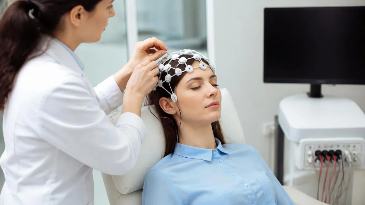
EEG Monitoring: What Is an EEG Test, How It Works, Cost, and What to Expect
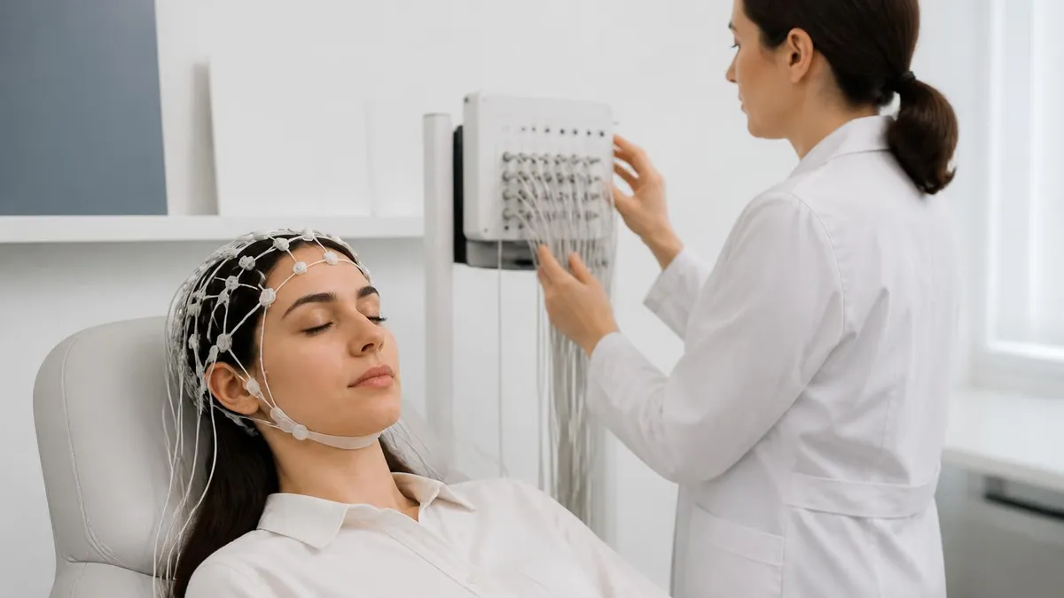
Ambulatory EEG: 24-72 Hour Home Recording for Suspected Seizures
How this page was made: written with AI tools and published by Nathan Beja, founder of Practice Test Geeks, who is responsible for its content. Exam facts are checked against the official sources listed below. Not reviewed by a subject-matter expert. Editorial policy
Sources & references
Exam facts on this page (fees, format, dates, passing scores) are checked against these official sources; the date is our last check. Editorial policy
- abret.org — Late registration, score validity, test delivery, fees, exam format, passing score, retake policy, score release, test dates · verified Oct 2026