
EEG Test and Alpha Activity: What Brain Waves Reveal, How Long It Takes, and What It Costs
🧠 EEG alpha activity explained: how an EEG test records brain waves, what 8-13 Hz rhythms mean, cost, length, side effects, and what to expect.
EEG alpha activity is the rhythmic 8 to 13 Hz signal that appears over the back of the head when a relaxed person closes their eyes, and it is the first thing a technologist looks for during an EEG test. Hans Berger recorded it in 1924, which is why it was once called the Berger rhythm. Alpha is simple to spot, easy to describe, and surprisingly informative, so it serves as a reference point for judging the rest of the recording. Practice with the free EEG - Electroencephalography practice test on PracticeTestGeeks (11 questions with answer explanations).
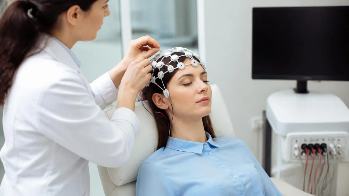
If you searched for an eeg test, you probably want plain answers first. An EEG, short for electroencephalogram, is a painless medical test that records the brain's electrical activity through small metal discs placed on the scalp. Nothing is sent into the brain. The machine only listens. Doctors order it to look into seizures, unexplained fainting, confusion, sleep disorders, and changes in alertness, and the whole picture is built from wave patterns like alpha, beta, theta, and delta.
This guide covers what is an eeg test, how long it takes, what it costs, and whether there are side effects. It also explains how to read the major frequency bands, with extra attention to alpha. You will not need a medical degree to follow it. Think of it as awareness content: a clear map of what happens in the room, what the squiggly lines represent, and what questions are worth asking your neurologist before and after the appointment.
Brain activity is measured in cycles per second, or hertz. Delta waves run from 0.5 to 4 Hz and dominate deep sleep. Theta runs 4 to 8 Hz and appears in drowsiness. Alpha sits at 8 to 13 Hz, beta covers 13 to 30 Hz during active thinking, and gamma goes above 30 Hz. To see how these rhythms are captured over hours or days, read our guide to eeg brain waves and monitoring.
Alpha is special because it is reactive. When your eyes are closed and your mind is idle, a smooth posterior dominant rhythm of roughly 9 to 11 Hz appears at the occipital electrodes, with an amplitude near 20 to 60 microvolts. The moment you open your eyes or solve a mental arithmetic problem, it flattens out. This simple on and off behavior, called alpha blocking, tells the reader that the cortex is awake and responsive.
A missing, asymmetric, or unusually slow alpha rhythm can point to problems, but context matters. A difference of more than 50 percent in amplitude between the left and right sides is usually flagged. A rhythm slower than 8 Hz in an awake adult may suggest diffuse dysfunction. Still, a single odd finding rarely means disease. Anxiety, medications, drowsiness, and poor electrode contact can all change alpha, so physicians interpret it alongside symptoms, history, and the remainder of the tracing.
The sections below walk through the stat snapshot, the step-by-step procedure, how alpha compares with other bands, pricing, preparation, and common mistakes. Short quizzes are placed along the way so you can test yourself on epileptiform patterns and activation procedures, two topics that often appear on technologist exams. Everything here is general education, not personal medical advice. Read straight through, or jump to the part that matches your question using the table of contents on this page.
EEG: upcoming dates and official changes
- Updated Oct 25, 2026: Exam fee (R. EEG T.) is now $700 application/exam fee (previously $700 application/exam fee).
- Updated Oct 25, 2026: Delivery (R. EEG T.) is now Prometric test center or live remote proctoring (previously Prometric test center or live remote proctoring).
- Updated Oct 25, 2026: Exam fee (R. EEG T.) is now $700 application/exam fee (previously $700 application/exam fee).
Facts verified Oct 25, 2026 against ABRET Neurodiagnostic Credentialing and Accreditation. Dates that have passed are removed automatically.
- The R. EEG T. exam is a computer-based multiple-choice exam presented in two sections; ABRET does not publish a question count. (ABRET Neurodiagnostic Credentialing and Accreditation)
- The R. EEG T. passing standard is set by the ABRET Board as the number of questions needed to be minimally competent; no numeric cut score is published. (ABRET Neurodiagnostic Credentialing and Accreditation)
- The ABRET R. EEG T. examination application fee is $700. (ABRET Neurodiagnostic Credentialing and Accreditation)
- After three unsuccessful R. EEG T. attempts within two years, candidates must wait six months and submit 20 more EEG-related ASET CEUs before retesting. (ABRET Neurodiagnostic Credentialing and Accreditation)
- The R. EEG T. credential is awarded for five years. (ABRET Neurodiagnostic Credentialing and Accreditation)
- The R. EEG T. exam can be taken at a Prometric test center or through live remote proctoring from home. (ABRET Neurodiagnostic Credentialing and Accreditation)
- R. EEG T. candidates get an unofficial result before leaving the test center, and the official result comes about 4–6 weeks after the exam. (ABRET Neurodiagnostic Credentialing and Accreditation)
Facts verified Oct 25, 2026 against ABRET Neurodiagnostic Credentialing and Accreditation. Dates that have passed are removed automatically.
EEG Exam at a Glance
| Item | Detail | Official source |
|---|---|---|
| Number of questions (R. EEG T.) | Computer-based multiple-choice, two sections | ABRET Neurodiagnostic Credentialing and Accreditation |
| Passing score (R. EEG T.) | Board-set passing standard (number not published) | ABRET Neurodiagnostic Credentialing and Accreditation |
| Exam fee (R. EEG T.) | $700 application/exam fee | ABRET Neurodiagnostic Credentialing and Accreditation |
| Retake policy (R. EEG T.) | After 3 fails in 2 years, wait 6 months + 20 ASET CEUs | ABRET Neurodiagnostic Credentialing and Accreditation |
| Validity (R. EEG T.) | 5 years | ABRET Neurodiagnostic Credentialing and Accreditation |
| Delivery (R. EEG T.) | Prometric test center or live remote proctoring | ABRET Neurodiagnostic Credentialing and Accreditation |
| Score release (R. EEG T.) | Unofficial result at test end; official in about 4–6 weeks | ABRET Neurodiagnostic Credentialing and Accreditation |
| Late registration deadline (R. EEG T. recertification) | Grace period to January 31, +$100 late fee | ABRET Neurodiagnostic Credentialing and Accreditation |
| Testing window (R. EEG T.) | 3 months from 'Eligible to Test'; tested Mon–Sat | ABRET Neurodiagnostic Credentialing and Accreditation |
Facts verified Oct 25, 2026 against ABRET Neurodiagnostic Credentialing and Accreditation. Dates that have passed are removed automatically.
Free EEG - Electroencephalography Study Tools
Interactive EEG - Electroencephalography resources on PracticeTestGeeks, free to start:
- Free EEG Routine Questions and Answers 1 — 11 questions with instant scoring and answer explanations
- EEG - Electroencephalography flashcards — review key terms and concepts card by card
- EEG - Electroencephalography study guide — topic-by-topic overview of what the exam covers
- EEG - Electroencephalography cheat sheet — one-page printable summary for last-minute review
EEG Alpha Activity by the Numbers
What Is an EEG Test? The Procedure Step by Step
Check-In and History
Measuring and Skin Prep
Electrode Placement
Baseline Recording
Activation and Wrap-Up
Understanding how alpha gets onto the screen starts with the electrodes. In a standard setup, a technologist places 19 to 21 recording electrodes according to the international 10-20 system, which spaces them at 10 or 20 percent of the distance between landmarks such as the nasion and inion. Letters mark the region, so O1 and O2 sit over the occipital lobes, exactly where the posterior alpha rhythm is strongest. Even spacing makes recordings comparable between hospitals.
Each electrode picks up tiny voltage fluctuations, usually measured in microvolts, which are millionths of a volt. The amplifier boosts them thousands of times, filters out slow drift and high-frequency noise, and displays them as lines. A standard display uses a 30 mm per second scale, so a rhythm of 10 Hz shows up as ten peaks in one second, about three centimeters of paper or screen width.
Readers do not look at one channel alone. They compare channels in arrangements called montages, such as longitudinal bipolar, transverse bipolar, and referential. Alpha is easiest to recognize in a referential montage because the voltage field spreads smoothly across the occipital area. If the pattern shifts from one side to the other or looks sharply localized, that observation becomes part of the final report. Montage changes are routine, not a sign of trouble.
For a deeper look at placement, caps, gel, and impedance checks, see our explainer on the eeg brain activity test hardware. Good contact matters more than most patients realize. Impedance is usually kept below 5 kilohms, and a loose electrode can fake slow waves or erase alpha entirely. Technologists recheck contact repeatedly because an unreliable signal wastes everyone's time and may require repeating the study.
The recording itself follows a script. The first several minutes are done with the patient relaxed and eyes closed, which is when alpha is at its fullest. Then the technologist asks the patient to open the eyes, and alpha should attenuate within a second or so. Opening and closing the eyes several times lets the reader confirm that the rhythm is reactive, symmetrical, and consistent in frequency from one cycle to the next.
Next come activation procedures. Hyperventilation for about three minutes can bring out generalized spike-and-wave discharges in absence epilepsy, and intermittent photic stimulation flashes a strobe light at rates from 1 to 30 flashes per second. A normal brain may show photic driving, where the occipital rhythm follows the flash frequency. These steps explain why a routine EEG can feel like a short checklist of simple tasks.
Sleep is sometimes added because many epileptiform abnormalities appear only in light sleep. A technologist may ask you to arrive sleep-deprived or may simply let you doze naturally during a 30 to 40 minute session. When alpha fades and slower theta waves take over, the reader knows you have drifted into stage N1. That transition is itself useful information, since it shows how the brain handles changing states of arousal.
EEG Test Cost, Length, and Side Effects
The eeg test cost in the United States varies widely by setting. A routine outpatient EEG at an independent lab commonly runs $200 to $800, hospital outpatient departments often bill $800 to $3,000, and a 24 to 72 hour ambulatory study can reach $1,000 to $3,500 or more. Inpatient video EEG monitoring costs the most because it bundles room, technologist, and physician time over several days. Regional pricing and facility fees explain much of the spread.
Most commercial plans and Medicare cover a medically necessary EEG, but you may still owe a deductible or coinsurance. Ask for the billing code, confirm the lab is in network, and compare the eeg test price at two facilities when your doctor allows flexibility. Self-pay discounts of 30 to 50 percent are sometimes offered, particularly at freestanding neurodiagnostic centers. Always ask whether the physician interpretation fee is included in the quote or billed separately afterward.
Is an EEG Medical Test Worth It? Benefits and Limits
- +Completely painless and noninvasive, with no radiation exposure
- +Captures brain activity in real time, down to fractions of a second
- +Helps diagnose and classify seizures and epilepsy syndromes
- +Useful for evaluating sleep disorders, encephalopathy, and altered consciousness
- +Widely available and far cheaper than many imaging studies
- +Safe for children, older adults, and during pregnancy
- −A single routine EEG can be normal even in people who have epilepsy
- −Scalp electrodes see mainly the cortex and miss deep brain structures
- −Artifacts from muscle, eye movement, or sweat can obscure the signal
- −Interpretation requires an experienced neurologist and benign variants can mislead
- −Setup takes time and the paste can be messy in the hair
- −Cost and insurance authorization vary widely between facilities
Pros and Cons at a Glance
| Pros | Cons |
|---|---|
| Completely painless and noninvasive, with no radiation exposure | A single routine EEG can be normal even in people who have epilepsy |
| Captures brain activity in real time, down to fractions of a second | Scalp electrodes see mainly the cortex and miss deep brain structures |
| Helps diagnose and classify seizures and epilepsy syndromes | Artifacts from muscle, eye movement, or sweat can obscure the signal |
| Useful for evaluating sleep disorders, encephalopathy, and altered consciousness | Interpretation requires an experienced neurologist and benign variants can mislead |
| Widely available and far cheaper than many imaging studies | Setup takes time and the paste can be messy in the hair |
| Safe for children, older adults, and during pregnancy | Cost and insurance authorization vary widely between facilities |
EEG Test Checklist: How to Prepare for Your Appointment
- ✓Wash your hair the night before and skip conditioner, gel, and oils.
- ✓Remove hair extensions or ask the lab whether braids can stay.
- ✓Avoid caffeine for the number of hours your lab specifies.
- ✓Eat a normal meal so low blood sugar does not alter the tracing.
- ✓Take prescribed medicines unless your doctor says to stop them.
- ✓Follow sleep-deprivation instructions exactly if the lab requests them.
- ✓Bring a list of medications and any prior EEG or MRI reports.
- ✓Wear a button-front shirt that will not pull over the electrodes.
- ✓Tell the technologist about seizures triggered by flashing lights.
- ✓Arrange a ride home if you are tired or may receive a sedative.
A normal EEG does not rule out epilepsy
A single routine EEG often catches epileptiform discharges in only about half of people who have epilepsy, because abnormal activity comes and goes. Repeat recordings, sleep, and longer monitoring raise the yield, so a normal first result is a starting point rather than a final answer.
Each frequency band tells a different story, and alpha only makes sense next to its neighbors. Delta waves under 4 Hz are normal in deep sleep but abnormal when they appear focally in an awake adult. Theta waves between 4 and 8 Hz are common in children and during drowsiness. Beta waves, 13 to 30 Hz, tend to be low in amplitude and are strongest over the frontal regions.
Gamma activity above 30 Hz is harder to capture on scalp recordings, so routine clinical reports rarely discuss it. Instead, clinicians focus on whether the background is organized, symmetrical, and appropriate for the person's age. A healthy adult has a clear posterior dominant rhythm, some frontal beta, and little else when awake. Anything that departs from that expected pattern gets described in the report, whether or not it turns out to matter.
Age changes the picture considerably. A three-month-old infant may show a posterior rhythm of only 3 to 4 Hz. By age three it reaches about 8 Hz, and by roughly 10 to 12 years old most children have an adult-like 9 to 11 Hz alpha. In older adults the rhythm may slow slightly, though a dominant rhythm below 8 Hz in a healthy elderly person still deserves a closer look.
Several benign variants can confuse beginners. The mu rhythm is an 8 to 13 Hz arch-shaped pattern over the central electrodes, and unlike alpha it persists with eye opening but fades with movement of the opposite hand. Lambda waves, posterior slow transients, and wicket spikes can also resemble pathology. Careful readers learn these mimics early, because over-reading normal variants is among the most common causes of a false epilepsy diagnosis.
Medications shape the background too. Benzodiazepines and barbiturates increase beta activity, often producing a fast, fuzzy tracing, while sedatives and some anti-seizure drugs can slow the alpha rhythm. Alcohol and caffeine have measurable effects as well. This is why reports often list current medications, and why you should never stop a prescribed drug before an EEG unless your doctor tells you to do so.
Alpha can also appear in abnormal settings. In alpha coma, a severely injured brain produces diffuse alpha that does not react to stimulation, and the outlook is often poor. Here the frequency is normal but the distribution and reactivity are not. That example shows why a rhythm should never be judged by frequency alone. Location, symmetry, and responsiveness are what separate a healthy pattern from a worrying one.
If you want to compare how an EEG differs from event-related testing, our overview of the eeg brain test comparison with ERP explains the distinction. In short, a routine EEG captures spontaneous background activity, while ERP averages time-locked responses to specific stimuli. Both use scalp electrodes, but they answer different clinical and research questions. Neither replaces the other, and doctors order them for different reasons.
An EEG is a scheduled diagnostic test, not an emergency service. If you or someone nearby has a seizure lasting more than five minutes, repeated seizures without recovery, trouble breathing, or sudden severe confusion, call 911 immediately rather than waiting for an appointment.
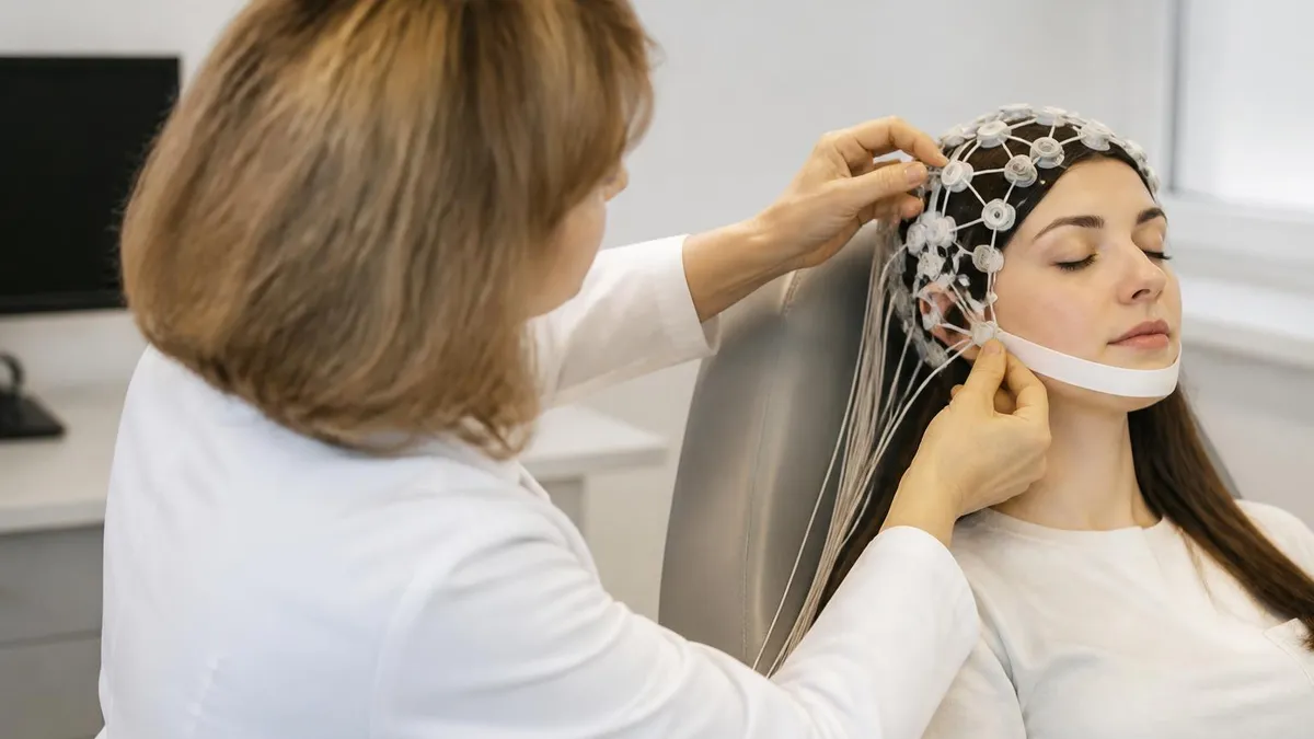
Not every wave on the screen comes from the brain. Artifacts are signals from other sources, and they are the biggest enemy of a clean recording. Eye blinks produce large frontal deflections, muscle tension creates fast spiky noise, sweat causes slow baseline sway, and a nearby power line adds a steady 60 Hz hum in the United States. Learning to spot them is a core skill for any EEG technologist.
Some artifacts imitate alpha itself. Rhythmic movement, a pulse beating under an electrode, or eyelid flutter can create repeating waveforms near 10 Hz that fool an untrained eye. Experienced readers check whether the activity reacts to eye opening, whether it has the right scalp distribution, and whether the same pattern appears on neighboring channels. Our guide to the eeg test for brain interference covers identification and fixes in detail.
Technologists reduce artifacts with simple habits. They ask patients to relax the jaw and forehead, keep the eyes gently closed, and avoid swallowing during recording when possible. They note events on the tracing, such as coughing or head movement, so the reader can discount them later. Clean skin prep, secure electrodes, and a quiet room matter just as much as sophisticated filters, because software cannot restore information that was never recorded.
Clinical use of alpha has limits worth knowing. A normal alpha rhythm does not rule out epilepsy, because many people with seizure disorders have normal routine EEGs between events. Studies commonly report that a single routine EEG captures epileptiform discharges in only around half of people with epilepsy, and repeat studies or sleep-deprived recordings raise that yield. A normal result means the abnormality was not seen today, not that it will never appear.
Likewise, an abnormal finding does not always mean a serious condition. Mild slowing can follow a concussion, a migraine, or a medication change. Small sharp transients appear in a fraction of healthy people with no seizures at all. Doctors therefore combine the report with a neurological exam, imaging such as MRI, blood tests, and your own description of events before deciding on a diagnosis or treatment.
Researchers and wellness companies also use alpha. Neurofeedback programs try to train people to increase alpha for relaxation, and consumer headbands estimate it with a handful of dry sensors. The evidence for medical benefit is mixed, and these devices are not substitutes for a clinical eeg medical test. They sit far below hospital systems in channel count, signal quality, and the expert review behind each diagnostic report.
Understanding these limits makes you a better partner in your own care. Ask your doctor what question the EEG is meant to answer, whether sleep will be recorded, and what a negative result would change. Those questions turn a vague appointment into a purposeful one. They also prepare you for the possibility that an ambulatory or video study may be recommended after a routine recording turns out normal.
Preparation is mostly simple. Wash your hair the night before and skip conditioner, gel, hairspray, and oils, since residue weakens electrode contact. Braids, extensions, and weaves can interfere, so ask the lab whether they can be left in or should be removed. Eat normally and take prescribed medicines unless told otherwise, because low blood sugar and abrupt medication changes can alter the tracing in misleading ways.
Caffeine is a common question. Many labs ask you to avoid coffee, tea, cola, and energy drinks for several hours before the study, since stimulants can change the background. If your lab requests sleep deprivation, you might be told to sleep only four or five hours the night before. Follow those instructions exactly, and never drive yourself home if you are exhausted or were given a sedative.
On the day, bring a list of medications, any previous EEG or MRI reports, and an insurance card. Wear a button-front shirt so nothing is pulled over the electrodes. Expect the visit to last about 60 to 90 minutes in total, with 20 to 40 minutes of actual recording. If you have a history of seizures triggered by flashing lights, tell the technologist beforehand so they can plan the photic step.
Cost deserves a plan. The eeg test price for a routine outpatient study often falls between $200 and $1,000 in an independent lab, while hospital billing can push the total to $1,500 or more once technical and professional fees are combined. Call your insurer, ask for the CPT code the facility will use, such as 95816 or 95819, and request a written estimate. Prior authorization is common, so schedule early.
After the test, the skin prep paste is easily washed out with warm water, though you may need a second shampoo. Mild redness at electrode sites fades within hours. Results usually take several days, because a neurologist must review the full recording rather than a summary. Ask when and how you will be told, and whether the report will be posted to a patient portal.
For students and trainees, the same discipline applies to studying. Learn the normal background first, because abnormalities only make sense against it. Practice identifying the posterior dominant rhythm, alpha blocking, sleep stages, and common artifacts. Then move on to epileptiform patterns, activation procedures, and montage reading using timed question sets. Repetition with feedback beats rereading notes, and short daily sessions help far more than one long weekend cram.
Finally, keep perspective. An EEG is one of the safest, most accessible tools in neurology, and for most people the hardest part is sitting still for half an hour. Come rested but not anxious, ask your questions, and treat the report as one piece of a larger puzzle. If anything remains unclear, request a follow-up conversation with the ordering physician instead of guessing from the wording of the report.
Sample EEG - Electroencephalography Practice Questions
Try these questions from our free EEG - Electroencephalography practice tests. The correct answer and an explanation follow each question.
Which of the following filter combinations would be most appropriate for routine clinical EEG recording to visualize cortical activity while minimizing slow-wave artifact and high-frequency noise?
- A. Low-Frequency Filter (LFF) at 0.1 Hz, High-Frequency Filter (HFF) at 15 Hz
- B. Low-Frequency Filter (LFF) at 5 Hz, High-Frequency Filter (HFF) at 70 Hz
- C. Low-Frequency Filter (LFF) at 1 Hz, High-Frequency Filter (HFF) at 35 Hz
- D. Low-Frequency Filter (LFF) at 1 Hz, High-Frequency Filter (HFF) at 70 Hz
Answer: D. Low-Frequency Filter (LFF) at 1 Hz, High-Frequency Filter (HFF) at 70 Hz
Standard settings for routine EEG are a Low-Frequency Filter (LFF) of 1 Hz and a High-Frequency Filter (HFF) of 70 Hz. This 'band-pass' allows the typical range of cerebral frequencies (1-70 Hz) to be displayed while attenuating very slow activity (like sweat artifact) and very high-frequency activity (like muscle artifact). Setting the LFF too high (e.g., 5 Hz) can filter out clinically relevant delta activity, and setting the HFF too low (e.g., 15 or 35 Hz) can filter out fast activity like spikes or beta.
The posterior dominant rhythm (alpha rhythm) in a normal awake adult is generated primarily by which brain structure interaction?
- A. Brainstem reticular formation alone
- B. Thalamocortical circuits involving the thalamus and occipital cortex
- C. Hippocampal-entorhinal connections
- D. Basal ganglia output to frontal cortex
Answer: B. Thalamocortical circuits involving the thalamus and occipital cortex
The alpha rhythm arises from synchronized oscillations in thalamocortical loops, with the thalamic relay nuclei (particularly the pulvinar and lateral geniculate) pacing cortical neurons in the occipital visual cortex.
In a comatose patient following an anoxic brain injury, the EEG shows periods of high-voltage, mixed-frequency activity lasting 1-3 seconds, which are separated by periods of profound voltage attenuation (<10 µV) lasting 4-8 seconds. This pattern is known as:
- A. Burst-suppression
- B. Hypsarrhythmia
- C. Alpha coma
- D. Slow spike-and-wave
Answer: A. Burst-suppression
The described pattern of alternating high-amplitude 'bursts' with periods of near-isoelectric 'suppression' is the definition of a burst-suppression pattern. It indicates profound cerebral dysfunction and is often seen in conditions like severe anoxia, deep anesthesia, or certain encephalopathies.
What is the typical voltage (amplitude) range of normal alpha waves recorded from the scalp?
- A. 5–10 µV
- B. 20–200 µV
- C. 200–500 µV
- D. 500–1000 µV
Answer: B. 20–200 µV
Alpha waves normally range from about 20 to 200 µV in amplitude when recorded from scalp electrodes.
EEG Questions and Answers
EEG Artifacts: How to Identify, Eliminate, and Document Common Recording Interference

ERP vs EEG: What Each Test Measures, How They Differ, and What to Expect

EEG Cap Electrodes: Everything You Need to Know About the EEG Test
EEG Monitoring: What Is an EEG Test, How It Works, Cost, and What to Expect
Ambulatory EEG: 24-72 Hour Home Recording for Suspected Seizures
How this page was made: written with AI tools and published by Nathan Beja, founder of Practice Test Geeks, who is responsible for its content. Exam facts are checked against the official sources listed below. Not reviewed by a subject-matter expert. Editorial policy
Sources & references
Exam facts on this page (fees, format, dates, passing scores) are checked against these official sources; the date is our last check. Editorial policy
- abret.org — Late registration, score validity, test delivery, fees, exam format, passing score, retake policy, score release, test dates · verified Oct 2026