White Areas on Brain MRI: What They Mean and Why They Appear 2026 July
⏳ White areas on brain MRI explained: what hyperintensities mean, common causes, T2/FLAIR appearance, and when to worry. Clear guide for patients & techs.
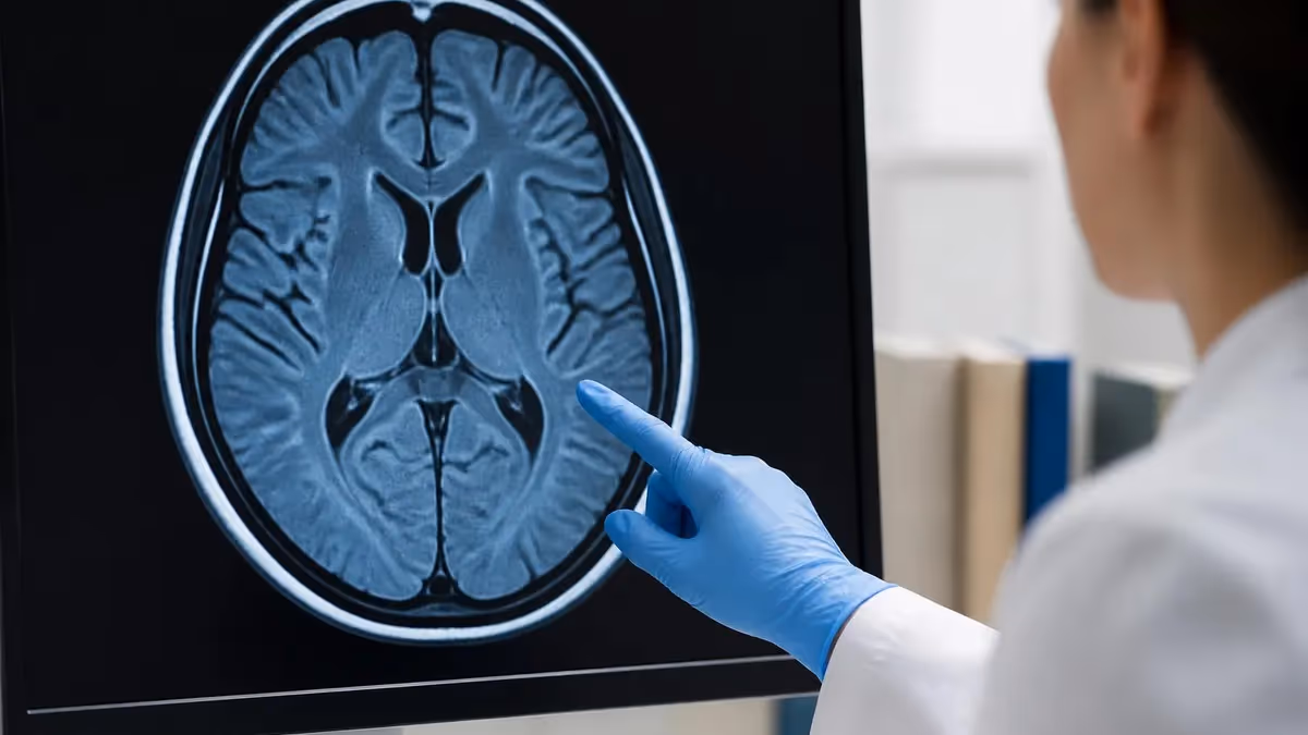
When a radiologist reports white areas on brain MRI, the description usually refers to bright spots, patches, or punctate foci that appear hyperintense on T2-weighted or FLAIR sequences. These white areas can range from harmless age-related changes to significant findings tied to multiple sclerosis, small vessel ischemic disease, migraine, infection, or tumor. Understanding what produces this brightness, where it sits in the brain, and how it behaves across pulse sequences is the key to interpreting the report accurately rather than panicking at the word lesion.
The brain MRI generates contrast by exploiting differences in water content, fat, protein binding, and tissue microstructure. On T2-weighted and FLAIR images, anything with extra free water glows white, which is why edema, demyelination, gliosis, and cerebrospinal fluid all appear bright. On T1-weighted images the relationship inverts, with fat and subacute blood appearing white while water turns dark. A radiologist cross-references these sequences to decide whether a white spot is fluid, demyelination, blood breakdown product, or something else entirely.
Patients often discover these findings after an MRI ordered for headaches, dizziness, memory complaints, or after a fall. The radiology report may use phrases like nonspecific white matter hyperintensities, T2 FLAIR foci, leukoaraiosis, or punctate subcortical lesions. Each phrase carries a slightly different clinical weight, and pairing the imaging description with patient age, vascular risk factors, and neurological symptoms is essential before assigning meaning. A 70-year-old hypertensive smoker and a 25-year-old with optic neuritis can have similar looking white spots that mean very different things.
For technologists and student radiographers, learning to recognize the appearance of these lesions across protocols is a frequent registry topic. You should know which sequences best demonstrate white matter disease, why FLAIR suppresses CSF, how diffusion-weighted imaging separates acute infarct from chronic gliosis, and when contrast is needed. Reviewing common MRI findings helps build the pattern recognition that experienced techs use to flag worrisome scans for prompt radiologist review.
This guide walks through the biology and physics behind white spots on brain MRI, the most common diagnoses associated with them, the imaging features that distinguish benign from concerning lesions, and what patients should ask after receiving an unclear report. Whether you are a patient trying to interpret your own scan, a tech preparing for the MRI registry, or a clinician explaining results to a worried family, the goal is to translate the radiology language into something practical and accurate.
The phrase white areas on brain MRI is not a diagnosis but a description. By the end of this article you should be able to match common appearances to likely causes, know which follow-up imaging or labs make sense, and understand why two reports with similar wording can lead to very different treatment paths. Imaging is one piece of the puzzle, and putting it in clinical context is what turns a frightening picture into useful information.
We will also cover what the bright spots look like on different field strengths, how 3 Tesla scanners reveal smaller lesions than 1.5 Tesla, and why repeat MRIs done on different machines sometimes show new findings that were not actually new. The technical side matters because it explains why a stable patient might appear to have changes that are really artifacts of resolution and protocol differences.
White Matter Hyperintensities by the Numbers
MRI Sequences That Reveal White Spots
Free water appears bright, making edema, demyelination, gliosis, and cysts hyperintense. The workhorse sequence for detecting pathology in white and gray matter throughout the brain.
Suppresses cerebrospinal fluid so periventricular and cortical lesions stand out against a dark CSF background. The most sensitive sequence for multiple sclerosis plaques and small vessel disease.
Restricted diffusion appears bright and distinguishes acute infarct from chronic lesions. A bright DWI with dark ADC map confirms acute ischemia within minutes to days of onset.
Active inflammation, neoplasm, and breakdown of the blood-brain barrier enhance with gadolinium. Helps separate active demyelinating plaques from old quiescent ones and identifies tumors.
Highlights blood products, calcium, and small vessel hemorrhages as dark foci. Useful for cerebral microbleeds in amyloid angiopathy and chronic hypertensive vascular disease.
The most common cause of white areas on brain MRI in adults is small vessel ischemic disease, also called chronic microvascular ischemia or leukoaraiosis. These lesions develop when tiny perforating arteries in the deep white matter become damaged by hypertension, diabetes, smoking, or aging itself. The result is patchy demyelination and gliosis that lights up on T2 and FLAIR. By age 60 most people have at least a few of these spots, and by age 80 they are nearly universal. They rarely cause symptoms in isolation but accumulate slowly over years.
Multiple sclerosis is the diagnosis patients fear most when they hear about white spots. MS plaques have a distinct pattern: they tend to be ovoid, oriented perpendicular to the ventricles like fingers (Dawson fingers), and frequently involve the corpus callosum, juxtacortical regions, brainstem, and spinal cord. The McDonald criteria use dissemination in space and time across multiple brain regions plus enhancement patterns to make the diagnosis. Looking at MRI with and without contrast is critical because active plaques enhance while chronic ones do not.
Migraine is another extremely common but underappreciated source of white matter spots, especially in women under 50. Patients with migraine, particularly migraine with aura, develop small punctate T2 hyperintensities in the deep and subcortical white matter at rates roughly double the migraine-free population. These spots do not typically grow or cause progressive deficits, but they are often the trigger for the patient to seek an opinion about whether they could indicate MS.
Infectious and post-infectious processes also cause white spots. Lyme neuroborreliosis, neurosyphilis, viral encephalitis, progressive multifocal leukoencephalopathy in immunocompromised patients, and post-viral acute disseminated encephalomyelitis all produce T2 hyperintensities. The distribution, patient history, and laboratory studies usually clarify the picture. Cerebral autosomal dominant arteriopathy with subcortical infarcts and leukoencephalopathy (CADASIL) is a genetic cause that produces striking anterior temporal pole white matter changes and should be considered in younger patients with family history of stroke or dementia.
Tumors and demyelinating tumefactive lesions can present as white masses, but they typically show mass effect, edema disproportionate to the lesion, and enhancement patterns that distinguish them from simple inflammation. Primary CNS lymphoma, glioma, and metastatic disease each have characteristic appearances. The radiologist examines whether the lesion crosses the corpus callosum, how it enhances, whether there is restricted diffusion, and how it relates to surrounding edema before suggesting biopsy or further workup.
Trauma and vascular malformations also contribute to bright signal on brain MRI. Diffuse axonal injury after head trauma produces punctate foci at the gray-white junction and in the corpus callosum that may persist for years. Old hemorrhagic contusions leave behind gliotic scars with surrounding hemosiderin. Cavernous malformations show a popcorn appearance with a dark rim. Each pattern offers clues that an experienced radiologist uses to suggest the most likely cause and the appropriate next step.
Finally, virchow-robin spaces, also called perivascular spaces, are normal fluid-filled channels around small penetrating vessels that can look like white spots but follow CSF signal on every sequence and have no surrounding signal abnormality. Recognizing these as normal anatomy prevents unnecessary workup. Their numbers increase with age and may be a marker of glymphatic dysfunction, but in isolation they are not pathologic.
MRI Practice Test Questions
Prepare for the MRI - Magnetic Resonance Imaging exam with our free practice test modules. Each quiz covers key topics to help you pass on your first try.
MRI Knowledge
MRI Exam Questions covering Knowledge. Master MRI Test concepts for certification prep.
MRI Physics
Free MRI Practice Test featuring Physics. Improve your MRI Exam score with mock test prep.
MRI Anatomy and Pathology
MRI Test Prep for MRI Anatomy and Pathology. Practice MRI Quiz questions and boost your score.
MRI Anatomy and Positioning
MRI Questions and Answers on MRI Anatomy and Positioning. Free MRI practice for exam readiness.
MRI Contrast Agents
Free MRI Quiz on MRI Contrast Agents. MRI Exam prep questions with detailed explanations.
MRI Patient Care and Positioning
MRI Practice Questions for MRI Patient Care and Positioning. Build confidence for your MRI certification exam.
How White Areas Behave on T2, FLAIR and DWI
On T2-weighted imaging the brain shows gray matter as moderately bright, white matter as moderately dark, and CSF as very bright. Anything with elevated free water content becomes hyperintense, which is why edema, demyelination, gliosis, cysts, and tumors all appear white. This is the most sensitive but least specific sequence because so many different processes produce similar signal increases.
The radiologist uses T2 to detect lesions but rarely to characterize them alone. A spot that is bright on T2 might be CSF, a vessel, edema, infarct, plaque, or tumor. Comparison with FLAIR, DWI, T1, and post-contrast images is essential before deciding what the white area represents. T2 is the screening sequence that triggers deeper inspection.

Strengths and Limitations of Brain MRI for White Matter Disease
- +Detects lesions as small as 3 mm on modern 3T scanners
- +No ionizing radiation, safe for repeated follow-up imaging
- +Multiparametric sequences narrow the differential diagnosis
- +FLAIR sequence highly sensitive to demyelination and small vessel disease
- +DWI distinguishes acute infarct from chronic lesions reliably
- +Contrast enhancement identifies active inflammation or tumor
- −Many findings are nonspecific and require clinical correlation
- −Older or 1.5T scanners may miss small lesions
- −Motion artifact in agitated or claustrophobic patients degrades images
- −Gadolinium contrast carries small risk of NSF in renal failure
- −Incidental findings can drive anxiety and unnecessary workup
- −Long scan times of 30-60 minutes limit access and throughput
Reading a Brain MRI Report About White Areas
- ✓Note the number of lesions and whether they are punctate, patchy, or confluent
- ✓Check the anatomic locations: periventricular, subcortical, juxtacortical, deep gray, brainstem
- ✓Look for descriptions of Dawson fingers or orientation perpendicular to ventricles
- ✓Confirm whether contrast was administered and whether any lesions enhance
- ✓Check the diffusion-weighted imaging comment for restricted diffusion
- ✓Compare with any prior MRI to assess stability versus interval change
- ✓Note the radiologist's differential diagnosis and recommended follow-up
- ✓Identify whether vascular risk factors are mentioned in the clinical history
- ✓Review for incidental findings unrelated to the original indication
- ✓Bring the report and images to a neurologist if findings are unclear
Nonspecific does not mean nothing
When a report calls white spots nonspecific, it means the appearance alone cannot pinpoint the cause. This is often reassuring in older patients with vascular risk factors but should still prompt discussion with a clinician who can correlate the findings with symptoms, labs, and exam.
The clinical significance of white areas on brain MRI depends heavily on the patient context. A few punctate T2 hyperintensities in the deep white matter of a 65-year-old with controlled hypertension are expected and usually require no further workup beyond cardiovascular risk management. The same lesions in a 30-year-old presenting with optic neuritis or limb weakness demand a thorough demyelinating workup including spinal cord MRI, lumbar puncture for oligoclonal bands, and evaluation by a neurologist familiar with MS criteria.
Lesion burden and distribution help guide concern. The Fazekas scale grades periventricular and deep white matter hyperintensities from 0 to 3 based on size and confluence. Higher Fazekas scores correlate with cognitive decline, increased stroke risk, and gait disturbance. Patients with extensive confluent white matter disease have measurable increases in dementia risk over the following five to ten years, which makes aggressive blood pressure and lipid control particularly important.
Enhancement after gadolinium is one of the most clinically actionable features. An enhancing white matter lesion suggests active inflammation, infection, demyelination, or tumor. A nonenhancing chronic lesion is much less likely to represent an actively evolving process. In multiple sclerosis the presence of both enhancing and nonenhancing lesions on the same scan fulfills dissemination in time, which is a diagnostic criterion under the 2017 McDonald guidelines.
The location of a lesion matters as much as its presence. Lesions in eloquent areas like the brainstem, internal capsule, or thalamus can produce symptoms out of proportion to their size. A 5 mm pontine infarct may cause hemiparesis, while a 2 cm subcortical frontal lesion might be entirely silent. Radiologists describe location precisely so clinicians can correlate symptoms with imaging and decide whether the white area on MRI is the cause of the presentation.
Comparison with prior imaging is essential whenever available. New lesions that were not present before raise concern for active disease and should prompt a clinical visit. Stable lesions over years are reassuring, particularly when the patient is asymptomatic. Apparent new lesions on different machines or with different protocols may not be truly new but rather reflect improved sensitivity on a newer scanner. Always ensure comparisons are made on equivalent sequences.
Specific patterns raise particular suspicions. Symmetric thalamic and basal ganglia hyperintensities suggest metabolic disease or osmotic demyelination. Bilateral medial temporal hyperintensities suggest limbic encephalitis or seizure-related changes. Bilateral occipital changes suggest posterior reversible encephalopathy syndrome. Reading these patterns is part of the radiologist's training and explains why a structured report often suggests a likely diagnosis even when individual spots look nonspecific in isolation.
Finally, age-related normal variation is worth emphasizing. The aging brain loses volume, accumulates microvascular damage, and develops Virchow-Robin spaces. None of these are diseases per se, but they reflect the cumulative effect of decades of vascular stress and aging biology. Distinguishing normal aging from early pathology is one of the harder parts of neuroradiology and one reason why these reports often end with the phrase clinical correlation recommended.
White areas on brain MRI combined with new neurological symptoms such as one-sided weakness, sudden vision loss, severe headache, confusion, seizure, or balance loss require urgent evaluation. Do not wait for a routine follow-up. Go to an emergency department or call your neurologist immediately for guidance.
After an MRI shows white areas, the appropriate next step depends on the clinical situation and the radiologist's recommendation. For an asymptomatic patient with a few age-appropriate spots and well-controlled cardiovascular risk factors, no further imaging may be needed. The primary care physician can manage blood pressure, cholesterol, and diabetes to slow the progression of small vessel disease. Lifestyle measures including exercise, smoking cessation, and dietary changes have evidence supporting reduced white matter lesion progression over time.
For patients with symptoms suggestive of demyelinating disease, the next steps usually include MRI of the cervical and thoracic spinal cord, lumbar puncture for cerebrospinal fluid analysis including oligoclonal bands and IgG index, blood work to exclude mimics such as neuromyelitis optica spectrum disorder, vitamin B12 deficiency, syphilis, Lyme disease, and HIV. Referral to a neurologist with MS expertise allows formal application of diagnostic criteria and discussion of disease-modifying therapy if MS is confirmed.
Patients undergoing follow-up MRIs for MS or other inflammatory diseases should ideally have their scans on the same machine with the same protocol to make comparisons reliable. Including MRI medical abbreviation reviews can help patients understand the recurring terms in their reports. Annual or semi-annual surveillance imaging is common in active demyelinating disease and helps detect subclinical progression that may warrant therapy escalation.
When new findings appear unexpectedly, repeat imaging in three to six months is often recommended to assess for evolution. Truly new and growing lesions warrant more aggressive workup. Lesions that remain stable on repeat imaging are usually less concerning. Communication between the patient, radiologist, and clinician about which lesions are old versus new prevents both undertreatment of progressive disease and overtreatment of benign incidental findings.
For tumor-like findings, the next step often involves contrast-enhanced MRI with perfusion and spectroscopy sequences, sometimes followed by biopsy. Multidisciplinary tumor board review brings together radiology, neurosurgery, neuro-oncology, and pathology to plan care. Patients should ask for an experienced neurosurgical opinion when masses are detected, especially in younger patients where survival depends heavily on early specialist care.
Patient anxiety after an MRI report is real and deserves acknowledgment. Internet searches for white spots on brain MRI return frightening results that often do not apply to the individual situation. A focused conversation with the ordering clinician, ideally with the actual images available, helps put findings in context. Many patients leave such conversations significantly reassured once the size, distribution, and clinical correlation are explained in plain language.
Documentation matters for future care. Patients should keep a copy of every brain MRI report and ideally a CD or digital copy of the images. If care moves to a new facility, having prior studies available eliminates the need for repeat imaging and allows direct comparison. Modern imaging platforms increasingly allow patient portal access to images, which makes second opinions and consultations much easier than they were a decade ago.
For technologists scanning patients who may have white matter disease, protocol optimization matters. A standard brain MRI for evaluating hyperintensities should include sagittal T1, axial T2, axial FLAIR (preferably 3D FLAIR on capable scanners), axial DWI with ADC map, axial T2*-weighted gradient echo or susceptibility-weighted imaging, and post-contrast T1 if contrast is administered. Adding 3D FLAIR allows multiplanar reformatting and improves detection of small juxtacortical lesions important for MS diagnosis.
Slice thickness should be 3 to 4 mm or less, with no gap when possible. Thicker slices miss small lesions and create partial volume effects that obscure the borders of lesions. On 3T scanners the higher signal-to-noise ratio supports thinner slices and improved resolution, which is one reason MS centers prefer 3T imaging. Knowing your scanner's capabilities and matching the protocol to the clinical question is part of providing high-quality imaging.
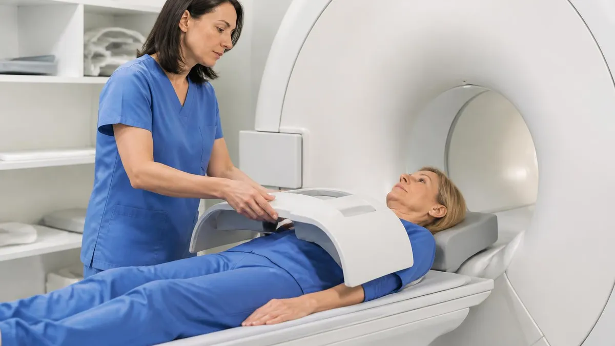
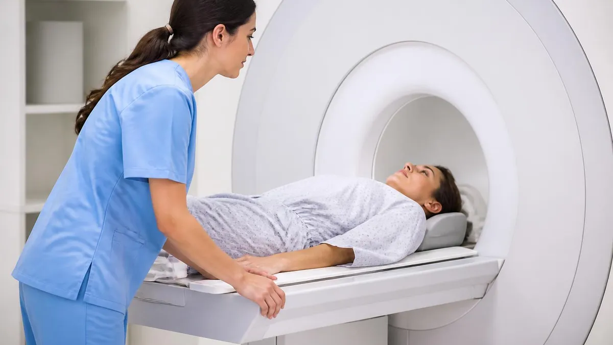
Patient positioning and motion control directly affect image quality. Padding the head, using prospective motion correction when available, and coaching patients to remain still for the duration of each sequence prevents motion artifact that can mimic or hide lesions. Patients with claustrophobia or movement disorders may need anxiolytic medication or even general anesthesia to obtain diagnostic images. The technologist's interaction with the patient often determines whether the scan succeeds.
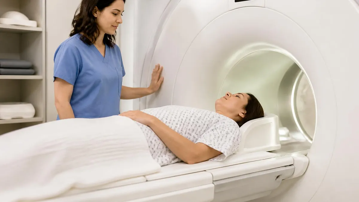
For patients, preparing for a brain MRI is straightforward. Wear loose clothing without metal, remove jewelry and dental appliances, and inform the technologist about any implants, pacemakers, or surgical hardware. Patients with reduced kidney function should discuss whether gadolinium contrast is necessary. Eating and drinking are usually fine before a noncontrast scan. The scan itself takes 30 to 60 minutes depending on the protocol and whether contrast is used.
Understanding what to expect during the scan reduces anxiety. The scanner is loud, with knocking and beeping sounds that vary by sequence. Earplugs or headphones are provided. Patients hold still and may need to hold their breath briefly for certain sequences, though most brain protocols do not require breath-holding. The MRI table moves into the bore feet-first or head-first depending on protocol and the patient's comfort with confined spaces.
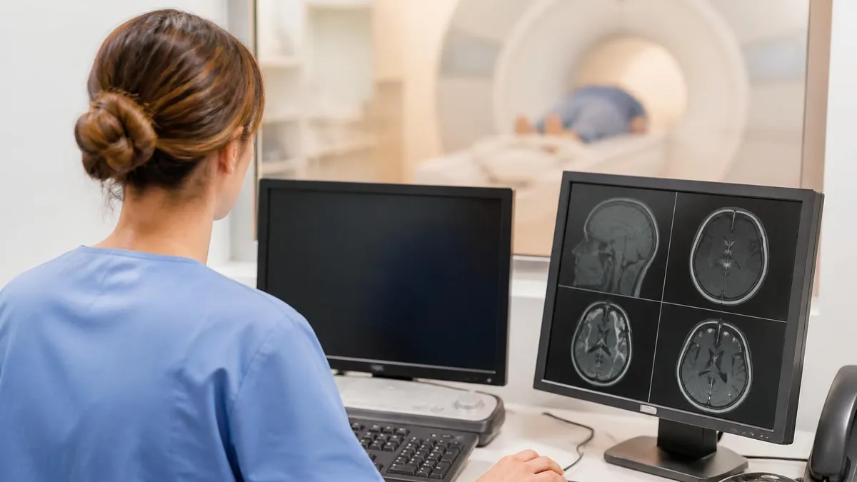
Reviewing your own MRI images with your clinician is valuable. Most picture archiving and communication systems allow side-by-side display of T1, T2, FLAIR, and DWI sequences. Seeing the actual location and appearance of the white areas often demystifies the report. Patients who understand their imaging tend to be more engaged in their care, more compliant with risk factor modification, and less anxious during follow-up surveillance. Tools like patient portals and DICOM viewers make this kind of review increasingly accessible.
Finally, remember that imaging is a snapshot in time. A single MRI captures one moment in the brain's complex life. Trends over multiple scans often matter more than any single finding. Stable findings over years are reassuring even when initially worrisome. Progressive findings demand attention even when individually small. Working with a consistent radiology team and clinician who reviews your serial imaging is the best way to interpret white areas on brain MRI accurately and act on them appropriately.
MRI Questions and Answers


About the Author
Medical Laboratory Scientist & Clinical Certification Expert
Johns Hopkins UniversityDr. Sandra Kim holds a PhD in Clinical Laboratory Science from Johns Hopkins University and is certified as a Medical Technologist (MT) and Medical Laboratory Scientist (MLS) through ASCP. With 16 years of clinical laboratory experience spanning hematology, microbiology, and molecular diagnostics, she prepares candidates for ASCP board exams, MLT, MLS, and specialist certification tests.
Join the Discussion
Connect with other students preparing for this exam. Share tips, ask questions, and get advice from people who have been there.
View discussion (6 replies)