
How Accurate Are MRI Scans for Prostate Cancer? Sensitivity, PI-RADS, and What to Expect
🧠 Learn how accurate MRI scans are for prostate cancer detection. Explore sensitivity rates, PI-RADS scoring, and what affects diagnostic accuracy in 2026 September.
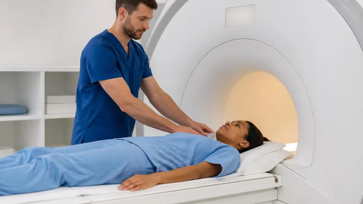
Understanding how accurate MRI scans are for prostate cancer is one of the most important questions facing patients and clinicians today. Multiparametric MRI has transformed prostate cancer diagnostics over the past decade, offering a non-invasive method to detect clinically significant tumors before biopsy. Modern studies consistently demonstrate that prostate MRI achieves sensitivity rates between 85 and 93 percent for detecting intermediate-to-high-grade cancers, making it one of the most reliable imaging tools in urological oncology available to patients across the United States.
The accuracy of prostate MRI depends on several interconnected factors including the strength of the magnetic field, the experience of the radiologist interpreting the images, the specific sequences used during scanning, and the biological characteristics of the tumor itself. A 3-Tesla scanner paired with a skilled radiologist can identify lesions as small as five millimeters in certain cases, though detection rates naturally vary based on tumor location within the gland and its Gleason grade at pathology.
Prostate MRI uses a standardized reporting system called PI-RADS, which stands for Prostate Imaging Reporting and Data System. This five-point scale helps radiologists communicate the likelihood that a suspicious area represents clinically significant cancer. PI-RADS scores of four or five indicate a high probability of significant disease, and studies show that targeted biopsies guided by these scores detect up to 30 percent more clinically significant cancers compared to traditional systematic biopsy approaches alone.
The negative predictive value of prostate MRI deserves particular attention because it directly impacts clinical decisions about whether to proceed with biopsy. When a high-quality MRI shows no suspicious lesions, the probability that the patient has clinically significant cancer ranges between three and ten percent depending on the study population. This high negative predictive value has led many urologists to recommend MRI as a triage tool that can safely reduce unnecessary biopsies in men with elevated PSA levels.
Despite its impressive diagnostic performance, prostate MRI is not perfect and has well-documented limitations that patients should understand. The technology performs less reliably for small anterior tumors, transition zone cancers, and low-grade Gleason six lesions that many experts now classify as indolent disease rather than true cancer requiring treatment. Additionally, previous prostate biopsies can create hemorrhagic artifacts that temporarily reduce image quality and interpretation accuracy for several weeks following the procedure.
Research published between 2022 and 2025 continues to refine our understanding of prostate MRI accuracy in specific clinical scenarios. Large multicenter trials including PRECISION, MRI-FIRST, and the 4M study have collectively enrolled thousands of patients and provide robust evidence supporting the integration of MRI into the diagnostic pathway. These trials demonstrate that MRI-targeted biopsy strategies detect similar or greater numbers of significant cancers while simultaneously reducing overdiagnosis of clinically insignificant disease by approximately 50 percent.
For patients navigating the diagnostic process, understanding the capabilities and limitations of prostate MRI empowers better conversations with urologists and radiologists. The technology continues advancing with artificial intelligence algorithms now assisting in lesion detection and characterization, potentially improving consistency across different institutions and reader experience levels. This comprehensive guide examines every aspect of prostate MRI accuracy to help you make informed decisions about your healthcare journey.
Prostate MRI Accuracy by the Numbers
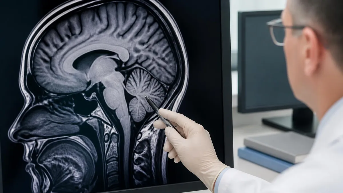
Key Factors That Determine Prostate MRI Accuracy
3-Tesla scanners provide significantly better spatial resolution and signal-to-noise ratio compared to 1.5T systems, improving detection of small lesions and peripheral zone abnormalities in prostate imaging protocols.
Fellowship-trained abdominal radiologists who read over 100 prostate MRI cases annually achieve consistently higher accuracy rates than generalists, with studies showing up to 15 percent improvement in detection.
Complete multiparametric protocols including T2-weighted, diffusion-weighted imaging with ADC maps, and dynamic contrast-enhanced sequences maximize diagnostic accuracy compared to abbreviated protocols.
Proper bowel preparation, antispasmodic medication, and avoiding biopsy for six weeks prior to scanning reduce artifacts that degrade image quality and lower diagnostic confidence significantly.
Tumor size, Gleason grade, and anatomic location within the prostate directly influence MRI visibility, with larger high-grade peripheral zone lesions being most readily detected on imaging.
The sensitivity and specificity of prostate MRI have been extensively studied across multiple large clinical trials, providing clinicians with reliable performance data for shared decision-making with patients. Sensitivity refers to the ability of MRI to correctly identify patients who actually have clinically significant prostate cancer, while specificity measures how well the test correctly identifies patients without significant disease. Understanding both metrics together gives the complete picture of diagnostic performance that neither measure alone can convey.
The landmark PRECISION trial published in the New England Journal of Medicine enrolled 500 biopsy-naive men and randomized them to either MRI-targeted biopsy or standard transrectal ultrasound-guided systematic biopsy. Results demonstrated that the MRI pathway detected clinically significant cancer in 38 percent of men compared to 26 percent in the standard biopsy arm, while simultaneously reducing detection of clinically insignificant cancers from 22 percent to 9 percent. These findings fundamentally changed clinical practice guidelines across North America and Europe.
Specificity values for prostate MRI typically range between 65 and 80 percent across published studies, meaning that approximately 20 to 35 percent of men without significant cancer will receive a positive MRI result suggesting possible malignancy. This false-positive rate translates to unnecessary biopsies in some patients, though targeted biopsies carry substantially lower complication rates than systematic approaches because fewer cores are obtained from precisely identified suspicious regions rather than sampling the entire gland randomly.
The negative predictive value represents perhaps the most clinically useful metric because it addresses the question patients most frequently ask: if my MRI is normal, can I safely avoid biopsy? Current evidence supports a negative predictive value exceeding 90 percent for clinically significant cancer when experienced radiologists interpret high-quality scans. This means that fewer than one in ten men with a negative MRI will harbor undetected significant disease, a rate many patients and clinicians find acceptable for active surveillance.
Detection rates vary substantially depending on the PI-RADS score assigned to any identified lesion. PI-RADS 5 lesions carry a cancer detection rate approaching 85 to 90 percent on subsequent targeted biopsy, while PI-RADS 4 lesions demonstrate cancer in approximately 50 to 70 percent of cases. PI-RADS 3 lesions, classified as equivocal, show significant cancer in only 15 to 30 percent of cases, creating a clinical gray zone where additional factors like PSA density and clinical history guide management decisions.
Subgroup analyses from major trials reveal important variations in accuracy based on patient demographics and tumor biology. MRI performs most reliably in detecting peripheral zone tumors with Gleason scores of seven or higher, achieving sensitivity exceeding 90 percent in many series. Conversely, transition zone cancers and anterior lesions present greater challenges because benign prostatic hyperplasia nodules can mimic malignancy on multiple MRI sequences, reducing specificity in these anatomic regions to approximately 55 to 65 percent.
Recent meta-analyses pooling data from over 10,000 patients confirm that multiparametric MRI followed by targeted biopsy represents the optimal diagnostic strategy for men with clinical suspicion of prostate cancer. The combined approach reduces overdiagnosis of indolent disease by approximately half while maintaining or improving detection of life-threatening cancers requiring treatment. These population-level benefits translate into fewer men experiencing the anxiety, complications, and side effects associated with unnecessary biopsies and overtreatment of low-risk disease.
MRI Practice Test Questions
Prepare for the MRI - Magnetic Resonance Imaging exam with our free practice test modules. Each quiz covers key topics to help you pass on your first try.
MRI Knowledge
MRI Exam Questions covering Knowledge. Master MRI Test concepts for certification prep.
MRI Physics
Free MRI Practice Test featuring Physics. Improve your MRI Exam score with mock test prep.
MRI Anatomy and Pathology
MRI Test Prep for MRI Anatomy and Pathology. Practice MRI Quiz questions and boost your score.
MRI Anatomy and Positioning
MRI Questions and Answers on MRI Anatomy and Positioning. Free MRI practice for exam readiness.
MRI Contrast Agents
Free MRI Quiz on MRI Contrast Agents. MRI Exam prep questions with detailed explanations.
MRI Patient Care and Positioning
MRI Practice Questions for MRI Patient Care and Positioning. Build confidence for your MRI certification exam.
Understanding PI-RADS Scoring and MRI Interpretation
The PI-RADS scoring system assigns each suspicious prostate lesion a score from one to five based on imaging characteristics across multiple MRI sequences. A score of one indicates highly unlikely clinically significant cancer, while a score of five indicates very high probability of significant malignancy. Radiologists evaluate T2-weighted imaging for transition zone lesions and diffusion-weighted imaging for peripheral zone lesions as the dominant sequence determining the final assessment category for each identified abnormality.
PI-RADS version 2.1, released in 2019, refined criteria for scoring transition zone and anterior lesions where previous versions showed inconsistency between readers. The updated guidelines emphasize that lesion morphology on T2 and restricted diffusion on ADC maps carry the most diagnostic weight. Dynamic contrast enhancement serves primarily as a tiebreaker for peripheral zone lesions scored as PI-RADS 3 on diffusion imaging, potentially upgrading equivocal cases to PI-RADS 4 when focal early enhancement is present with corresponding washout patterns.
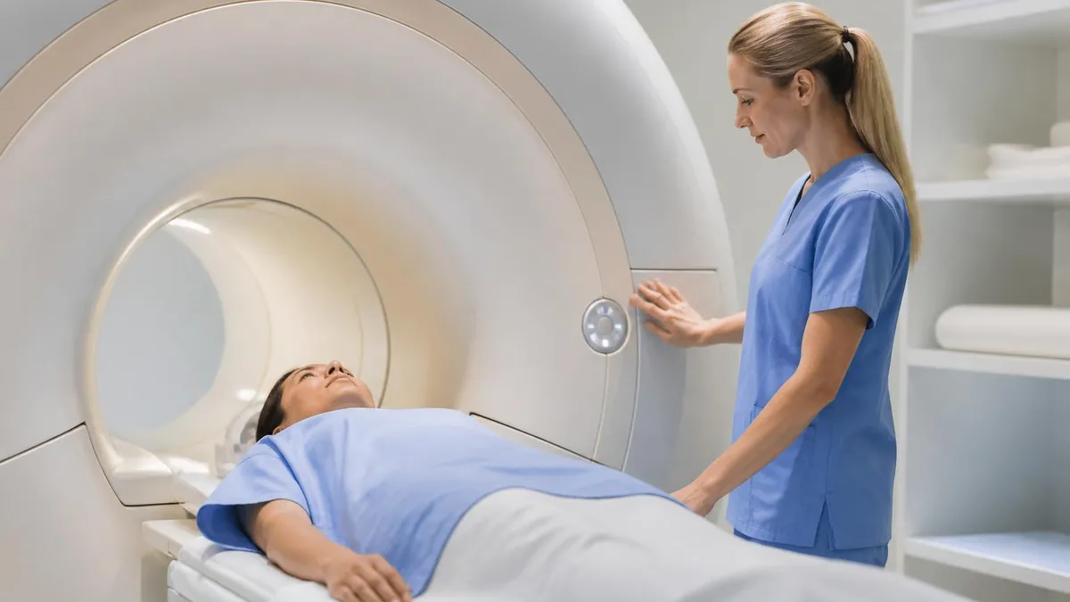
Advantages and Limitations of Prostate MRI for Cancer Detection
- +Detects 85-93% of clinically significant cancers with Gleason score 7 or higher
- +Reduces unnecessary biopsies by over 25% through high negative predictive value
- +Enables targeted biopsy approaches that improve cancer grade accuracy
- +Non-invasive imaging without ionizing radiation exposure to patients
- +Identifies extracapsular extension and seminal vesicle invasion for staging
- +Reduces overdiagnosis of clinically insignificant low-grade disease by 50%
- −False-positive rate of 20-35% leads to some unnecessary biopsies
- −Reduced accuracy for transition zone and anterior prostate tumors
- −Requires experienced radiologist with high-volume prostate MRI practice
- −Post-biopsy hemorrhage artifacts degrade image quality for 6-8 weeks
- −Limited sensitivity for small tumors under 5 millimeters in diameter
- −Gadolinium contrast carries small risk for patients with kidney disease
Prostate MRI Patient Preparation Checklist
- ✓Confirm at least 6 weeks have passed since any previous prostate biopsy procedure
- ✓Complete bowel preparation as directed to reduce rectal gas artifacts on imaging
- ✓Abstain from ejaculation for 48 to 72 hours before the scheduled MRI appointment
- ✓Arrive with a comfortably full bladder unless otherwise instructed by the imaging center
- ✓Inform the technologist about any metallic implants including hip replacements or cardiac devices
- ✓Remove all metal objects including belt buckles, jewelry, and clothing with zippers before scanning
- ✓Report any history of kidney disease or prior reactions to gadolinium contrast agents
- ✓Take prescribed antispasmodic medication 30 minutes before scan to reduce bowel motion artifacts
- ✓Bring previous PSA results and any prior imaging reports for radiologist reference during interpretation
- ✓Expect the examination to last 30 to 45 minutes with instructions to remain still throughout scanning
When MRI Shows No Suspicious Lesions
A negative multiparametric prostate MRI has a 90-95% negative predictive value for clinically significant cancer. This means that only 5-10% of men with a clean MRI will have undetected significant disease, making it a powerful triage tool that can safely defer biopsy in appropriately selected patients with low PSA density values.
Despite its strong overall performance, prostate MRI has well-documented limitations that patients and referring physicians must understand to set appropriate expectations and make informed clinical decisions. No imaging test achieves perfect accuracy, and recognizing the specific scenarios where MRI underperforms helps contextualize individual results within the broader diagnostic pathway. False-negative and false-positive results both carry clinical consequences that warrant careful discussion between patients and their healthcare teams before and after scanning.
False-negative results occur when MRI fails to identify a cancer that is actually present within the prostate gland. This happens most frequently with small tumors measuring less than five millimeters, diffuse low-grade cancers that lack the focal restriction pattern typical of higher-grade disease, and tumors located in the anterior fibromuscular stroma or extreme apex where imaging quality diminishes. Studies estimate that approximately 7 to 15 percent of clinically significant cancers are missed on initial multiparametric MRI, necessitating continued clinical surveillance even after a reassuring scan result.
False-positive results represent suspicious MRI findings that prove benign on subsequent biopsy, causing patient anxiety and potentially unnecessary invasive procedures. Common mimickers of prostate cancer on MRI include granulomatous prostatitis, post-inflammatory scarring, stromal benign prostatic hyperplasia nodules, and hemorrhagic changes from prior biopsies. The specificity challenge is particularly pronounced in the transition zone where approximately 30 to 40 percent of suspicious findings ultimately prove non-malignant on histologic examination.
Inter-reader variability remains a significant challenge affecting prostate MRI accuracy across different clinical settings. Studies comparing interpretations between general radiologists and subspecialty-trained prostate MRI experts reveal discordance rates of 20 to 30 percent for PI-RADS scoring, with the greatest disagreement occurring at the PI-RADS 3 threshold where clinical management changes most dramatically. This variability underscores the importance of seeking imaging centers with dedicated genitourinary radiologists who maintain high case volumes.
Artificial intelligence applications are rapidly addressing some of these accuracy limitations by providing computer-aided detection and characterization of prostate lesions. Deep learning algorithms trained on thousands of annotated prostate MRI cases can highlight suspicious regions for radiologist review, potentially reducing miss rates for subtle lesions and improving scoring consistency across readers with varying experience levels. Several FDA-cleared AI tools are now commercially available in the United States, though they function as decision-support rather than autonomous diagnostic systems.
The impact of scanner technology on accuracy cannot be overstated when patients are choosing where to undergo prostate MRI. Research consistently demonstrates that 3-Tesla scanners outperform 1.5-Tesla systems for prostate imaging, providing higher signal-to-noise ratios and better spatial resolution that translate into improved lesion detection sensitivity. However, even on 3T systems, the use of an endorectal coil versus surface coil alone remains debated, with most high-volume centers now achieving diagnostic-quality images using external phased-array coils without the discomfort of endorectal placement.
Prior treatments and interventions further complicate MRI interpretation accuracy in specific patient populations. Men who have undergone radiation therapy, hormonal therapy, or focal ablation treatments show altered tissue signal characteristics that can mask residual or recurrent cancer. Similarly, patients on active surveillance who undergo serial MRI monitoring may develop subtle progressive changes that fall below the detection threshold of current imaging technology, requiring integration of MRI findings with PSA kinetics and clinical judgment for optimal management decisions.
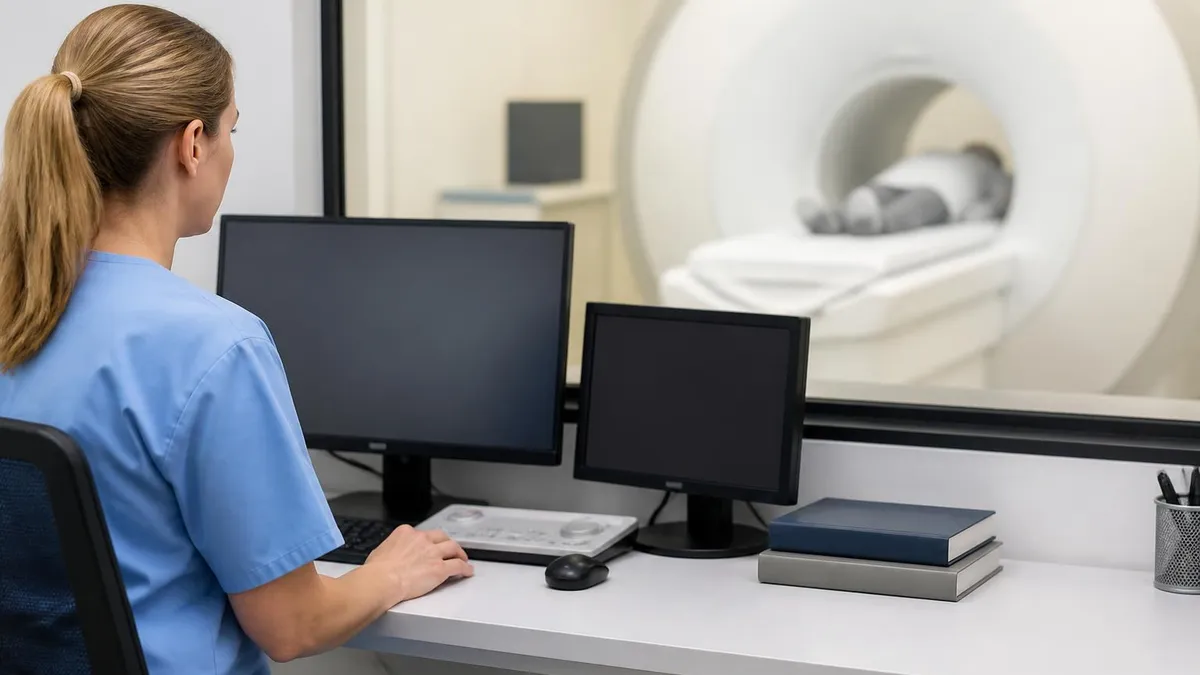
If you have had a prostate biopsy within the past six weeks, residual blood products create T1-bright artifacts that significantly degrade MRI image quality and reduce diagnostic accuracy. Most radiology guidelines recommend waiting a minimum of six to eight weeks after biopsy before scheduling a prostate MRI to ensure optimal imaging conditions and reliable interpretation results.
Comparing prostate MRI to other available diagnostic methods reveals its unique position in the cancer detection pathway and helps patients understand why physicians increasingly recommend imaging before proceeding to invasive tissue sampling. Traditional diagnostic approaches relied almost exclusively on PSA blood testing followed by systematic transrectal ultrasound-guided biopsy, an approach that detected many cancers but suffered from both overdiagnosis of indolent tumors and underdetection of significant anterior lesions missed by standard posterior sampling patterns.
PSA testing alone demonstrates limited specificity for prostate cancer, with only approximately 25 to 30 percent of men with PSA levels between 4 and 10 nanograms per milliliter actually harboring cancer on subsequent biopsy. This means that roughly 70 percent of men undergoing biopsy based solely on elevated PSA experience an unnecessary invasive procedure with associated risks of bleeding, infection, and anxiety. Adding MRI as an intermediate step between PSA testing and biopsy reduces this unnecessary biopsy rate by 25 to 40 percent while maintaining high detection rates for significant disease.
Transrectal ultrasound, the traditional imaging guidance for prostate biopsy, demonstrates poor sensitivity for cancer detection averaging only 40 to 50 percent for identifying hypoechoic lesions that may represent malignancy. Ultrasound cannot reliably distinguish between cancer and benign conditions, which is precisely why systematic sampling of the entire gland became standard practice despite its inherent limitations. MRI offers dramatically superior soft-tissue contrast that enables actual visualization of suspicious lesions before any needles enter the prostate.
PET imaging with prostate-specific membrane antigen radiotracers represents the newest competitor to MRI in prostate cancer detection, though these modalities serve complementary rather than competing roles in most clinical scenarios. PSMA-PET excels at detecting metastatic disease and biochemical recurrence after treatment but carries radiation exposure and higher cost compared to MRI. For initial cancer detection in the intact prostate gland, multiparametric MRI remains the preferred imaging modality recommended by major urological guidelines including those from the American Urological Association and European Association of Urology.
Micro-ultrasound technology operating at 29 megahertz represents an emerging alternative that some institutions are exploring as a potential real-time imaging option during biopsy. Early studies suggest micro-ultrasound may approach the sensitivity of MRI for detecting significant cancers while offering the convenience of real-time guidance without requiring a separate prior imaging appointment. However, the technology lacks the extensive multicenter validation that supports multiparametric MRI and does not yet appear in major society clinical practice guidelines as a standard recommendation.
Cost-effectiveness analyses consistently favor incorporating MRI into the diagnostic pathway despite the higher per-scan cost compared to proceeding directly to biopsy. When accounting for reduced biopsy rates, fewer complications from unnecessary procedures, decreased overtreatment of indolent disease, and improved detection of clinically significant cancers requiring timely intervention, the MRI-first approach demonstrates favorable cost-per-quality-adjusted-life-year ratios that meet accepted thresholds for healthcare value in United States economic models.
The integration of multiple diagnostic modalities continues evolving as research identifies optimal combinations for different clinical scenarios and risk profiles. Many leading centers now employ risk calculators incorporating PSA density, MRI findings, patient age, family history, and genomic biomarkers to generate individualized probability estimates that guide shared decision-making about biopsy. This multimodal approach represents the future of prostate cancer diagnosis, with MRI serving as the central imaging pillar around which other diagnostic information is organized and interpreted.
Patients preparing for a prostate MRI can take several practical steps to maximize the diagnostic quality of their examination and ensure the most accurate results possible from their imaging study. Preparation begins well before the appointment day and involves coordination with both the ordering urologist and the imaging facility to address logistical and clinical factors that influence scan quality. Taking an active role in preparation helps patients feel more confident and contributes directly to obtaining reliable diagnostic information.
Choosing the right imaging facility represents perhaps the single most impactful decision patients can make regarding their prostate MRI accuracy. Look for centers that specifically advertise multiparametric prostate MRI as a specialty service, utilize 3-Tesla scanners, and employ fellowship-trained abdominal or genitourinary radiologists who interpret high volumes of prostate cases. Asking the facility about their radiologist's prostate MRI experience and whether they follow PI-RADS version 2.1 reporting standards provides valuable quality indicators before committing to an appointment.
During the examination itself, remaining as still as possible throughout the 30 to 45 minute scan directly impacts image sharpness and diagnostic quality. Motion artifacts from breathing, bowel peristalsis, or patient movement degrade spatial resolution and can create false signals that mimic or obscure pathology. Using relaxation techniques such as slow deep breathing, keeping eyes closed, and focusing on remaining comfortable reduces involuntary movement that the scanner cannot correct through post-processing algorithms alone.
Understanding what happens after the scan helps patients manage expectations about the timeline for receiving results and planning next steps in their diagnostic journey. Most radiology reports require 24 to 72 hours for completion as the radiologist carefully evaluates all sequences, measures lesion characteristics, assigns PI-RADS scores, and generates a comprehensive structured report. Some complex cases may benefit from additional review at multidisciplinary tumor board meetings where urologists, radiologists, and pathologists discuss findings collaboratively.
If your prostate MRI reveals suspicious findings warranting biopsy, understanding the targeted biopsy process helps reduce anxiety about the next procedural step. MRI-fusion targeted biopsy uses software to overlay the MRI images onto real-time ultrasound, guiding needles precisely into identified lesions with millimeter accuracy. This approach typically requires only two to four cores per target compared to twelve systematic cores, reducing procedure time, discomfort, and infection risk while improving the diagnostic yield for significant cancers within the identified suspicious regions.
Patients receiving PI-RADS 3 equivocal results face the most challenging decision point because this category sits at the boundary between reassurance and concern. In these cases, additional factors including PSA density calculations, lesion size, prior biopsy history, and patient risk tolerance guide management decisions that should be discussed thoroughly with your urologist. Some patients with PI-RADS 3 findings and low PSA density may safely undergo short-interval surveillance MRI rather than immediate biopsy, while others with concerning clinical profiles may benefit from proceeding directly to tissue sampling.
Following up appropriately after a negative prostate MRI ensures that the small percentage of cancers potentially missed by imaging do not progress undetected over time. Most guidelines recommend continued PSA monitoring at regular intervals following a reassuring MRI, with repeat imaging considered if PSA velocity increases beyond established thresholds or if clinical suspicion changes based on digital rectal examination findings. Active communication with your urologist about ongoing surveillance plans provides the safety net that accounts for the inherent imperfection of any single diagnostic test in isolation.
MRI Questions and Answers
Side Effects of MRI: What Patients Need to Know About Risks, Reactions, and Recovery

MRI of Cervical Spine: Complete Guide to Imaging, Anatomy, Pathology, and Patient Preparation
MRI Medical Abbreviation: What MRI Stands For and Why It Matters

Knee MRI Images: A Complete Guide to Reading, Understanding, and Interpreting Knee Scans
Noise of MRI Machine: Why MRI Scanners Are So Loud and What to Expect
About the Author

Medical Laboratory Scientist & Clinical Certification Expert
Johns Hopkins UniversityDr. Sandra Kim holds a PhD in Clinical Laboratory Science from Johns Hopkins University and is certified as a Medical Technologist (MT) and Medical Laboratory Scientist (MLS) through ASCP. With 16 years of clinical laboratory experience spanning hematology, microbiology, and molecular diagnostics, she prepares candidates for ASCP board exams, MLT, MLS, and specialist certification tests.
Join the Discussion
Connect with other students preparing for this exam. Share tips, ask questions, and get advice from people who have been there.
View discussion (8 replies)