MRI vs CAT Scan: Key Differences, Costs, and When Each Is Used
MRI vs CAT scan compared head-to-head: imaging tech, costs, scan time, radiation, and when doctors order each. Plain-English guide for patients.
Walking into a radiology department for the first time can feel like stepping onto a movie set. Massive machines hum behind glass walls. Technologists slide into rooms wearing badges and serious expressions. Somewhere in your head a tiny voice asks the obvious question: is this thing safe, and is it even the right test for what my doctor suspects? If your physician handed you a slip that says "CT scan" or "MRI," you probably nodded along. Now you are at home, googling, and the differences are not as obvious as you hoped.
Here is the honest answer most people never get during a five-minute consultation. MRI and CAT scans look similar from the outside, but they work in completely different ways. One uses radio waves and a giant magnet. The other shoots X-rays through your body at hundreds of angles per second. Both produce cross-section images that radiologists can study slice by slice. Yet the situations that call for one over the other are very specific, and picking the wrong scan can mean missed diagnoses, unnecessary radiation, or a bill that surprises you weeks later.
This guide breaks the whole topic down. You will learn the physics in plain language, the price ranges, the actual experience of lying in each machine, and the medical situations where doctors lean one way or the other. By the end you should feel ready to ask smarter questions at your appointment and maybe even challenge a recommendation that does not sit right.
What Each Scan Actually Does
A CAT scan (computed axial tomography, often shortened to CT) is essentially a souped-up X-ray. The machine sends a thin fan of radiation around your body while detectors on the opposite side record what gets through. A computer stitches those readings into cross-sectional slices. Think of slicing a loaf of bread and laying each piece flat: that is what your insides look like to a radiologist scrolling through CT images.
An MRI (magnetic resonance imaging) skips radiation entirely. It uses a powerful magnet, usually 1.5 or 3 Tesla, roughly 30,000 to 60,000 times Earth's magnetic field, plus pulses of radio waves. Hydrogen atoms in your body's water molecules respond to those pulses, and the machine measures the signals as they relax back. Different tissues give off different signals, which is why MRI is so good at showing soft tissue contrast.
So one is fast and uses X-rays. The other is slow and uses magnets. That single sentence covers about 80% of what most patients need to remember.
MRI vs CT By the Numbers
The One-Sentence Difference
CT uses X-rays and finishes in minutes. MRI uses magnets and radio waves and takes much longer but shows soft tissue in far greater detail. That single distinction drives almost every other choice your doctor makes when ordering an imaging study, from cost to scan length to whether you can have one at all with a pacemaker or implant.
Speed, Cost, and the Patient Experience
Speed is where CT crushes MRI. A typical chest CT finishes in under five minutes, including positioning. The actual scanning takes seconds. Emergency departments rely on this when someone arrives with a possible stroke, head injury, or internal bleeding. You cannot wait 45 minutes for an MRI when a clot is forming.
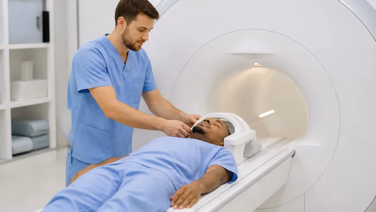
MRI, by contrast, is a marathon. A standard brain MRI runs 30 to 45 minutes. Adding contrast or extra sequences can push past an hour. The machine is also louder, imagine someone slamming a hammer near your ear for a few minutes at a time. Most facilities offer earplugs or headphones with music, but claustrophobic patients still struggle. Open MRI machines exist, though they typically produce lower-resolution images.
Cost reflects all of this. In the United States, a CT scan ranges from roughly $300 to $1,500 without insurance, depending on body part and contrast. MRI runs $400 to $3,500, with brain and spine studies on the higher end. Some outpatient imaging centers charge a fraction of hospital rates for the exact same scan, so it pays to call around if you have time before scheduling.
When Doctors Order Each One
CT scans dominate emergency medicine. They are the first call for trauma, suspected stroke (specifically to rule out bleeding before giving clot-busting drugs), pulmonary embolism, kidney stones, appendicitis, and most chest or abdominal pain that needs urgent answers. CT also shows bone fractures with remarkable clarity and is the standard for lung cancer screening in high-risk patients.
MRI shines wherever soft tissue detail matters most. Brain tumors, multiple sclerosis lesions, spinal cord problems, torn ligaments and cartilage, prostate cancer staging, and many cardiac conditions all get worked up with MRI. Orthopedic surgeons rely on it before knee and shoulder operations. Neurologists order it to map out anything from seizures to memory loss.
There is overlap. Both can image the abdomen, both can guide biopsies, both can detect cancer. The choice often comes down to what the doctor specifically suspects, how urgent the situation is, and whether the patient can tolerate the scan. Knowing how to read cross-sectional anatomy helps technologists position patients correctly for either modality.
Strengths and Best Uses
Fast, bone-detailed, widely available emergency imaging.
- ▸Speed under 5 minutes
- ▸Excellent bone detail
- ▸Lung imaging gold standard
- ▸Emergency trauma workup
- ▸Stroke bleeding assessment
- ▸Kidney stone detection
- ▸Lung cancer screening
Unmatched soft tissue contrast without radiation.
- ▸Brain tumor evaluation
- ▸Spinal cord imaging
- ▸Ligament and cartilage tears
- ▸Prostate cancer staging
- ▸Cardiac soft tissue
- ▸Repeat imaging safety
- ▸Pediatric follow-up
Speed-critical or radiation-tolerant scenarios.
- ▸Acute trauma in ER
- ▸Suspected internal bleeding
- ▸Pulmonary embolism workup
- ▸Severe abdominal pain
- ▸Patients with MRI contraindications
- ▸Rapid triage decisions
Soft tissue questions or radiation avoidance.
- ▸Neurological symptoms
- ▸Sports injuries to joints
- ▸Soft tissue cancer staging
- ▸Pediatric repeat imaging
- ▸Pregnant patients after first trimester
- ▸Chronic disease monitoring
Radiation, Contrast, and Safety
This is the part patients ask about most, and rightly so. A single chest CT delivers roughly 7 millisieverts of radiation, about the same as 70 chest X-rays or two to three years of natural background exposure. Abdominal and pelvic CTs can hit 10 to 20 mSv. The cancer risk from any one scan is small, but doctors weigh repeated scans carefully, especially in children and young adults.
MRI uses zero ionizing radiation. That alone makes it the preferred option for pregnant patients (after the first trimester), children needing repeat imaging, and anyone with conditions requiring frequent follow-up. The trade-off is the magnet itself: implanted pacemakers, certain aneurysm clips, cochlear implants, and metal fragments in the eye can make MRI dangerous or impossible. Newer pacemakers are MRI-conditional, but every patient gets screened with a long checklist before entering the room.
Both scans can use contrast agents. CT contrast is usually iodine-based and can stress the kidneys. MRI contrast is typically gadolinium-based, which carries a small risk of nephrogenic systemic fibrosis in patients with severe kidney disease. Allergic reactions to either are uncommon but documented.
Comparing Each Factor Side by Side
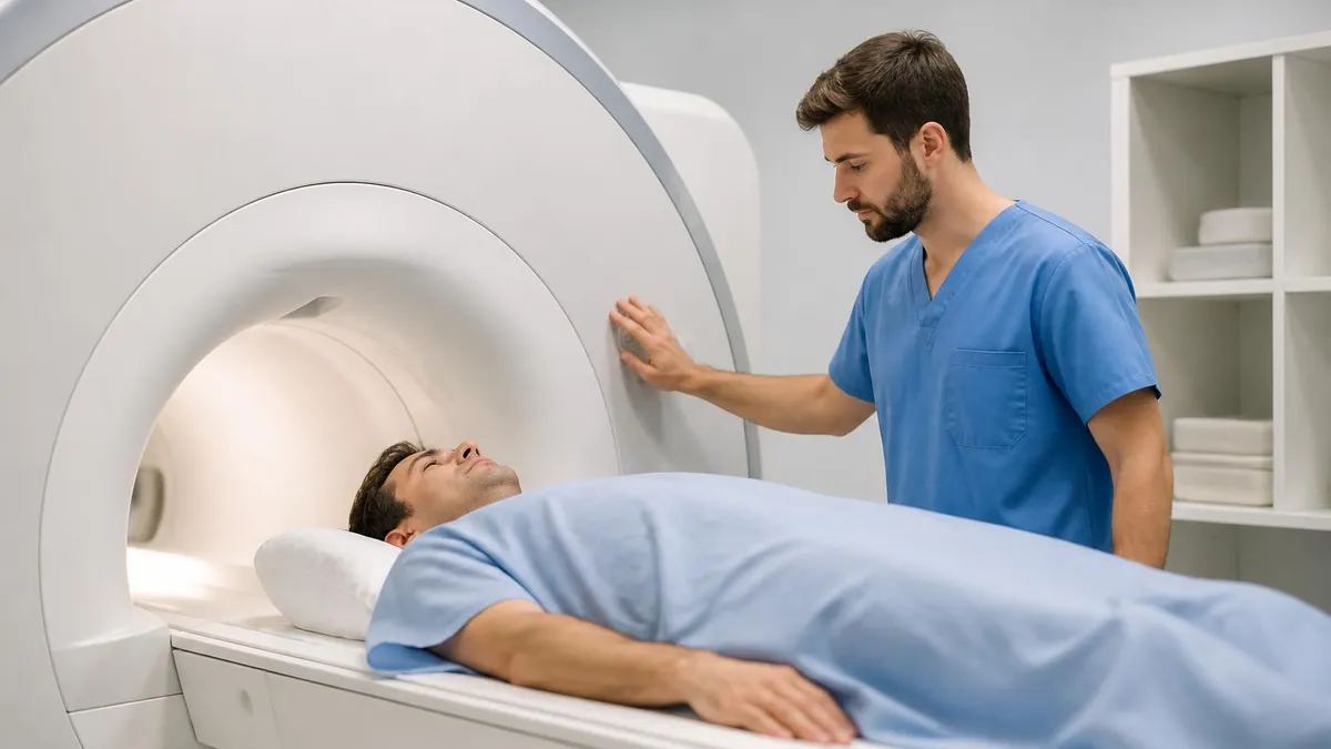
Image Quality and Detail
If you have ever seen both scans side by side, the difference is striking. CT renders bone in bright white, air in deep black, and soft tissue in shades of gray. The contrast between soft tissues, say, distinguishing a tumor from surrounding muscle, is limited. CT excels when the structures of interest have very different densities.
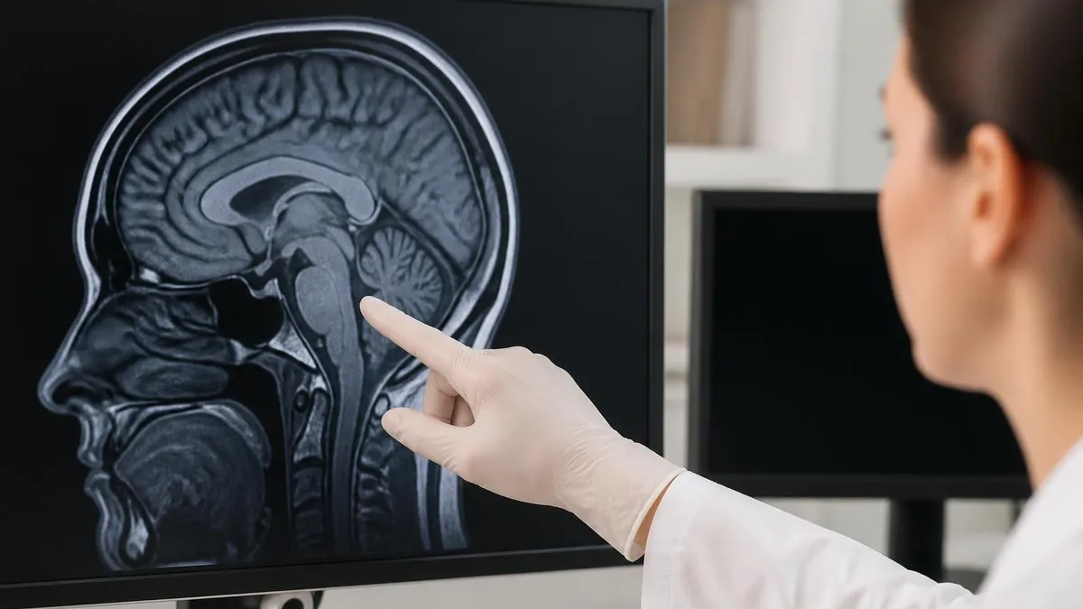
MRI shows soft tissue gradations that CT simply cannot match. A radiologist can tell muscle from fat from edema from blood, often within the same anatomical region. That sensitivity is why neurologists prefer MRI for almost any brain question beyond acute bleeding, and why orthopedic surgeons rely on it to assess ligament tears the moment they are no longer urgent.
Resolution itself depends on the machine. A 3 Tesla MRI produces sharper images than 1.5 Tesla. Modern multidetector CT scanners can capture submillimeter slices in a single breath-hold. Technology keeps advancing on both sides, which is why imaging recommendations shift every few years.
How Doctors Actually Choose
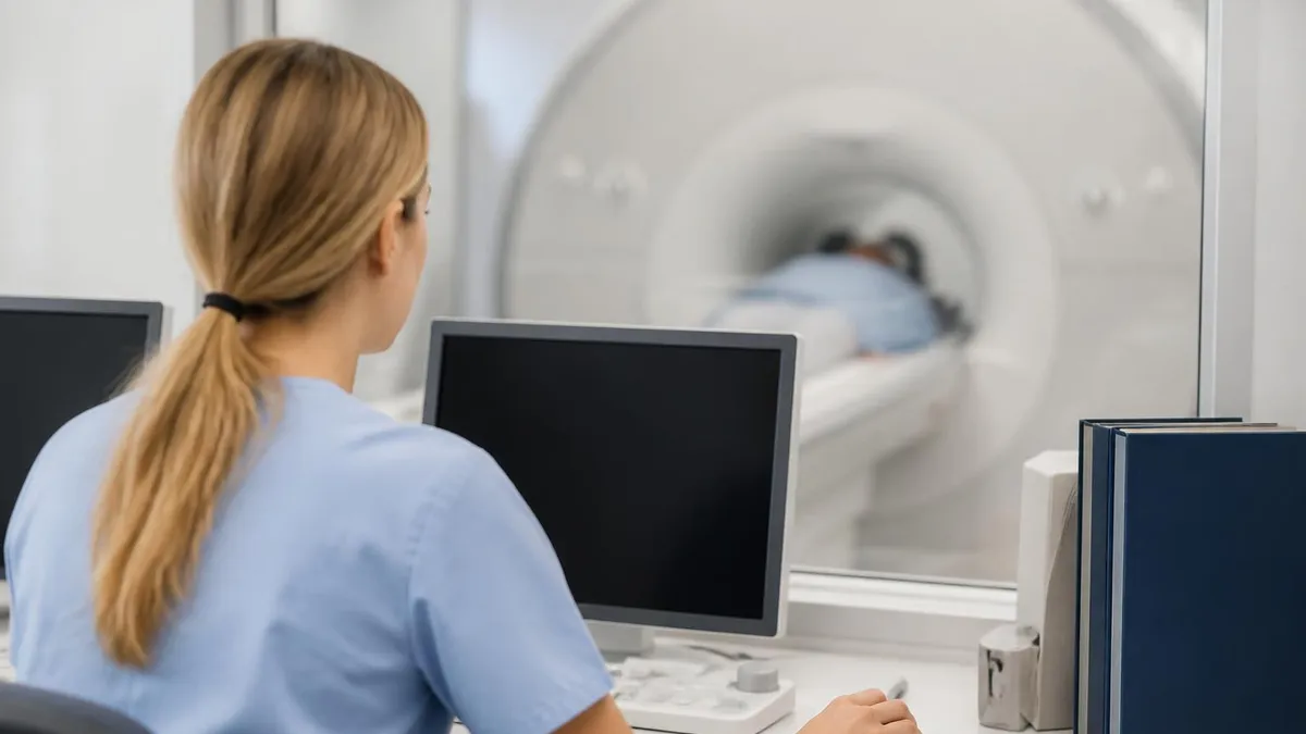
The textbook answer is "based on clinical suspicion." The real-world answer is messier. Availability matters: small hospitals may have one MRI and three CT scanners, so the faster machine wins by default. Patient factors matter: someone with severe claustrophobia may need sedation for MRI, while someone with metal hardware cannot have one at all. Insurance pre-authorization adds another layer, especially for outpatient MRI studies.
Good clinicians explain the trade-offs. If your doctor recommends a CT for a chronic, non-urgent problem, it is fair to ask whether MRI would give a clearer answer without radiation. Conversely, if you are sitting in an emergency department with sudden chest pain, do not push for MRI. CT is faster and safer for that moment. Familiarity with MRI physics helps clinicians explain why one test trumps the other.
Second opinions are reasonable for big decisions. Cancer staging, pre-surgical planning, and unexplained neurological symptoms all justify careful imaging choices.
Tell the technologist about every implant, surgery, and metal exposure. Pacemakers, cochlear implants, certain aneurysm clips, and metal eye fragments can make MRI dangerous. The screening form exists for your safety, so answer every question honestly. If you have worked with metal grinding or welding, you may need an orbital X-ray to rule out fragments in your eyes before the scan can proceed.
Smart Questions Before Any Scan
- ✓Why is this specific scan being ordered?
- ✓Would the other modality show the same information?
- ✓Will contrast be used, and what are the risks?
- ✓How much radiation does this involve, if any?
- ✓When will results be available and how will I be notified?
- ✓What is the out-of-pocket cost with my insurance?
- ✓Are there cheaper outpatient imaging centers I could use?
- ✓Should I bring prior images for comparison?
- ✓How long will the scan take, including prep?
- ✓Can I drive home afterward, or will I need a ride?
How to Prepare for Either Scan
For a CT, preparation depends on the body part and whether contrast is used. Abdominal CTs often require fasting for four to six hours and drinking an oral contrast that highlights the digestive tract. You will lie on a flat table that slides through a doughnut-shaped machine. The whole thing is fast and quiet compared to MRI.
MRI prep is heavier on screening. Expect a detailed questionnaire about implants, surgeries, tattoos (older inks sometimes contain iron), and metal exposure. You will change into a gown to eliminate any zippers, underwires, or jewelry. Inside the scanner, you cannot move. Even slight motion ruins images and forces repeat sequences. The technologist will hand you a squeeze ball connected to an alarm in case you need to call for help.
Hydration helps before either scan if contrast is involved. Bring something soothing, music, an audiobook, a meditation app, for MRI. Some facilities allow streaming through MRI-safe headphones, which makes a 45-minute brain study feel less like a sensory deprivation tank. Reviewing MRI safety guidelines in advance answers many last-minute worries.
After the Scan: Results, Costs, and Next Steps
Radiologists typically read scans within 24 to 48 hours, though emergency studies are often interpreted in minutes. Results land in your patient portal or arrive via a phone call from your referring physician. Reports use specific language, "unremarkable" is good, while "nodule" or "lesion" may need follow-up.
Billing can be confusing. You may receive separate charges for the facility (the technical fee) and the radiologist (the professional fee). Always ask for an itemized bill if anything looks off. Many imaging centers offer cash discounts that beat insurance copays for high-deductible plans.
Follow-up imaging is sometimes recommended at three, six, or twelve months. Keep digital copies of every scan, DICOM files on a CD or a cloud link, so future providers can compare directly rather than ordering another study.
Patient portals make accessing past results easier than ever, but they are not perfect. Reports sometimes lag behind images, and the technical language can be confusing without context. Schedule a follow-up appointment to walk through results with your referring physician rather than self-interpreting. The same finding can mean very different things depending on age, family history, and clinical picture, and a five-minute call beats hours of internet anxiety.
If you receive a report that mentions findings requiring additional imaging, do not panic. Recommendations for follow-up scans are common and usually conservative. Radiologists flag anything that could possibly need attention because missing something is worse than over-calling it. Most "follow-up recommended" notes turn out to be nothing.
Common Myths Patients Believe
Reality check on radiation fears.
- ▸Speed and availability matter just as much
- ▸CT is the right answer for acute trauma
- ▸Suspected stroke needs CT first to rule out bleeding
- ▸Lung imaging is far clearer on CT
- ▸Bone fractures show up sharper on CT
Dose context every patient should know.
- ▸Radiation doses are small relative to diagnostic value
- ▸Modern scanners use dose-reduction software
- ▸Pediatric protocols cut dose by 30 to 50 percent
- ▸Cumulative dose matters more than any single scan
- ▸Skipping a needed CT carries its own risks
What the magnet actually does and does not do.
- ▸Magnetic field does not harm body tissue
- ▸Risk comes from ferromagnetic objects being pulled in
- ▸Screening protocols eliminate almost all risk
- ▸MRI-conditional pacemakers exist for many patients
- ▸Tattoo concerns apply mostly to old inks
Most implants are safer than you think.
- ▸Old surgical clips are usually MRI-safe
- ▸Dental fillings cause no MRI safety issue
- ▸Most orthopedic hardware is MRI-compatible
- ▸Cochlear implants may be a true contraindication
- ▸Metal eye fragments require X-ray clearance first
MRI at a Glance
- +MRI: zero radiation exposure
- +MRI: superior soft tissue contrast
- +MRI: safer for repeat imaging
- +MRI: no iodine contrast required
- +MRI: preferred for brain and spine
- −MRI: 30-60 minutes per study
- −MRI: contraindicated with many implants
- −MRI: louder, more claustrophobic
- −MRI: typically more expensive than CT
- −MRI: limited availability in small hospitals
Specialized Variants You Might Hear About
Beyond standard CT and MRI, hospitals run several specialized variants. A CT angiogram uses iodine contrast to map blood vessels in detail, often replacing older catheter angiography for stroke or pulmonary embolism workups. A CT perfusion study measures blood flow through brain tissue, which guides treatment decisions in stroke patients within the critical first hours.
On the MRI side, functional MRI (fMRI) tracks blood oxygenation changes to show which brain regions activate during tasks. Neurosurgeons use this before tumor removal to map speech and motor areas. Diffusion-weighted MRI highlights areas where water molecule movement is restricted, which often signals fresh stroke or aggressive tumor. Cardiac MRI measures heart muscle function and scarring with detail no other test matches.
Each variant changes scan time, cost, and preparation. If your doctor orders something more specific than a "routine" CT or MRI, ask what the variant looks for and why it was chosen. The extra few minutes of conversation prevents confusion when results come back with unfamiliar terms.
If You Are Studying to Work in Imaging
Radiologic technologists, MRI technologists, and CT technologists all train through accredited programs and pass national certification exams. Each modality has its own scope. CT techs learn cross-sectional anatomy, contrast protocols, and dose optimization. MRI techs study magnet safety, pulse sequences, and patient positioning in ways CT techs never touch.
Many programs start with general radiography, then specialize. Continuing education credits keep certifications active. Test prep matters: the ARRT exam and the related ARRT MRI exam both require deep familiarity with safety zones, scan parameters, and patient care scenarios that show up in practice questions year after year.
Salaries reflect the specialization. MRI and CT technologists in the United States typically earn between $65,000 and $95,000 annually, with cardiac and interventional specialists higher. Demand stays strong as imaging volumes grow.
The Bottom Line
If you remember nothing else, remember this. CT is fast, uses X-rays, and shines for bones, lungs, and emergencies. MRI is slow, uses magnets, and shines for brain, spine, and soft tissue. Neither is universally better. Your doctor picks the one that answers your specific question with the lowest risk.
Asking smart questions at your appointment changes everything. Why this scan? What will it show that the other would not? Is contrast necessary? Are there alternatives? Good physicians welcome curious patients because curious patients catch errors and stick with treatment plans.
Imaging is a tool, not an answer in itself. The radiologist reads, the referring clinician interprets in context, and you decide what to do with the findings. Walk in informed and you walk out with a clearer path forward. The technology behind these machines is genuinely impressive, but the people running them and reading the images are the ones who turn pixels into answers. Treat them like partners in your care, not vendors of a service, and you will get better outcomes every time.
MRI Questions and Answers
About the Author
Medical Laboratory Scientist & Clinical Certification Expert
Johns Hopkins UniversityDr. Sandra Kim holds a PhD in Clinical Laboratory Science from Johns Hopkins University and is certified as a Medical Technologist (MT) and Medical Laboratory Scientist (MLS) through ASCP. With 16 years of clinical laboratory experience spanning hematology, microbiology, and molecular diagnostics, she prepares candidates for ASCP board exams, MLT, MLS, and specialist certification tests.