MRI Sedation: A Complete Guide to Sedated MRI Scans, Medications, Safety and What to Expect
MRI sedation guide covering medications, safety, recovery, pediatric and adult protocols, NPO rules, and what to expect before, during, and after a 🎯

MRI sedation is a medically supervised process used to help patients remain still, calm, and comfortable during a magnetic resonance imaging exam. Because MRI scans require absolute stillness for image quality, and because the scanner environment can be loud, confined, and lengthy, many patients benefit from medication that reduces anxiety, suppresses movement, or induces sleep. Understanding how mri sedation works, who needs it, and what the safety considerations are can help you prepare for a smoother, safer, and more diagnostically successful imaging experience.
Sedation for MRI ranges from minimal anxiolysis with oral medications such as lorazepam or diazepam to deep sedation and even general anesthesia using propofol, dexmedetomidine, or inhaled agents. The level chosen depends on patient age, medical history, claustrophobia severity, scan duration, and the body region being imaged. A pediatric brain MRI lasting 45 minutes usually demands deeper sedation than a 15-minute adult knee scan, where a single oral anxiolytic may be sufficient.
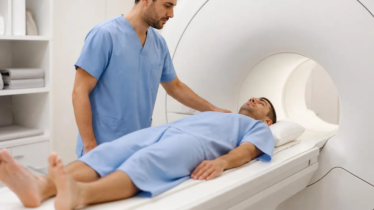
Adults frequently request sedation when claustrophobia, chronic pain, movement disorders, or post-traumatic stress make lying motionless in the bore intolerable. Children, particularly those under seven, almost always need sedation because they cannot reliably hold still for the long acquisition times required. Patients with developmental disabilities, dementia, or severe anxiety also commonly receive pharmacologic support to complete an exam that would otherwise be impossible or yield non-diagnostic images.
The decision to sedate is never automatic. Anesthesiologists, radiologists, and referring physicians weigh the diagnostic necessity of the scan against medication risks, airway concerns, and the logistical complexity of monitoring a sedated patient inside a strong magnetic field. MRI-compatible monitoring equipment, trained sedation nurses, and emergency airway carts must all be available, and patients must meet fasting and screening requirements before any sedative is administered. MRI with and without contrast studies often add time and complexity, which can influence sedation planning.
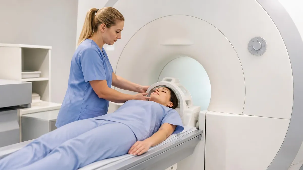
For families and patients, understanding the workflow reduces fear. The process typically begins with a pre-procedure phone screening, followed by an in-person assessment on the day of the scan. IV access is established, monitoring leads are placed, and the chosen agent is titrated to effect. Once imaging is complete, the patient recovers in a dedicated area until they meet discharge criteria, which usually requires stable vitals, a clear airway, and the ability to drink fluids without nausea.
This guide explains every aspect of MRI sedation in plain language: the medications used, the safety protocols that protect patients in the magnet, the difference between pediatric and adult approaches, the fasting and consent process, recovery expectations, and the practical questions you should ask before the day of your scan. Whether you are a parent preparing a child, a caregiver helping an elderly relative, or an adult patient managing claustrophobia, you will leave with a clear picture of what to expect and how to advocate for safe, effective imaging.
By the end, you will understand why some patients need only headphones and a blanket while others require an anesthesiologist and an endotracheal tube, and what the modern standard of care looks like in hospitals and outpatient imaging centers across the United States.
MRI Sedation by the Numbers
Levels of MRI Sedation Explained
A light pharmacologic state where the patient remains fully awake and able to respond normally. Oral lorazepam or alprazolam is typical. Breathing and cardiovascular function are unaffected, making this option ideal for mild claustrophobia or short scans.
Often called conscious sedation, the patient is drowsy but arousable to verbal stimuli. Midazolam combined with fentanyl is common. Airway reflexes are preserved, but pulse oximetry and capnography monitoring are required throughout the entire MRI examination.
The patient is not easily aroused but responds to repeated or painful stimulation. Propofol or dexmedetomidine infusions are typical. Airway support may be needed, and an anesthesia provider must be present with full resuscitation equipment available at bedside.
Complete loss of consciousness with no response to stimulation. Used for long pediatric scans, complex protocols, or patients who cannot tolerate deep sedation. Requires endotracheal intubation or laryngeal mask airway and continuous anesthesiologist supervision throughout the procedure.
The pharmacology of MRI sedation has evolved considerably over the past two decades. Where chloral hydrate once dominated pediatric imaging, modern centers now favor propofol, dexmedetomidine, midazolam, and combinations tailored to scan length and patient profile. Each agent has a distinct onset, duration, side effect profile, and reversal strategy. Choosing the right drug is a clinical decision based on age, weight, comorbidities, airway anatomy, and whether contrast will be administered during the study.
Midazolam, a short-acting benzodiazepine, is one of the most widely used sedatives because it can be given intravenously, intranasally, or orally. It produces anxiolysis, amnesia, and mild sedation within five to ten minutes. The amnestic effect is particularly valuable because patients often have little memory of the scan afterward, reducing future anxiety. Flumazenil reverses its effects if respiratory depression occurs, though reversal is rarely needed at standard doses.
Propofol has become the workhorse of deep sedation in MRI suites. Its rapid onset within 30 seconds, brief duration, and quick recovery profile make it ideal for scans lasting 30 to 90 minutes. It is delivered by continuous infusion, titrated to maintain stillness while preserving spontaneous breathing when possible. However, propofol can cause hypotension and apnea, so it must always be administered by trained anesthesia providers with full airway equipment available.
Dexmedetomidine, an alpha-2 agonist, has gained popularity for pediatric MRI because it produces sedation that resembles natural sleep without significant respiratory depression. The patient can usually be roused and breathes spontaneously throughout the scan. Onset is slower than propofol, often 15 to 20 minutes, and bradycardia is a known side effect. It is frequently combined with midazolam for induction and maintained as a continuous infusion. For information on what radiologists are looking for, see common MRI findings.
Oral benzodiazepines such as lorazepam, diazepam, and alprazolam are the mainstay of adult anxiolysis. A patient with moderate claustrophobia may take 1 to 2 milligrams of lorazepam 30 to 60 minutes before the appointment, arriving relaxed but fully awake. These oral agents do not require IV access or anesthesia monitoring, which makes them practical for outpatient imaging centers that lack on-site sedation teams. A driver home is always required.
Inhaled agents such as sevoflurane are reserved for general anesthesia cases, typically in children who cannot tolerate IV placement while awake. Induction by mask in a pre-anesthesia bay is followed by IV insertion, airway management, and transfer into the MRI suite using non-ferromagnetic equipment. This approach is more resource intensive but produces reliable stillness for long, technically demanding studies such as cardiac MRI or full spine imaging in young children.
Ketamine, ketofol mixtures, and chloral hydrate remain in selective use, particularly in resource-limited environments or for patients with specific contraindications to first-line drugs. Regardless of the agent chosen, the underlying principle is the same: use the minimum effective dose to achieve adequate stillness while preserving the patient's airway, hemodynamics, and recovery profile.
MRI Practice Test Questions
Prepare for the MRI - Magnetic Resonance Imaging exam with our free practice test modules. Each quiz covers key topics to help you pass on your first try.
MRI Knowledge
MRI Exam Questions covering Knowledge. Master MRI Test concepts for certification prep.
MRI Physics
Free MRI Practice Test featuring Physics. Improve your MRI Exam score with mock test prep.
MRI Anatomy and Pathology
MRI Test Prep for MRI Anatomy and Pathology. Practice MRI Quiz questions and boost your score.
MRI Anatomy and Positioning
MRI Questions and Answers on MRI Anatomy and Positioning. Free MRI practice for exam readiness.
MRI Contrast Agents
Free MRI Quiz on MRI Contrast Agents. MRI Exam prep questions with detailed explanations.
MRI Patient Care and Positioning
MRI Practice Questions for MRI Patient Care and Positioning. Build confidence for your MRI certification exam.
Pediatric vs Adult MRI Sedation Approaches
Children under three years rarely tolerate MRI without pharmacologic support. The combination of separation anxiety, unfamiliar surroundings, loud gradient noise, and the need for prolonged stillness makes general anesthesia or deep sedation the standard approach. Most pediatric centers use propofol infusions or sevoflurane induction followed by propofol maintenance, with continuous capnography and pulse oximetry monitoring throughout the scan.
Feed-and-swaddle techniques are sometimes used for infants under six months for short scans. The baby is fed, swaddled tightly, fitted with hearing protection, and placed in the scanner during natural sleep. This avoids medications entirely but requires precise timing and a tolerant scan protocol. When feed-and-swaddle fails, the team transitions to formal sedation with anesthesia support.
Should You Choose Sedation for Your MRI?
- +Eliminates anxiety and panic attacks during the scan
- +Guarantees stillness, producing higher-quality diagnostic images
- +Makes long or complex protocols tolerable for sensitive patients
- +Reduces the need for repeat scans due to motion artifact
- +Allows children and special-needs patients to complete imaging safely
- +Provides amnesia, so future scans cause less anticipatory fear
- +Permits combined procedures such as MR-guided biopsies in one visit
- −Requires fasting, IV access, and longer total appointment time
- −Adds medication risks including respiratory depression and hypotension
- −Demands a driver home and same-day activity restrictions
- −Increases cost, often adding several hundred to several thousand dollars
- −Requires pre-procedure screening and clearance for some patients
- −May cause nausea, grogginess, or paradoxical agitation in recovery
- −Limits availability to centers with trained sedation or anesthesia teams
MRI Sedation Pre-Procedure Checklist
- ✓Follow NPO instructions strictly: no solids 6–8 hours, clear liquids 2 hours before
- ✓Disclose all medications including over-the-counter, herbal, and recreational substances
- ✓Report any allergies to sedatives, contrast agents, or latex
- ✓Notify the team of sleep apnea, asthma, or breathing problems
- ✓Arrange a responsible adult driver for the trip home
- ✓Wear comfortable, metal-free clothing or change into a gown
- ✓Remove all jewelry, piercings, hearing aids, and dentures before entry
- ✓Leave credit cards, phones, and electronics outside the magnet room
- ✓Inform staff of pregnancy, recent surgeries, or implanted devices
- ✓Bring a list of current prescriptions and emergency contact information
- ✓Plan to rest at home for the remainder of the day after discharge
- ✓Avoid alcohol and operating machinery for 24 hours after sedation
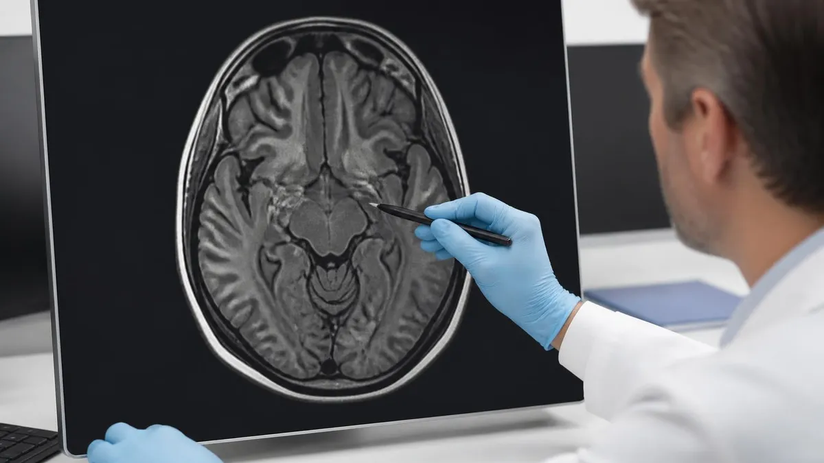
Every millimeter of motion degrades MRI images
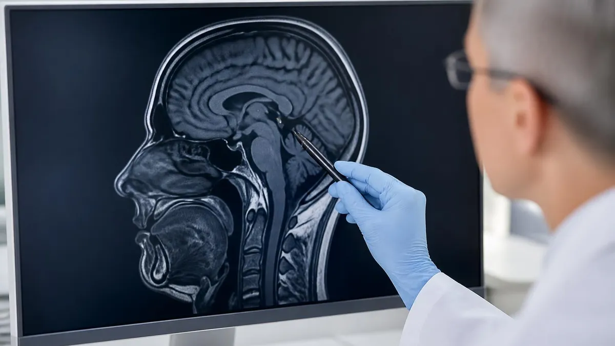
MRI is exquisitely sensitive to movement. Even small head shifts during a brain scan can blur cortical detail and obscure subtle lesions. This is why sedation is not just about patient comfort — it directly determines whether the radiologist can make an accurate diagnosis. A successfully sedated scan often prevents a repeat study and the cumulative risks that come with it.
Safety inside the MRI suite begins with the recognition that the magnetic field is always on. Unlike a CT scanner that can be powered down, a 1.5 or 3 Tesla magnet maintains its field continuously, which means every piece of equipment brought into the room must be MRI conditional or MRI safe. This requirement transforms sedation logistics: standard monitors, infusion pumps, oxygen tanks, and laryngoscopes cannot enter Zone IV. Specialized non-ferromagnetic equipment is mandatory, and personnel must be trained in MRI-specific emergency protocols.
Monitoring a sedated patient inside the bore is technically challenging. The patient is often partially or fully enclosed, making visual assessment difficult. Pulse oximetry, capnography, ECG, and non-invasive blood pressure must all function reliably in a high-field environment. Fiber-optic cables transmit signals to a console outside the magnet room, where the sedation provider watches waveforms continuously. Any change in respiratory rate, oxygen saturation, or end-tidal carbon dioxide triggers immediate intervention, including stopping the scan and rapidly extracting the patient if necessary.
The most common adverse events during MRI sedation are respiratory depression, airway obstruction, hypotension, and paradoxical agitation. Respiratory depression is most often caused by opioid-benzodiazepine combinations or propofol overdose. Airway obstruction typically results from positioning, sleep apnea, or excess soft tissue collapse. Both are managed with jaw thrust, chin lift, supplemental oxygen, or if needed, advanced airway placement. Hypotension responds to fluid bolus and dose reduction.
Pre-sedation screening identifies patients at elevated risk before any drug is given. The American Society of Anesthesiologists physical status classification helps stratify risk: ASA I and II patients tolerate sedation well, while ASA III, IV, and V patients require more intensive monitoring and often an anesthesiologist rather than a sedation nurse. Mallampati score, neck mobility, body mass index, and a history of difficult intubation all factor into the airway plan.
Drug interactions deserve special attention. Patients on chronic opioids, benzodiazepines, or sleep medications often need adjusted doses because of tolerance. Conversely, frail or elderly patients may experience profound effects from small doses. SSRIs, MAOIs, and certain antihypertensives can interact with sedation agents in unpredictable ways. A thorough medication reconciliation is mandatory, and any recent dose changes should be communicated to the sedation team during the pre-procedure interview.
Contrast administration during sedation adds another layer of complexity. Gadolinium-based contrast agents are generally well tolerated, but allergic reactions, although rare, can be catastrophic in a patient who cannot communicate symptoms. Premedication with steroids and antihistamines is standard for patients with prior contrast reactions, and emergency medications including epinephrine must be immediately available. The sedation provider monitors for tachycardia, hypotension, urticaria, or bronchospasm throughout the scan.
Finally, the team itself is a critical safety element. National guidelines recommend a dedicated sedation provider whose sole responsibility is monitoring the patient, separate from the radiologic technologist running the scan. For deep sedation and general anesthesia, an anesthesiologist or certified registered nurse anesthetist must be present. Hospital and outpatient centers undergo regular accreditation reviews to confirm staffing ratios, equipment readiness, and emergency drill performance meet current standards.
Eating or drinking before sedation can cause stomach contents to enter the lungs if the airway reflexes are suppressed. This complication, called aspiration pneumonitis, can be fatal. Follow fasting instructions exactly: no solid food for 6–8 hours, breast milk 4 hours, clear liquids 2 hours. If you accidentally eat or drink, call the imaging center immediately — the scan will likely be rescheduled.
Recovery from MRI sedation begins the moment the scan ends and continues until the patient meets formal discharge criteria. The patient is transferred out of the magnet room to a post-anesthesia care area, where nurses monitor vital signs every five to fifteen minutes. The pace of awakening depends on the agent used: propofol patients often open their eyes within minutes, while midazolam recipients may remain drowsy for an hour or longer. Dexmedetomidine produces a particularly natural-feeling wake-up, with patients describing the experience as a long nap.
Standard discharge criteria include stable oxygen saturation on room air, return to baseline mental status, ability to tolerate oral fluids without nausea, and adequate pain control. The Aldrete score or a modified version is commonly used to objectively evaluate readiness. Pediatric patients also need to demonstrate appropriate motor function for their age, and infants must be feeding normally before going home. Most centers require a minimum recovery period of 30 to 60 minutes before discharge.
Common post-sedation effects include drowsiness, mild headache, sore throat if an airway was placed, nausea, and temporary unsteadiness. These symptoms usually resolve within four to six hours. Some patients experience emotional lability or vivid dreams as benzodiazepines wear off, and a small percentage report partial amnesia for the entire day, which is expected and not concerning. Hydration, light meals, and rest are the cornerstones of home recovery.
Safe transport home is non-negotiable. Patients cannot drive themselves, take public transportation alone, or operate machinery for at least 24 hours after sedation. A responsible adult must accompany them, and ideally stay with them for the first several hours at home. Discharge instructions explicitly forbid signing legal documents, making important decisions, or consuming alcohol during the post-sedation period because cognition remains subtly impaired even after the patient feels normal.
Parents of sedated children should expect their child to sleep more than usual for the rest of the day. Light foods such as toast, applesauce, or crackers are introduced first, with full diet resumed if no nausea develops. Younger children may be clingy, irritable, or emotionally unsettled as the medications clear, and parents are encouraged to provide a calm, low-stimulation environment. Most children return to normal activity by the following morning. For follow-up planning, see how to find an MRI scan near you for any future imaging needs.
Follow-up communication varies by facility. Many centers call the patient or family the next day to confirm a smooth recovery and answer questions. Imaging results are typically reviewed by a radiologist within 24 hours and reported to the referring physician, who then contacts the patient. If any unusual symptoms develop — persistent vomiting, difficulty breathing, severe headache, or behavior changes — patients are instructed to call the imaging center or visit an emergency department immediately.
Most patients recover from MRI sedation uneventfully and report a much more tolerable experience than they expected. The combination of careful pre-screening, modern short-acting medications, and skilled monitoring has made sedated MRI one of the safest procedures performed in outpatient medicine. Knowing what to expect after the scan empowers patients and families to participate confidently in their care.
Practical preparation makes the difference between a stressful sedated MRI and a smooth experience. Start by scheduling the scan early in the day if possible, because morning slots align well with overnight fasting and leave the afternoon for rest. Confirm the appointment time, arrival window, and any special instructions during the call from the imaging center, and write down questions to ask during the pre-procedure interview. Bring a list of medications, including doses and timing of the last administration.
Pack thoughtfully. Wear loose, comfortable clothing without metal zippers, snaps, or underwire. Leave valuables, jewelry, and electronics at home. Bring a phone charger, a book, and a snack for after the scan. If the patient is a child, bring a favorite blanket or stuffed animal, but check first whether it can enter the scan room — some centers allow comfort items that have been screened for metal. A change of clothing is helpful in case of post-sedation nausea.
The night before the scan, follow NPO instructions exactly and avoid alcohol, recreational drugs, and any new medications. Get a full night of sleep because fatigue increases sensitivity to sedation effects. For pediatric patients, keep bedtime routines normal and avoid telling the child the scan is painful or scary; instead, describe it as a special picture-taking machine where they get to take a nap. Reading age-appropriate books about MRI can dramatically reduce anticipatory anxiety.
On the day of the scan, arrive 30 to 60 minutes early to allow time for paperwork, screening, IV placement, and any pre-sedation interviews. The sedation provider will ask about medical history, allergies, recent illnesses, and previous reactions to anesthesia. Be honest about substance use, including marijuana and alcohol, because these affect dosing. The screening team will also confirm there are no MRI safety contraindications such as pacemakers, cochlear implants, or retained metal fragments.
During the scan, the sedation team manages everything. The patient is positioned, monitors are placed, ear protection is fitted, and medication is administered. The scan begins once stillness is confirmed. Family members typically wait in a designated lounge, although some centers allow one parent to remain in the control room for pediatric cases. The radiologic technologist communicates with the sedation provider continuously, pausing the scan if any safety concern arises.
After discharge, follow written instructions carefully. Hydrate, eat light, and rest. Do not return to work, school, or driving until the next day. If you are caring for a sedated patient, observe them for the first several hours and call the center if you have concerns. Mark your calendar for the follow-up appointment with the referring physician, which is usually scheduled within one to two weeks to review the imaging results and plan next steps.
Finally, advocate for yourself. If you have had difficult sedation experiences in the past, request a consult with the anesthesia team before the scan day. Ask about alternatives such as wide-bore or open MRI scanners, which can reduce or eliminate the need for sedation in mildly claustrophobic patients. The best outcomes happen when patients, families, and the imaging team plan together with realistic expectations and clear communication.
MRI Questions and Answers


About the Author
Medical Laboratory Scientist & Clinical Certification Expert
Johns Hopkins UniversityDr. Sandra Kim holds a PhD in Clinical Laboratory Science from Johns Hopkins University and is certified as a Medical Technologist (MT) and Medical Laboratory Scientist (MLS) through ASCP. With 16 years of clinical laboratory experience spanning hematology, microbiology, and molecular diagnostics, she prepares candidates for ASCP board exams, MLT, MLS, and specialist certification tests.
Join the Discussion
Connect with other students preparing for this exam. Share tips, ask questions, and get advice from people who have been there.
View discussion (4 replies)