MRI and Metal in Body: Complete Safety Guide for Patients and Technologists
Learn everything about MRI and metal in body — implants, screening, contraindications, and what's safe. Expert safety guide for patients and MRI technologists.
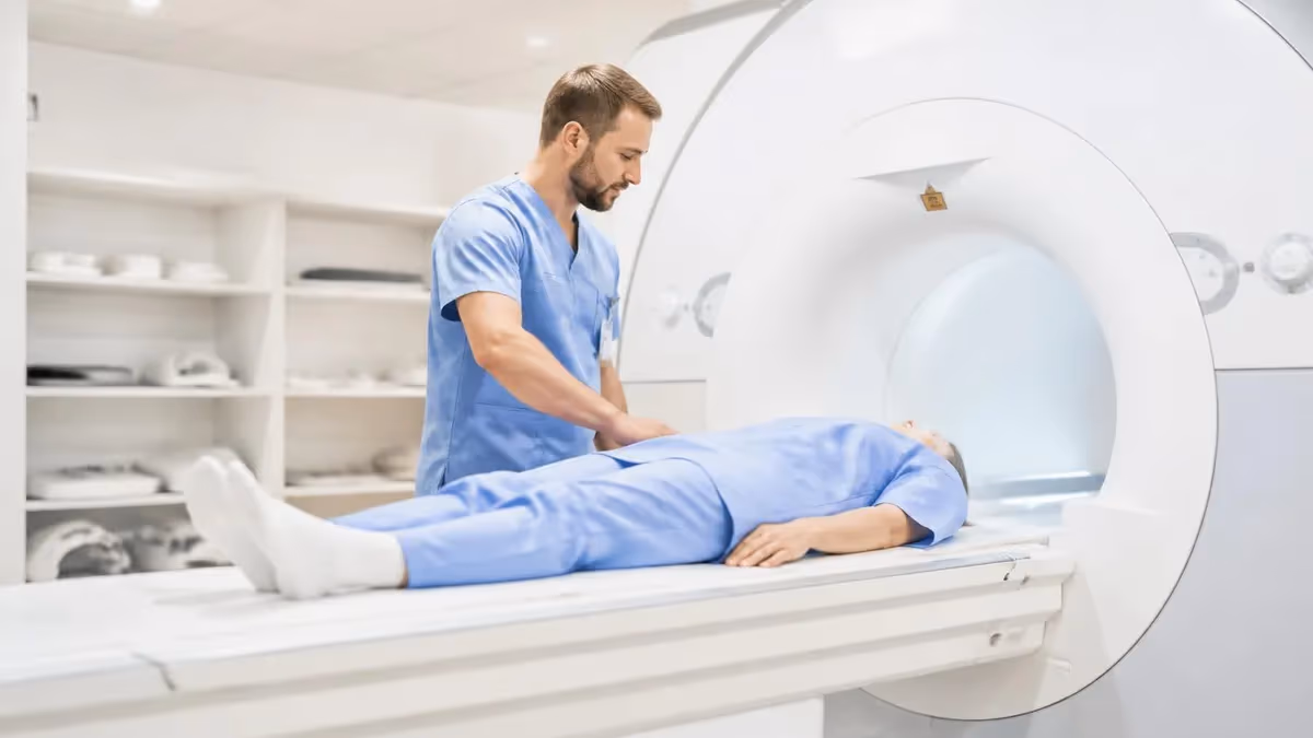
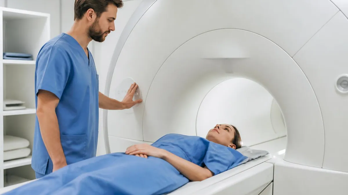
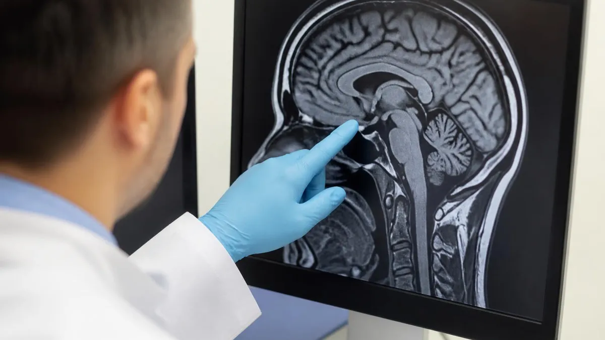
Understanding the relationship between MRI and metal in body is one of the most critical safety considerations in modern medical imaging. MRI scanners generate extraordinarily powerful magnetic fields — typically between 1.5 and 3 Tesla in clinical settings — that can exert tremendous force on ferromagnetic materials. When metal objects interact with these fields, the consequences range from minor image distortion to life-threatening projectile events. Every patient undergoing MRI must be carefully screened for metallic implants, foreign bodies, and medical devices before entering the scan room.
The physics behind MRI metal interactions involve three distinct mechanisms: translational force, torque, and radiofrequency heating. Translational force pulls ferromagnetic objects toward the center of the magnetic field with a strength that increases dramatically as the object approaches the bore. Torque causes elongated metal objects to rotate and align with field lines. Radiofrequency heating occurs when conductive materials absorb RF energy and convert it to heat, which can cause burns at implant sites. Understanding all three mechanisms is essential for anyone working in or around MRI environments.
Not all metals respond identically to magnetic fields. The critical distinction in MRI safety is between ferromagnetic materials (iron, nickel, cobalt and their alloys) and non-ferromagnetic materials (titanium, aluminum, gold, platinum, and surgical-grade stainless steel). Ferromagnetic metals are strongly attracted to magnets and pose the greatest risk. Weakly magnetic or paramagnetic materials may still cause image artifact but typically present much lower safety risks. Most modern medical implants are manufactured from titanium or other MRI-conditional materials precisely because of these safety considerations.
Patients with a history of surgery, trauma, occupational metal exposure, or any implanted medical device require thorough pre-MRI screening. This screening process includes a detailed questionnaire covering all known implants, a review of operative reports and implant identification cards, and sometimes plain radiographs to identify unknown metallic foreign bodies. The stakes are high: skipping or rushing this process has resulted in serious injuries and fatalities, making systematic screening protocols non-negotiable in accredited MRI facilities across the United States.
MRI safety terminology has evolved significantly over the past two decades. The American College of Radiology and the device industry now use standardized labels: MR Safe, MR Conditional, and MR Unsafe. An MR Safe device poses no known hazard in any MRI environment. An MR Conditional device is safe only under specific conditions — particular field strengths, specific SAR limits, or defined scan parameters.
An MR Unsafe device should never enter the MRI environment under any circumstances. These labels replaced older, less precise terms like "MRI compatible," which implied both safety and absence of image artifact but was often misunderstood by clinical staff.
For MRI registry candidates and practicing technologists, mastering mri safety metal protocols is not just a regulatory requirement — it is a core professional competency. The ARRT MRI examination includes a substantial section on patient safety and screening, reflecting how central these concepts are to safe MRI practice. A thorough understanding of which implants are safe, how to interpret manufacturer conditioning statements, and what screening steps to follow is the foundation upon which all advanced MRI knowledge rests. This guide covers everything you need to know to protect patients and pass your certification exam.
Beyond implants, the MRI environment poses risks from external metal objects brought into the scan room. Oxygen tanks, wheelchairs, IV poles, and even small items like scissors or keys become dangerous projectiles when introduced near the scanner. The "zone" system established by the ACR divides MRI facilities into four areas of increasing magnetic field exposure, with progressively stricter access controls. Understanding this framework helps technologists create and enforce safe environments, preventing the "projectile effect" accidents that continue to occur in facilities where safety protocols are not rigorously enforced.
MRI Metal Safety by the Numbers
Types of Metal and Their MRI Risk Levels
Titanium, gold, platinum, and surgical-grade non-magnetic stainless steel pose no known risk in any MRI environment. Most modern orthopedic hardware, joint replacements, and surgical clips are manufactured from these materials. Image artifact may still occur but safety risk is negligible.
These devices are safe only under defined conditions: specific field strengths (e.g., ≤1.5T only), maximum SAR limits, or restricted scan modes. Cardiac pacemakers, cochlear implants, and some neurostimulators fall here. Always verify manufacturer guidelines before scanning any MR Conditional patient.
Ferromagnetic materials including traditional iron-based surgical clips, certain older aneurysm clips, some cardiac valves, and external metal objects like oxygen tanks are MR Unsafe. These must never enter Zone III or IV. Attempting to scan patients with MR Unsafe implants can result in serious injury.
Workers in machining, welding, metal grinding, or military occupations may have embedded ferromagnetic fragments from prior injuries. Orbital foreign bodies are especially dangerous, as magnetic torque can cause blindness or globe rupture. Pre-MRI orbital X-rays are required for patients with this exposure history.
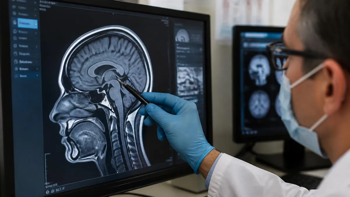
The landscape of MRI-safe implants has changed dramatically over the past twenty years, driven by both engineering advances and the explosion of implantable medical devices. Orthopedic hardware — including hip and knee replacements, spinal fusion hardware, and fracture fixation plates — is now almost universally manufactured from titanium alloy or cobalt-chrome, both of which are non-ferromagnetic. Patients with these modern implants can typically undergo MRI without restriction, though significant image distortion may occur near the implant site due to susceptibility artifact. Radiologists must account for this when interpreting images.
Cardiac implantable electronic devices (CIEDs), including pacemakers and implantable cardioverter-defibrillators (ICDs), represent the most complex and clinically significant category of MRI-conditional devices. For many years, all pacemakers were considered absolute contraindications to MRI. That changed in 2011 with the FDA approval of the first MR-conditional pacemaker system. Today, many modern CIED systems are labeled MR Conditional at 1.5T with specific programming requirements. However, the scanning process requires coordination between cardiology and radiology, specific device reprogramming before and after the scan, and continuous cardiac monitoring throughout the procedure.
Cochlear implants present a unique challenge because they incorporate both a surgically implanted internal component and an external processor. The internal component contains a magnet that can be displaced or demagnetized by the MRI field. Many newer cochlear implant systems are MR Conditional at 1.5T when the external processor is removed, but some require surgical explantation of the internal magnet before scanning. Patients considering cochlear implantation who may need future MRI should discuss implant selection with their surgeon to choose a system optimized for MRI compatibility.
Neurostimulator systems — including deep brain stimulators (DBS), spinal cord stimulators, and peripheral nerve stimulators — carry significant MRI risk due to the long conductor leads that can act as antennas and concentrate RF energy. This heating can occur along the entire lead length, potentially causing tissue damage at distances far from the implant site. Some DBS systems are MR Conditional under highly restrictive conditions, such as head-only coil use and very specific SAR limits. Patients with these devices require multi-disciplinary consultation and often cannot receive certain types of MRI scans even with conditional approval.
Intravascular stents, including coronary artery stents, carotid stents, and peripheral vascular stents, are generally considered safe for MRI after a post-implantation waiting period of six to eight weeks. During this initial healing period, the stent is not yet incorporated into the vessel wall and torque forces present a theoretical risk. After full endothelialization, the stent is anchored and the risk of displacement is negligible. Most stents in current clinical use are constructed from stainless steel, nitinol, or cobalt-chrome alloys that are either non-magnetic or only weakly paramagnetic.
Older aneurysm clips represent one of the most dangerous categories of intracranial implants in MRI. Clips made before the 1990s — particularly those from manufacturers such as Drake and Heifetz — used ferromagnetic alloys that can generate enough torque at 1.5T to rupture a vessel or cause catastrophic hemorrhage. Any patient with an intracranial aneurysm clip requires exhaustive documentation: the exact manufacturer, model number, and alloy composition must be verified against a comprehensive database such as the MRIsafety.com implant reference before scanning. When documentation is unavailable, the study must not proceed without neurosurgical consultation.
Tattoos and permanent makeup occasionally contain metallic pigments — particularly red and flesh-toned inks that may contain iron oxide or other compounds. In rare cases, patients have reported burning sensations or skin irritation over tattooed areas during MRI. The risk is generally low, but technologists should inform patients with large or multi-colored tattoos about this possibility and instruct them to alert the technologist immediately if they experience any discomfort during the scan. Cooling measures such as a damp cloth placed over the tattoo area can reduce the likelihood of skin reactions.
MRI Screening Protocols Explained
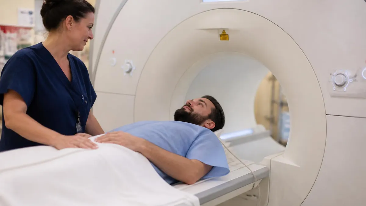
The MRI safety questionnaire is the first and most fundamental screening tool. A comprehensive questionnaire covers surgical history, presence of implanted devices, history of metal injuries or shrapnel, occupational exposure to metal fragments, and prior MRI experiences. Patients must complete the form themselves when possible, and the MRI technologist or radiologist must review every answer before the patient enters Zone III or IV. Forms should be available in multiple languages at facilities serving diverse populations.
One common failure mode is relying solely on a questionnaire completed during registration without technologist review. Patients frequently misunderstand questions or fail to recognize relevant implants — for example, not realizing that a cardiac monitor implanted during a hospitalization counts as a metallic device. Technologists must verbally confirm key answers, particularly regarding implanted electronic devices, intracranial clips, and any history of eye or metal injury. This two-step process of written questionnaire plus verbal verification significantly reduces screening failures.
MRI With Metal Implants: Benefits vs. Risks
- +Most modern implants are titanium or non-ferromagnetic and fully compatible with MRI scanning
- +MR Conditional device labeling provides clear, actionable safety parameters for clinical teams
- +MRI remains the gold standard for soft tissue imaging even in patients with orthopedic hardware
- +Newer pacemaker systems allow MRI access that was previously impossible for cardiac device patients
- +Metal artifact reduction sequences (MARS) can substantially improve image quality near implants
- +Systematic pre-screening catches contraindications before any risk is introduced to the patient
- −Ferromagnetic implants and older devices can cause serious injury or death if MRI is attempted
- −MR Conditional devices require complex, time-consuming protocol verification and coordination
- −Metallic implants always cause some degree of susceptibility artifact, potentially obscuring pathology
- −Lack of implant documentation (lost cards, unknown manufacturers) can make safe scanning impossible
- −RF heating risk along neurostimulator leads may prevent MRI even with conditional device approval
- −Emergency situations create pressure to scan without full screening, increasing risk of accidents
Pre-Scan Metal Safety Checklist for MRI Technologists
- ✓Provide and collect the MRI safety screening questionnaire before the patient enters Zone III.
- ✓Verbally confirm all questionnaire answers with the patient, clarifying any ambiguous responses.
- ✓Identify every implanted device and obtain manufacturer, model number, and implant card if available.
- ✓Look up each implant in an authoritative MRI safety database before proceeding.
- ✓For MR Conditional devices, read the full conditioning statement and verify all parameters are achievable.
- ✓Obtain orbital plain radiographs for any patient with reported occupational metal fragment exposure.
- ✓Instruct the patient to remove all external metal: jewelry, piercings, hearing aids, glasses, dentures.
- ✓Have the patient change into a facility gown and perform a pat-down to confirm no metal remains.
- ✓Verify that all equipment entering the scan room is labeled MR Safe or MR Conditional for your field strength.
- ✓Document the screening process in the patient record, including device database lookup and staff verification.
"MRI Conditional" Is Not a Blanket Green Light
An MR Conditional label means safety is guaranteed ONLY when every specified parameter is met. Field strength, SAR limits, scan duration, and coil selection must all comply with the manufacturer's conditioning statement. Scanning a conditional device outside its approved parameters is equivalent to scanning an unsafe device — the conditional label no longer applies and the risk of patient harm rises sharply.
Several clinical scenarios present particularly complex MRI metal safety challenges that require careful multidisciplinary decision-making. One of the most common is the patient who arrives for an urgent or emergent MRI with an unverifiable implant. When a patient is unconscious, cognitively impaired, or otherwise unable to provide history, and no surgical records are immediately available, the clinical team must weigh the diagnostic urgency of the MRI against the unknown implant risk. In true emergencies where the MRI result will directly guide life-saving treatment, the decision may be made to proceed with continuous monitoring and emergency response capability standing by.
Pregnant patients represent another important special case. MRI does not use ionizing radiation and is generally preferred over CT in pregnant patients who require cross-sectional imaging. However, gadolinium-based contrast agents are avoided during pregnancy whenever possible, as gadolinium crosses the placenta and its effects on the fetus are not fully established.
The high magnetic field itself poses no known risk to the developing fetus based on available evidence, and MRI is performed throughout all trimesters when clinically necessary. The first trimester, during organogenesis, is treated with the most caution, and MRI is deferred until the second or third trimester when alternatives are available.
Patients with implanted drug infusion pumps — including insulin pumps and intrathecal drug delivery systems — require specific consideration. External insulin pumps must be removed before entering the MRI environment, as the magnetic field can damage the pump electronics and potentially alter drug delivery. Implanted intrathecal pumps may be MR Conditional, but the drug reservoir may require aspiration and replacement before and after scanning at some field strengths. Failure to manage implanted pumps correctly can result in dangerous over- or under-delivery of medications including opioids, baclofen, and chemotherapy agents.
Patients who have undergone recent neurosurgical procedures face an additional timing consideration. Surgical staples and clips placed during craniotomy or spine surgery are often ferromagnetic in older patients or in cases where titanium was not used.
Beyond the implant identification challenge, there is a theoretical concern about tissue healing around new implants: a clip that has not yet been fully incorporated by granulation tissue may have more freedom to move under magnetic force than one that has been in place for months. Most MRI facilities observe a minimum waiting period of four to six weeks after neurovascular clip placement before performing MRI, absent urgent clinical need.
The intraocular foreign body scenario deserves special emphasis because of the severity of potential consequences and the subtlety of the patient history that raises concern. A patient who worked in a metal shop twenty years ago and suffered a minor eye splash may not spontaneously report this as relevant to MRI screening.
The technologist must specifically ask about any history of metal entering the eyes, any prior eye injury, or any work in environments where metal fragments were generated. A retained intraocular ferromagnetic fragment as small as 0.1mm can cause retinal detachment or perforation when subjected to MRI gradient forces. This is one of the few scenarios where even a small risk demands radiographic clearance before proceeding.
Patients with certain cardiovascular conditions require additional consideration beyond the device itself. For example, a patient with a mechanical heart valve made of a ferromagnetic alloy would be at risk even if the valve were technically MR Conditional, because the hemodynamic consequences of valve displacement or dysfunction would be immediately catastrophic. Mechanical valves implanted since the 1980s are generally made from MRI-safe materials, but careful verification remains essential. Similarly, patients with coronary artery bypass grafts using sternal wires should be assessed for wire type, though most sternal wires are non-ferromagnetic and pose no significant risk.
The management of patients with external fixators — orthopedic devices that stabilize fractures from outside the body using pins that pass through the skin into bone — is another practical challenge. External fixators typically contain a mix of materials, and the large conductive loops formed by the frame components can concentrate RF heating. Many manufacturers provide specific guidance for MRI with external fixators in place, including positioning requirements and SAR restrictions. When external fixators cannot be safely accommodated, the orthopedic team may be asked to consider whether the fixator can be temporarily modified or removed for the imaging procedure.
Intracranial aneurysm clips made before the mid-1990s may be ferromagnetic and can generate sufficient torque to rupture a vessel at 1.5T. Never assume a clip is MRI safe because the patient has had prior MRI elsewhere — field strength, clip age, and documentation standards vary. Obtain and verify the exact manufacturer and model before every scan in a patient with a known intracranial clip history.
For candidates preparing for the ARRT MRI certification examination, MRI safety — and metal safety in particular — is one of the highest-yield topic areas. The exam blueprint allocates significant weight to patient care and safety, and within that domain, screening procedures, implant identification, and contraindication management are consistently represented. Understanding the ACR MRI safety zones, the standard terminology (MR Safe, MR Conditional, MR Unsafe), and the specific risks associated with major implant categories will directly translate to correct answers on examination day.
One area where exam candidates frequently struggle is distinguishing between the different mechanisms of metal-related MRI risk. Translational attraction, torque, and RF-induced heating are three distinct physical phenomena, each relevant to different types of metallic objects and implants. Translational attraction is most relevant to large ferromagnetic objects and the projectile effect. Torque is particularly important for elongated implants like aneurysm clips. RF heating is the primary concern with long conductive leads in neurostimulators. Being able to identify which mechanism applies to a given clinical scenario is a higher-order reasoning skill that the ARRT exam rewards.
The MR Conditional concept is another area requiring careful study. The conditioning statement for any given device specifies the maximum static field strength (expressed in Tesla), the maximum spatial gradient field (expressed in Tesla per meter or Gauss per centimeter), and the maximum whole-body averaged SAR (expressed in watts per kilogram). Some devices also specify maximum local SAR, maximum scan time, and required positioning. Exam questions often present a clinical scenario where one parameter falls outside the conditioning range and ask whether scanning can proceed — the correct answer is always no, regardless of which parameter is violated.
Radiofrequency safety and the specific absorption rate (SAR) concept merit particular attention because SAR interacts with metal in ways that are not always intuitive. SAR is determined by the pulse sequence, the patient's weight, and the operating field strength. Higher field strengths generate higher SAR for equivalent sequences.
Implants with conductive leads can experience local SAR many times higher than the whole-body average, because the lead acts as an antenna that concentrates RF energy. This is why the whole-body SAR limit specified in a conditioning statement may seem conservative — it is designed to keep local lead tip heating within a safe range even under worst-case antenna coupling conditions.
The ACR zones framework is directly testable and worth memorizing in detail. Zone I is the publicly accessible area outside the MRI suite. Zone II is the interface between the public area and the controlled MRI environment, where patient screening takes place. Zone III is the region where the fringe field begins to pose a risk to unscreened individuals; access must be physically restricted and supervised by MR-trained personnel.
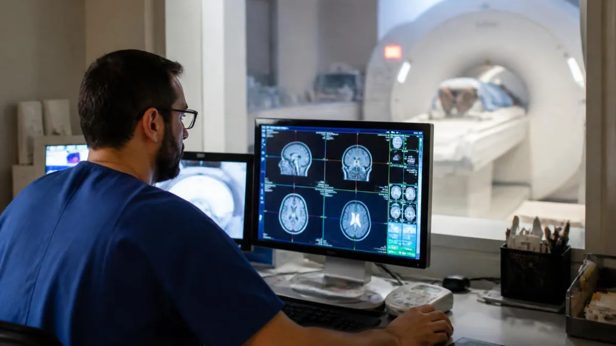
Zone IV is the MRI scanner room itself, the highest-risk area, where the static field is always present even when the scanner is not actively imaging. The static field cannot be turned off in a superconducting MRI system without a quench — an emergency that involves significant cost and scanner downtime.
Registry candidates should also understand the concept of the five-Gauss line, which defines the boundary of the MRI fringe field at which cardiac pacemakers can begin to be affected. Historically, the five-Gauss line was used to establish safety perimeters around MRI suites.
Modern actively shielded magnets confine the fringe field more tightly, but the concept remains important for understanding why controlled access to Zone III and IV areas is necessary even when the scanner is not actively running. Pacemakers that are not MR Conditional can be inhibited or permanently damaged by field exposure at levels well below those in Zone IV.
Practice questions are among the most effective tools for consolidating MRI safety knowledge. Working through clinical vignettes that require you to apply screening protocols, interpret device labels, and identify contraindications builds both the factual knowledge and the clinical reasoning skills that the ARRT exam assesses.
Many candidates find that safety questions, while initially intimidating due to the breadth of device types involved, become a reliable source of correct answers once the underlying framework is mastered. Use the practice resources linked throughout this guide, and focus your review on the three MRI safety labels, the ACR zones, the major implant categories, and the mechanisms of metal-field interaction.
Practical preparation for working in a clinical MRI environment goes beyond memorizing safety labels and protocols. Experienced MRI technologists develop a systematic habit of mind — a mental checklist that runs automatically with every patient encounter. This professional reflex is built through deliberate practice, mentorship, and the internalization of near-miss events and case reports. Reviewing documented MRI accidents, such as the well-publicized 2001 case in which a child was killed by a ferromagnetic oxygen tank that became a projectile, helps technologists understand viscerally why every step of the screening protocol matters.
Communication skills are as important as technical knowledge in MRI safety. Technologists must be able to explain the screening process to anxious patients in plain language, ask questions in ways that elicit accurate answers from patients who may be minimally educated or cognitively impaired, and advocate for proper screening with referring physicians who may pressure staff to abbreviate the process for time efficiency.
A technologist who can clearly explain why an implant must be verified before scanning — and who knows how to escalate to a radiologist or MRI medical director when a referring physician pushes back — is practicing safety at the highest level.
Ongoing education is essential because the landscape of implantable devices changes continuously. New devices receive FDA clearance regularly, and MR Conditional labeling may be updated as manufacturers gather post-market safety data. MRI technologists should subscribe to updates from professional organizations such as the Society for MR Radiographers and Technologists (SMRT) and review new additions to implant safety databases periodically. Facilities should maintain a current version of their implant reference database and ensure that all scanning staff know how to access it.
Quality assurance programs at MRI facilities typically include regular audits of the screening process. These audits may review questionnaire completion rates, documentation of implant verification lookups, and adherence to zone access controls. Facilities accredited by the American College of Radiology must demonstrate compliance with ACR MRI safety practice parameters. Technologists who understand the reasoning behind these parameters, rather than following them mechanically, are better equipped to handle novel situations and edge cases that fall outside the written protocol.
Team-based safety culture is the final and perhaps most important element of MRI metal safety. No individual technologist, no matter how knowledgeable, can maintain perfect vigilance in every case without the support of a culture that prioritizes safety over throughput.
This means that anyone on the MRI team — from the receptionist who first asks about implants during scheduling to the radiology nurse or anesthesiologist who accompanies sedated patients — understands their role in the screening chain. It also means that speaking up about a safety concern is encouraged and that technologists are empowered to delay or cancel a study when proper screening has not been completed.
For students and new graduates entering the MRI field, developing comfort with the uncertainty that sometimes surrounds implant decisions is part of professional growth. Not every situation will have a clear, database-confirmed answer. In those cases, the correct response is to escalate to a supervising radiologist or MRI medical director, document the decision-making process thoroughly, and err on the side of caution when patient safety and diagnostic urgency are in equipoise. The MRI registry examination tests knowledge, but real-world MRI safety ultimately rests on judgment, communication, and a genuine commitment to the patients in your care.
Whether you are studying for your ARRT MRI certification or working to strengthen your clinical protocols, building expertise in MRI and metal safety is one of the most valuable investments you can make in your career. The principles are stable, the stakes are high, and the patients who depend on your knowledge deserve nothing less than your full professional attention. Use the practice tests and resources on this site to assess your current knowledge level, identify gaps, and build the confident mastery that safe MRI practice demands.
MRI Questions and Answers
About the Author
Medical Laboratory Scientist & Clinical Certification Expert
Johns Hopkins UniversityDr. Sandra Kim holds a PhD in Clinical Laboratory Science from Johns Hopkins University and is certified as a Medical Technologist (MT) and Medical Laboratory Scientist (MLS) through ASCP. With 16 years of clinical laboratory experience spanning hematology, microbiology, and molecular diagnostics, she prepares candidates for ASCP board exams, MLT, MLS, and specialist certification tests.
Join the Discussion
Connect with other students preparing for this exam. Share tips, ask questions, and get advice from people who have been there.
View discussion (4 replies)