What Does a Hand MRI Look Like? A Complete Guide to MRI Images of the Hand and Wrist
What does a hand MRI look like? See real hand MRI image examples, learn anatomy, sequences, and how radiologists read scans of bones, tendons, and 🆕
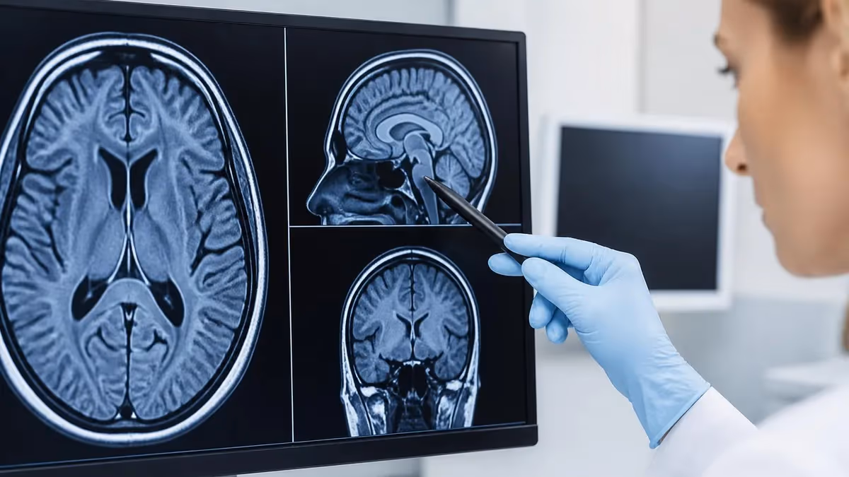
If you have ever wondered what does a hand MRI look like, the short answer is that it appears as a stack of grayscale cross-sections showing every bone, tendon, ligament, nerve, vessel, and pocket of fluid in the hand and wrist. Unlike a flat X-ray, an MRI of the hand produces dozens of thin slices in three planes, allowing radiologists to scroll through the anatomy layer by layer and spot tiny tears, cysts, fractures, or inflammation invisible on other imaging tests.
A typical hand MRI study includes axial, coronal, and sagittal images, plus specialized sequences like T1, T2, proton density, and fat-suppressed acquisitions. Each sequence highlights different tissue properties. T1 images make fat appear bright and fluid appear dark, giving a crisp anatomical map. T2 and mri stir sequences flip that contrast so that water, edema, and inflammation glow brightly against darker background tissue, making subtle pathology pop out for the interpreting radiologist.
The hand contains 27 bones, more than 30 muscles, dozens of tendons, and a delicate web of nerves and vessels packed into a small space. Because the structures are so small, hand MRIs are usually performed with a dedicated wrist or extremity coil and a high field strength magnet, often 3 Tesla, to maximize spatial resolution. Slice thickness commonly drops to 2 or 3 millimeters, which is far thinner than slices used for the brain or abdomen.
On the images themselves, cortical bone appears as a dark black rim because it contains very little water or fat to generate signal. Bone marrow inside the metacarpals and phalanges appears bright on T1 because of its fatty content. Tendons such as the flexor and extensor groups appear as dark, well-defined cords, while articular cartilage at the joint surfaces shows up as a thin gray line. Muscles fall somewhere in the middle gray range.
Fluid is the radiologist's best friend on a hand MRI. Joint effusions, tendon sheath fluid, ganglion cysts, and bone marrow edema all light up brightly on fluid-sensitive sequences. This is why a patient with unexplained hand pain often gets a definitive diagnosis from MRI when X-rays and ultrasound looked normal. Even a stress fracture invisible on plain film will show a telltale bright signal in the marrow.
This guide will walk you through what radiologists actually see when they open a hand MRI, how to recognize normal versus abnormal structures, what each sequence reveals, and what common findings look like on the screen. If you want to test your knowledge as you go, try our MRI Medical Abbreviation: What MRI Stands For and Why It Matters guide for the terminology that will appear throughout this article.
By the end, you will be able to look at a hand MRI image and identify the carpal bones, the major tendon groups, the median and ulnar nerves, the triangular fibrocartilage complex, and the common pathologies that drive most hand MRI referrals. You will also understand why the same hand can look dramatically different on T1 versus T2 sequences and why that contrast difference is the whole point of magnetic resonance imaging.
Hand MRI Imaging by the Numbers
Standard Hand MRI Sequence Protocol
Provides the best anatomical overview of the carpal bones, metacarpals, and phalanges. Fat appears bright and fluid dark, making bone marrow and soft tissue planes easy to identify across the entire hand in one plane.
Suppresses fat signal so that any edema, fluid, or inflammation glows white. Critical for detecting occult fractures, bone bruises, tendon tears, and synovitis that would be hidden on T1 imaging.
Proton density with fat suppression in the axial plane is the workhorse for evaluating individual tendons in cross section, the median nerve in the carpal tunnel, and small ligament structures.
Allows scrolling finger by finger to see the flexor and extensor tendons along their length. Essential for evaluating tendon ruptures, pulley injuries, and joint alignment in trigger finger or mallet finger cases.
Optional high-resolution isotropic sequence that allows reconstruction in any plane. Often used for cartilage assessment, small ligament evaluation, and pre-surgical planning where multiplanar review is essential.
Normal hand anatomy on MRI begins with the carpal bones. There are eight of them, arranged in two rows of four. The proximal row contains the scaphoid, lunate, triquetrum, and pisiform, while the distal row holds the trapezium, trapezoid, capitate, and hamate. On a coronal T1 image, each bone appears as a brightly marrowed island surrounded by a thin dark cortical rim, with hairline joint spaces separating them in a predictable geometric pattern.
The metacarpals extend distally from the carpals and form the framework of the palm. Each metacarpal has a base, shaft, neck, and head. On sagittal sequences, the metacarpal heads articulate with the proximal phalanges, and the smooth articular cartilage appears as a thin intermediate gray line between two darker cortical surfaces. Beyond the metacarpals come the proximal, middle, and distal phalanges of each finger, with the thumb being the only digit that has just two phalanges.
Tendons are arguably the most striking feature of a hand MRI because they appear pitch black against the brighter surrounding tissues. The flexor digitorum profundus and superficialis tendons run along the palmar side, passing through the carpal tunnel beneath the transverse carpal ligament. The extensor tendons travel along the dorsal side in six distinct compartments at the wrist. On axial images, you can count each tendon as a small dark dot, which is essential when checking for tears or tenosynovitis.
The median nerve is one of the most clinically important structures on any hand or wrist MRI because of carpal tunnel syndrome. On axial T2 images at the level of the proximal carpal row, the median nerve appears as an oval intermediate signal structure sitting superficial to the flexor tendons and deep to the transverse carpal ligament. A normal nerve is round to slightly oval. A flattened, swollen, or brightly signaling median nerve suggests compression neuropathy.
Vascular structures are usually visible without contrast because flowing blood produces signal voids on most spin echo sequences. The radial and ulnar arteries appear as small dark circles on axial images, accompanied by their paired veins. The superficial and deep palmar arches can sometimes be traced on coronal images. When contrast is administered, vessels brighten dramatically, and any abnormal enhancement in surrounding tissues becomes a red flag for tumor, infection, or active synovitis.
The triangular fibrocartilage complex, or TFCC, sits between the distal ulna and the proximal carpal row on the ulnar side of the wrist. On coronal images it appears as a dark triangular wedge of fibrocartilage. Tears of the TFCC are one of the most common reasons for a wrist MRI, and they appear as bright linear fluid signals cutting through the normally dark cartilage. Understanding the Knee MRI Images: A Complete Guide to Reading, Understanding, and Interpreting Knee Scans approach can help, since the principles of cartilage and ligament evaluation transfer directly.
Finally, the skin and subcutaneous fat form the outermost layers on every image. Subcutaneous fat is bright on T1 and intermediate on T2, while the skin itself shows as a thin intermediate gray border. The fat planes between muscles, tendons, and bones serve as natural contrast agents, and any obliteration of these planes by edema, blood, or mass tissue is a clue that something abnormal is happening beneath the surface.
MRI Practice Test Questions
Prepare for the MRI - Magnetic Resonance Imaging exam with our free practice test modules. Each quiz covers key topics to help you pass on your first try.
MRI Knowledge
MRI Exam Questions covering Knowledge. Master MRI Test concepts for certification prep.
MRI Physics
Free MRI Practice Test featuring Physics. Improve your MRI Exam score with mock test prep.
MRI Anatomy and Pathology
MRI Test Prep for MRI Anatomy and Pathology. Practice MRI Quiz questions and boost your score.
MRI Anatomy and Positioning
MRI Questions and Answers on MRI Anatomy and Positioning. Free MRI practice for exam readiness.
MRI Contrast Agents
Free MRI Quiz on MRI Contrast Agents. MRI Exam prep questions with detailed explanations.
MRI Patient Care and Positioning
MRI Practice Questions for MRI Patient Care and Positioning. Build confidence for your MRI certification exam.
Understanding MRI Sequences on a Hand Scan
T1-weighted images are the anatomical maps of MRI. On a hand T1 sequence, fat appears bright white, water and fluid appear dark, and muscle sits in the middle gray zone. The bone marrow of the carpals, metacarpals, and phalanges glows brightly because of its fatty content, while cortical bone forms a sharp black outline. This makes T1 ideal for identifying the precise location and shape of every anatomical structure.
Radiologists rely on T1 to spot bone marrow replacement, which is one of the earliest signs of fracture, infection, or tumor. A normal bright marrow signal that suddenly turns dark on T1 is a major red flag. T1 is also the sequence of choice for evaluating fat planes between tendons, muscles, and neurovascular bundles in the palm and fingers, where loss of fat clarity often signals underlying inflammation or mass.
Hand MRI: Strengths and Limitations Compared to Other Imaging
- +Superior soft tissue contrast reveals tendons, ligaments, and nerves invisible on X-ray
- +No ionizing radiation, making it safe for repeated imaging and younger patients
- +Multiplanar imaging in axial, coronal, and sagittal views from a single scan
- +Detects occult fractures and bone bruises before they appear on plain radiographs
- +Excellent for evaluating the triangular fibrocartilage complex and small ligaments
- +Identifies early rheumatoid arthritis changes years before erosions appear on X-ray
- +Can characterize soft tissue masses, cysts, and tumors with high specificity
- −Longer scan times of 30 to 45 minutes can be difficult for claustrophobic patients
- −Metal implants and pacemakers may be contraindicated or cause artifact
- −Higher cost compared to X-ray, ultrasound, or CT for the same body part
- −Patient must keep hand perfectly still to avoid motion artifact ruining images
- −Cortical bone detail is inferior to CT for complex fracture mapping
- −Loud acoustic noise during scanning can be uncomfortable without ear protection
Hand MRI Image Interpretation Checklist
- ✓Confirm patient name, hand side, and date are correct on every series
- ✓Scroll through coronal T1 to assess all 27 bones for marrow signal abnormality
- ✓Check each carpal bone for normal alignment and intact cortical contour
- ✓Evaluate the scaphoid carefully on every plane for occult waist fractures
- ✓Review STIR or fat-sat sequences for any bright bone marrow edema
- ✓Trace each flexor and extensor tendon along its full length in two planes
- ✓Measure the median nerve at the proximal carpal tunnel for size and signal
- ✓Inspect the triangular fibrocartilage complex for bright linear tear signal
- ✓Look for joint effusions, synovitis, and erosions at every joint level
- ✓Document any soft tissue masses, ganglion cysts, or abnormal enhancement
Always check the scaphoid first
The scaphoid is the most commonly fractured carpal bone and the most commonly missed on initial imaging. A subtle bright signal on STIR with a faint dark line on T1 across the waist can be the only sign of an occult fracture that, if missed, leads to avascular necrosis. Make scaphoid evaluation your first habit on every hand or wrist MRI.
Common pathology on hand MRI follows predictable visual patterns once you know what to look for. Carpal tunnel syndrome, for example, shows up as a swollen, flattened median nerve at the level of the pisiform, with the nerve becoming brighter on T2 sequences than it should be. The transverse carpal ligament may appear bowed forward, and the flexor tendons can show surrounding fluid in their sheaths. Severe cases also demonstrate atrophy of the thenar muscles, visible as fatty replacement on T1 images.
Triangular fibrocartilage complex tears are another classic finding. A normal TFCC appears as a uniformly dark triangle on coronal images between the distal ulna and the lunate. A tear appears as a bright linear or wedge-shaped fluid signal cutting through the dark cartilage, often associated with a small joint effusion. Tears are graded by location and pattern, with central perforations behaving differently than peripheral ulnar-sided tears that may be repairable surgically.
Scaphoid fractures, especially nondisplaced ones, are notoriously difficult to see on X-ray but obvious on MRI. The classic appearance is a low signal line on T1 crossing the scaphoid waist, accompanied by bright bone marrow edema on STIR or fat-sat T2 surrounding the fracture line. MRI within 72 hours of injury catches nearly all clinically suspected scaphoid fractures and prevents the dreaded complication of nonunion and avascular necrosis of the proximal pole.
Rheumatoid arthritis produces some of the most striking findings on hand MRI. Early disease shows synovitis as bright enhancing tissue lining the joints, often before any X-ray changes appear. Bone marrow edema adjacent to joint surfaces is a strong predictor of future erosions. As disease progresses, frank cortical erosions appear as focal dark defects on T1 with bright fluid signal on T2, and tendon ruptures, especially of the extensor tendons over the ulnar styloid, become visible.
Ganglion cysts are extremely common and have a characteristic appearance. They are sharply circumscribed, lobulated, and fluid bright on T2, with dark or intermediate signal on T1. Most arise from the dorsal scapholunate ligament or the volar wrist joint capsule. A ganglion does not enhance internally with contrast, which helps distinguish it from solid soft tissue masses that require further workup or biopsy for definitive diagnosis.
Tendon injuries appear in several distinct patterns. A complete rupture shows discontinuity of the dark tendon with a fluid-filled gap and often retraction of the proximal stump. A partial tear shows linear bright signal extending into but not through the tendon. Tenosynovitis appears as bright fluid surrounding an intact tendon within its sheath, while tendinosis without tear shows thickening and intermediate signal within the tendon substance itself, especially common in the flexor pollicis longus and extensor pollicis brevis.
Infection of the hand, including osteomyelitis and abscess, requires urgent recognition. Bone infection shows marrow edema bright on STIR, low on T1, with surrounding soft tissue swelling and often a sinus tract. Abscesses appear as fluid bright collections with thick enhancing rims after contrast administration. Diabetic patients are particularly prone to deep palmar space infections, and MRI is the gold standard for mapping the extent of infection before surgical drainage and debridement.
Always screen patients for retained metal in the hand before scanning. Surgical hardware, embedded glass, metal slivers from machining work, and even some tattoo inks can cause significant image artifact or, rarely, heating and movement in the magnetic field. A pre-scan radiograph is often required for patients with a history of metal work or eye trauma to ensure safety inside the MRI suite.
When a musculoskeletal radiologist sits down to read a hand MRI, the workflow is highly systematic. The first step is always to verify the technical adequacy of the study. Are all the required sequences present? Did the patient hold still? Is the field of view centered correctly? A high quality hand MRI should show sharp cortical margins, no motion ghosting, and uniform fat suppression across the entire image. Any technical compromise can hide pathology and may require repeating sequences before interpretation can proceed safely.
Next comes the bone marrow survey. Radiologists scroll through the coronal T1 from one side of the hand to the other, checking that every carpal, metacarpal, and phalanx has uniformly bright marrow signal. Then they repeat the survey on STIR or fat-sat T2 looking for any focus of bright edema. This two-sequence comparison catches occult fractures, bone bruises, early avascular necrosis, and marrow replacement from infection or tumor. A focal abnormality on both sequences in the right pattern is almost always significant.
The tendon review follows. Each major tendon group is traced in both axial and longitudinal planes. The flexor tendons are counted at the carpal tunnel level, and any thickening, fluid surrounding them, or discontinuity is noted. The extensor compartments at the dorsal wrist are evaluated one through six, with special attention to the extensor pollicis longus which is prone to rupture after distal radius fractures. Finger tendons are followed all the way to their insertions to exclude mallet finger or jersey finger injuries.
Ligament assessment requires careful attention to specific structures. The scapholunate and lunotriquetral ligaments are the most clinically important intrinsic ligaments. They appear as small dark triangular bands between adjacent carpal bones on coronal images. A tear shows bright fluid signal disrupting the band, often with widening of the joint space. The extrinsic ligaments and the TFCC complete the ligamentous workup before moving on to the soft tissues of the palm and fingers.
Nerve evaluation focuses primarily on the median and ulnar nerves. The median nerve cross-sectional area is measured at the proximal carpal tunnel, and any value above ten square millimeters is suggestive of carpal tunnel syndrome. The ulnar nerve is followed through Guyon's canal medial to the pisiform. Increased signal intensity, swelling, or compression by a mass like a ganglion can produce ulnar neuropathy symptoms that imaging will clarify when clinical exam is ambiguous.
The final pass evaluates the joints, soft tissues, and any contrast enhancement if intravenous gadolinium was administered. Synovial thickening, joint effusions, cartilage defects, and erosions are documented at every joint. Soft tissue masses are characterized by their signal pattern, margins, and enhancement. The radiologist then synthesizes all findings into a coherent report, correlating with the clinical history. For context on alternative imaging, see our MRI Alternatives: When CT, Ultrasound, X-Ray, and PET Make More Sense Than an MRI article.
Finally, the radiologist writes an impression that answers the clinical question directly. If the referring physician suspected a TFCC tear, the report explicitly addresses that structure. Differential diagnoses are offered when findings are nonspecific, and recommendations for additional imaging, follow up, or specialist referral are included when appropriate. A well organized hand MRI report saves the referring clinician time and gives the patient a clear path forward toward treatment or reassurance.
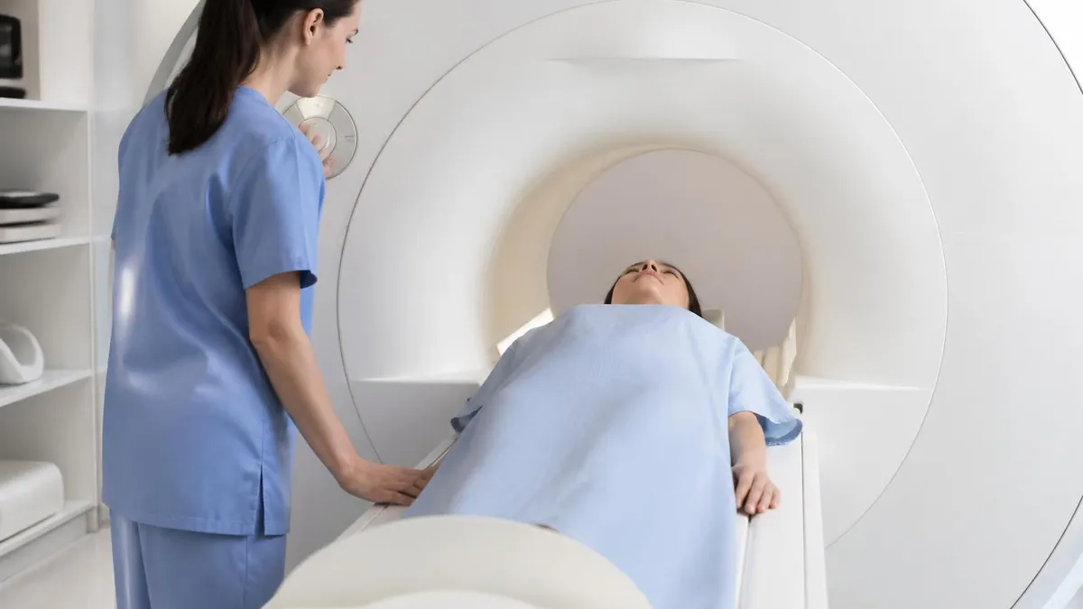
For patients preparing for a hand MRI, a few practical tips can make the experience smoother and the images sharper. First, remove all jewelry, watches, and accessories well before arrival. Even small items like rings and bracelets can cause significant artifact in the imaging field. Wear comfortable clothing without metal zippers or snaps near the hand, and be ready to change into a gown if the technologist requests it. Arrive about thirty minutes early to complete safety screening forms thoroughly and honestly.
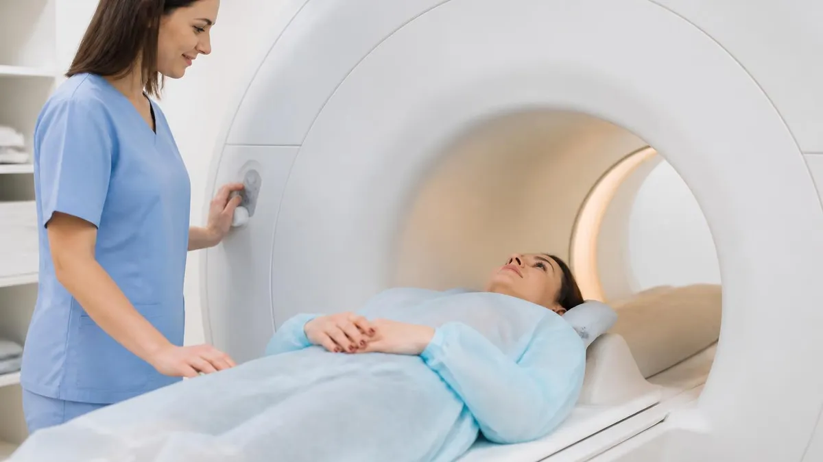
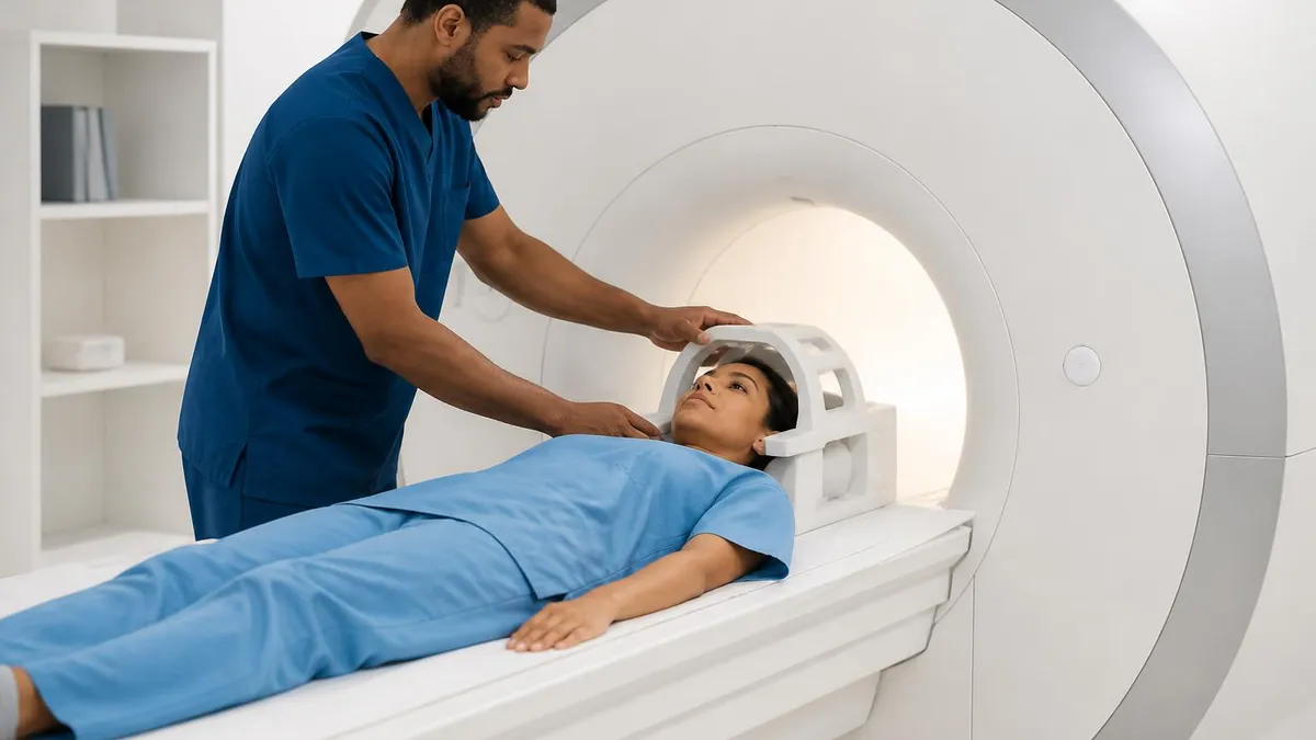
The hand will be positioned in a dedicated extremity coil. Most centers use a superman position where the patient lies prone with the affected arm extended overhead into the bore. Others use a position with the arm at the side, which is more comfortable but sometimes sacrifices image quality. Either way, the hand will be padded and secured to minimize motion. Patients who struggle to hold still should mention this in advance so the technologist can use additional padding or arrange for mild sedation if appropriate.
During the scan, expect to hear loud knocking, buzzing, and hammering sounds. These come from the gradient coils switching rapidly inside the magnet. Hearing protection in the form of earplugs and headphones will be provided. Some centers offer music through the headphones to make the time pass more pleasantly. The most important instruction is to keep the hand absolutely still. Even small movements can blur the high resolution images and require repeating sequences, extending total scan time considerably.
If intravenous contrast is needed, an IV will be placed before the scan begins. Gadolinium based contrast agents are generally safe but require checking kidney function in patients with renal disease. Contrast is typically used when evaluating tumors, infection, or inflammatory arthritis where enhancement patterns provide critical diagnostic information. The injection happens midway through the study, and the patient may feel a brief cool sensation in the arm but should not feel pain.
For radiologic technologists and students learning hand MRI, the key skills are coil selection, positioning, and protocol customization. Choosing the correct coil for hand size and the area of interest dramatically affects signal-to-noise ratio. Positioning must place the structure of interest at the magnet isocenter. Protocols should be tailored to the clinical question rather than running a one-size-fits-all approach. A suspected scaphoid fracture needs different sequences than a suspected ganglion or rheumatoid arthritis evaluation.
Understanding the typical noise levels and patient experience helps technologists prepare patients better. Knowing the difference between the Noise of MRI Machine: Why MRI Scanners Are So Loud and What to Expect on different sequences allows you to warn patients in advance. The pulse sequences with the loudest acoustic noise are typically the gradient echo and 3D acquisitions, while spin echo sequences tend to be quieter. Setting accurate expectations reduces patient anxiety and improves cooperation throughout the study.
Finally, for anyone preparing for the ARRT MRI registry exam, hand and wrist imaging is high yield content. Expect questions on coil selection, sequence parameters, anatomy identification, and common pathology appearances. Practice reading actual cases whenever possible, since pattern recognition is the core skill being tested. Use practice quizzes regularly to build your visual vocabulary, and review every wrong answer thoroughly to understand the underlying physics or anatomy concept being tested. Consistent daily practice beats cramming every time.
MRI Questions and Answers

MRI Medical Abbreviation: What MRI Stands For and Why It Matters

The History of MRI: From Discovery to Modern Medicine

Knee MRI Images: A Complete Guide to Reading, Understanding, and Interpreting Knee Scans

Noise of MRI Machine: Why MRI Scanners Are So Loud and What to Expect
MRI Alternatives: When CT, Ultrasound, X-Ray, and PET Make More Sense Than an MRI
About the Author

Medical Laboratory Scientist & Clinical Certification Expert
Johns Hopkins UniversityDr. Sandra Kim holds a PhD in Clinical Laboratory Science from Johns Hopkins University and is certified as a Medical Technologist (MT) and Medical Laboratory Scientist (MLS) through ASCP. With 16 years of clinical laboratory experience spanning hematology, microbiology, and molecular diagnostics, she prepares candidates for ASCP board exams, MLT, MLS, and specialist certification tests.
Join the Discussion
Connect with other students preparing for this exam. Share tips, ask questions, and get advice from people who have been there.
View discussion (6 replies)