
MRI Brain With Contrast: Complete Patient and Technologist Guide to Gadolinium-Enhanced Brain Imaging
MRI brain with contrast explained: gadolinium safety, what it detects, scan duration, costs, side effects, and prep tips from imaging experts. ✅
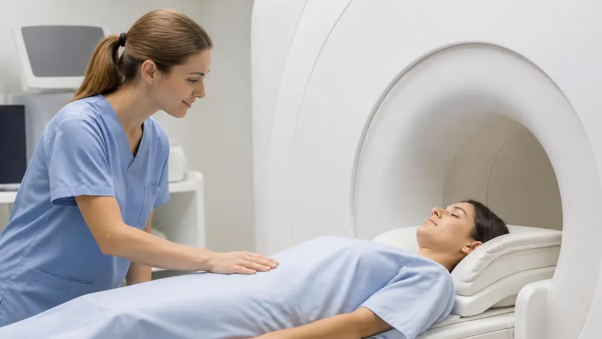
An MRI mri brain with and without contrast cpt contrast is one of the most powerful diagnostic tools in modern neuroradiology, capable of revealing tumors, infections, demyelinating disease, and vascular abnormalities that non-contrast scans simply cannot detect. The procedure combines a standard magnetic resonance scan of the head with an intravenous injection of a gadolinium-based contrast agent (GBCA) that highlights areas where the blood-brain barrier has broken down. For patients facing unexplained headaches, neurological symptoms, or known central nervous system conditions, this enhanced study often provides answers that change management.
Radiologists rely on contrast-enhanced sequences to distinguish between active and chronic disease, to grade tumors, and to monitor treatment response over months and years. A glioblastoma, for instance, may look like a vague area of edema on T2-weighted images but light up dramatically after gadolinium administration, revealing its true size and aggressive nature. Similarly, multiple sclerosis plaques that are actively inflamed will enhance, while older scars remain dark, giving clinicians a real-time window into disease activity.
The contrast agent itself is a chelated form of gadolinium, a rare-earth metal with unique paramagnetic properties that shorten T1 relaxation times in nearby tissues. When injected through a peripheral IV, it circulates throughout the body within seconds, crossing into brain tissue only where the normally tight blood-brain barrier has been compromised by tumor, infection, or inflammation. This selective enhancement is what makes the study so diagnostically valuable for neurological conditions.
Patients often arrive at the imaging center with questions about safety, what to expect, and whether contrast is truly necessary. While modern macrocyclic gadolinium agents have an excellent safety profile, understanding the procedure, its indications, and potential side effects helps patients feel prepared and informed. Technologists and radiologists work together to ensure each scan is tailored to the clinical question, balancing diagnostic yield with patient comfort and safety considerations.
The scan itself typically takes 30 to 60 minutes inside the bore of the magnet, with the contrast injection occurring partway through the examination. Pre-contrast and post-contrast sequences are compared side by side, allowing the radiologist to identify subtle areas of enhancement that might otherwise be missed. The resulting images are reviewed in axial, sagittal, and coronal planes, providing a comprehensive three-dimensional view of brain anatomy and pathology.
This guide walks through every aspect of MRI brain with contrast, from the physics of gadolinium enhancement to practical preparation tips, safety considerations, and what your results actually mean. Whether you are a patient scheduled for your first scan, a caregiver supporting a loved one, a technologist preparing for board exams, or a clinician brushing up on contrast protocols, the following sections deliver the depth and clarity you need. For broader background on the technology itself, see our overview of What Is an MRI Test? How Magnetic Resonance Imaging Scans Diagnose Disease in 2026.
By the end of this article, you will understand exactly why contrast is added, how the gadolinium works at a molecular level, what conditions the study can detect, what side effects to watch for, and how to prepare for the most accurate, comfortable examination possible. We will also cover renal function screening, the difference between macrocyclic and linear agents, and the latest guidelines on gadolinium retention in brain tissue.
MRI Brain With Contrast by the Numbers
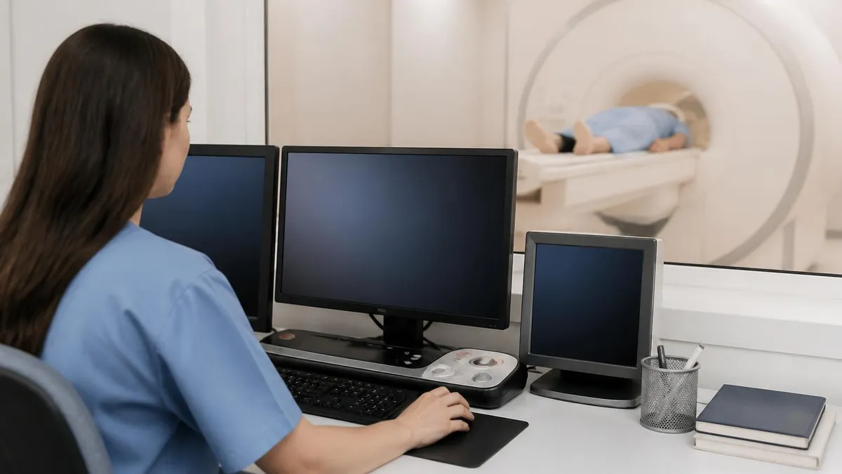
What Happens During Your Scan
Check-In and Screening
IV Placement
Pre-Contrast Imaging
Gadolinium Injection
Post-Contrast Sequences
Review and Discharge
An MRI brain with contrast is ordered when clinicians need to detect or characterize pathology that disrupts the blood-brain barrier, a tightly regulated interface between cerebral blood vessels and brain tissue. Under normal conditions, this barrier prevents gadolinium and most other large molecules from entering brain parenchyma. When disease processes such as tumors, abscesses, demyelination, or vascular malformations damage the barrier, gadolinium leaks into the affected tissue and appears bright on T1-weighted images, creating the characteristic enhancement pattern radiologists use for diagnosis.
brain tumor mris are among the most common indications for contrast-enhanced MRI. Primary tumors like gliomas, meningiomas, and pituitary adenomas each have distinctive enhancement patterns that help narrow the differential diagnosis. Metastatic disease from lung, breast, or melanoma primaries often appears as multiple ring-enhancing lesions at the gray-white matter junction. The pattern, location, number, and intensity of enhancement all provide critical clues that guide biopsy planning, surgical resection, and radiation therapy decisions for neuro-oncology patients.
Multiple sclerosis evaluation is another major application. Active demyelinating plaques enhance with gadolinium because inflammation disrupts the blood-brain barrier, while older inactive lesions appear as bright T2/FLAIR signals without enhancement. This distinction allows neurologists to assess disease activity and treatment response, often dictating whether to escalate disease-modifying therapy. The McDonald criteria for MS diagnosis specifically incorporate enhancing versus non-enhancing lesion patterns to establish dissemination in time, a cornerstone of modern MS diagnosis.
Infectious conditions including bacterial abscesses, viral encephalitis, and fungal infections often demonstrate dramatic enhancement patterns. A pyogenic brain abscess typically shows a thin, smooth ring of enhancement around a central area of low signal intensity representing pus. Encephalitis from herpes simplex virus classically enhances in the temporal lobes, while toxoplasmosis in immunocompromised patients produces multiple small ring-enhancing lesions. Contrast is essential for distinguishing these infections from tumors and guiding appropriate antimicrobial therapy.
Vascular pathology benefits enormously from contrast administration. Aneurysms, arteriovenous malformations, dural arteriovenous fistulas, and venous sinus thromboses are all better characterized with contrast-enhanced sequences and MR angiography. Stroke evaluation may use contrast to assess collateral circulation, identify mass effect from hemorrhagic transformation, or detect underlying tumors masquerading as infarcts. Post-stroke imaging at subacute timepoints frequently shows gyriform enhancement, a normal finding that can mimic disease if not recognized.
Pituitary and skull base pathology requires dedicated thin-section contrast imaging. Microadenomas as small as 2 to 3 millimeters can be detected on dynamic post-contrast sequences that capture the differential enhancement between normal pituitary tissue and tumor over time. Schwannomas of the eighth cranial nerve, meningiomas of the cerebellopontine angle, and craniopharyngiomas all show characteristic enhancement patterns that guide neurosurgical planning. For technologists studying these protocols, our guide to MRI Medical Abbreviation: What MRI Stands For and Why It Matters covers the terminology you will encounter on requisitions and reports.
Post-operative and post-treatment surveillance imaging relies heavily on contrast to monitor for residual or recurrent disease. After tumor resection, baseline post-operative scans within 48 to 72 hours establish a reference for future comparisons. Radiation necrosis can be difficult to distinguish from tumor recurrence on conventional sequences, sometimes requiring perfusion imaging or MR spectroscopy as adjuncts. Serial enhancement patterns over months help neuro-oncologists determine whether treatment is working or whether disease progression has occurred.
MRI Practice Test Questions
Prepare for the MRI - Magnetic Resonance Imaging exam with our free practice test modules. Each quiz covers key topics to help you pass on your first try.
MRI Knowledge
MRI Exam Questions covering Knowledge. Master MRI Test concepts for certification prep.
MRI Physics
Free MRI Practice Test featuring Physics. Improve your MRI Exam score with mock test prep.
MRI Anatomy and Pathology
MRI Test Prep for MRI Anatomy and Pathology. Practice MRI Quiz questions and boost your score.
MRI Anatomy and Positioning
MRI Questions and Answers on MRI Anatomy and Positioning. Free MRI practice for exam readiness.
MRI Contrast Agents
Free MRI Quiz on MRI Contrast Agents. MRI Exam prep questions with detailed explanations.
MRI Patient Care and Positioning
MRI Practice Questions for MRI Patient Care and Positioning. Build confidence for your MRI certification exam.
Gadolinium Contrast Agents Compared
Macrocyclic gadolinium-based contrast agents like gadobutrol (Gadavist), gadoteridol (ProHance), and gadoterate meglumine (Dotarem) feature a cage-like molecular structure that tightly binds the gadolinium ion. This stable architecture dramatically reduces the risk of free gadolinium being released into tissues, making these agents the preferred choice for most clinical applications in 2026. The FDA classifies them as Group II agents with the lowest reported risk of nephrogenic systemic fibrosis.
These agents are particularly favored for patients requiring repeated contrast studies, such as MS patients on long-term surveillance or oncology patients receiving serial tumor monitoring. Studies have shown minimal gadolinium retention in brain tissue with macrocyclic agents compared to linear formulations. The American College of Radiology recommends macrocyclic agents as first-line choice for routine contrast-enhanced MRI examinations of the central nervous system in most modern imaging centers.
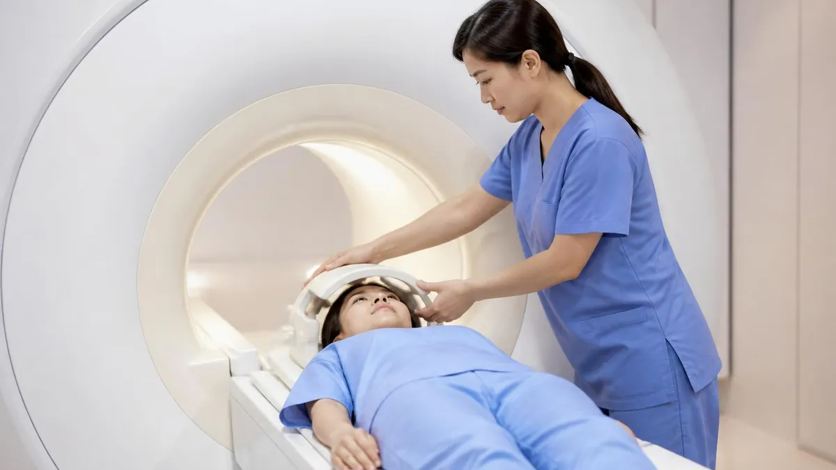
Should You Get MRI Brain With or Without Contrast?
- +Detects tumors, infections, and inflammation that non-contrast scans may miss entirely
- +Distinguishes active from chronic disease, especially in multiple sclerosis
- +Characterizes enhancement patterns that narrow the differential diagnosis
- +Essential for post-treatment monitoring of brain tumors and surgical resections
- +Visualizes vascular malformations, aneurysms, and venous thromboses with greater clarity
- +Allows detection of small pituitary microadenomas and skull base tumors
- −Requires IV placement which some patients find uncomfortable
- −Adds 15 to 20 minutes to total scan time and increases overall cost
- −Cannot be used in patients with severe kidney disease (eGFR <30)
- −Carries small risk of allergic reaction or contrast extravasation
- −Gadolinium retention in brain tissue remains under ongoing study
- −Not necessary for many indications including routine headache evaluation
MRI Brain With Contrast Preparation Checklist
- ✓Inform your doctor of any kidney disease, diabetes, or history of contrast reactions
- ✓Complete required lab work, including creatinine or eGFR, if requested by the facility
- ✓Remove all jewelry, hair accessories, hearing aids, and removable dental work before scan
- ✓Wear comfortable clothing without metal zippers, snaps, or underwire (gown often provided)
- ✓Continue routine medications unless specifically instructed otherwise by your physician
- ✓Eat lightly and stay well hydrated in the hours leading up to your appointment
- ✓Bring a list of all metal implants, surgical history, and previous imaging studies
- ✓Arrive 30 minutes early to complete screening forms and IV placement
- ✓Notify the technologist immediately if you experience claustrophobia or anxiety
- ✓Plan for a companion to drive you home if you receive sedation for the examination
Post-contrast images must be acquired within minutes of injection
Gadolinium begins to wash out of brain tissue within minutes of intravenous administration, making timing of post-contrast sequences critical. Most enhancing lesions show peak conspicuity 1 to 5 minutes after injection, with subtle leptomeningeal enhancement sometimes requiring imaging up to 10 minutes post-injection. Technologists must coordinate sequence acquisition precisely to capture maximum diagnostic information before contrast clears.
Safety screening for MRI brain with contrast involves two distinct considerations: standard MRI safety related to the magnetic field, and contrast-specific safety related to gadolinium administration. Both must be addressed before any patient enters the scanner room. The screening questionnaire covers implanted devices, prior surgeries, metal foreign bodies, kidney function, pregnancy status, prior contrast reactions, and current medications. Technologists trained in MR safety review every form and clarify ambiguous answers before granting room access.
Kidney function is the most important contrast-specific safety screen. Patients with severely reduced glomerular filtration rate (eGFR less than 30 mL/min/1.73m²) face increased risk of nephrogenic systemic fibrosis (NSF), a rare but potentially devastating complication characterized by progressive skin thickening, joint contractures, and organ involvement. Modern macrocyclic agents have dramatically reduced NSF incidence to near zero in recent years, but most institutions still require recent creatinine testing for patients over 60, diabetics, hypertensive patients, and those with known kidney disease.
Acute allergic reactions to gadolinium occur in approximately 0.04 to 0.7 percent of administrations, with most being mild reactions like hives, itching, or nausea. Moderate reactions involving bronchospasm or facial swelling occur in roughly 1 in 10,000 doses, while severe anaphylactoid reactions requiring epinephrine and emergency intervention are extremely rare at approximately 1 in 100,000. Imaging centers maintain crash carts, oxygen, and trained personnel ready to manage reactions should they occur during or after contrast injection.
Patients with a prior history of contrast reaction may receive premedication with corticosteroids and antihistamines before repeat examinations. A typical premedication protocol includes 50 mg of prednisone at 13, 7, and 1 hour before contrast administration, plus 50 mg of diphenhydramine one hour before. Patients with severe prior reactions or true gadolinium allergy may require alternative imaging modalities like non-contrast MRI, CT with iodinated contrast, or referral for allergy consultation before proceeding.
Pregnancy presents a special situation. Gadolinium crosses the placenta and is excreted into fetal urine, where it can persist in amniotic fluid for indeterminate periods. The ACR and FDA recommend against routine gadolinium administration during pregnancy unless the diagnostic benefit clearly outweighs potential risks to the fetus. Non-contrast MRI is generally safe at any trimester and is often substituted for contrast-enhanced studies in pregnant patients with neurological symptoms. Breastfeeding mothers may continue nursing after gadolinium administration based on current evidence.
Gadolinium retention in brain tissue has emerged as a significant area of research and patient concern. T1 signal hyperintensity in the dentate nucleus and globus pallidus has been documented after repeated administrations of certain linear agents, with smaller amounts retained after macrocyclic agent exposure. While no definitive clinical consequences have been established, the FDA requires a Medication Guide and patient counseling for all gadolinium-based agents. Most institutions limit unnecessary repeat contrast studies and document medical justification for each examination.
Extravasation of contrast into soft tissue around the IV site occurs in less than 1 percent of injections but can cause local pain, swelling, and rarely skin necrosis if large volumes leak. Technologists monitor the injection site during administration and stop immediately if extravasation is detected. Treatment includes elevation, cold or warm compresses, and surgical consultation for large extravasations. Patients should report any pain, swelling, or skin changes at the IV site even hours after the examination concludes for prompt evaluation.
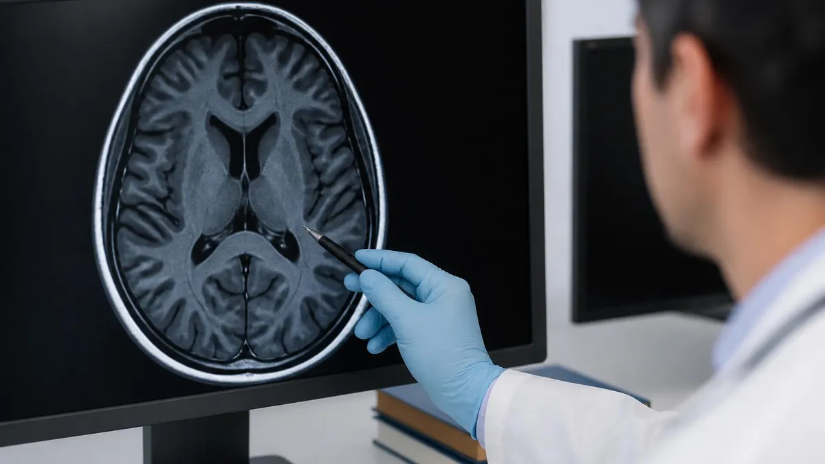
Patients with eGFR below 30 mL/min/1.73m² should not receive gadolinium contrast except in life-threatening circumstances where no alternative exists. Always disclose kidney disease, diabetes, hypertension, or recent contrast exposure to your imaging team. Acute kidney injury within the past 30 days is also a contraindication. Recent creatinine testing within 30 to 60 days is typically required for at-risk patients before scheduling contrast-enhanced examinations.
The cost of an MRI brain with contrast varies dramatically based on geographic location, facility type, and insurance coverage. National averages range from $1,200 at outpatient imaging centers to over $4,000 at hospital-based radiology departments. Self-pay patients often qualify for discounted rates, sometimes 40 to 60 percent below billed charges, by asking about cash prices upfront. Insurance pre-authorization is usually required, and denied claims commonly cite lack of medical necessity documentation rather than the procedure itself being uncovered.
Medicare typically covers MRI brain with contrast when ordered by a physician for an approved indication, with patients responsible for the Part B deductible and 20 percent coinsurance. Commercial insurance plans vary widely in coverage, copays, and out-of-pocket maximums. High-deductible health plans may leave patients facing the full negotiated rate until their deductible is met. Health savings accounts (HSAs) and flexible spending accounts (FSAs) can offset costs with pre-tax dollars for qualifying examinations.
The radiologist who interprets your scan generates a separate professional fee bill in addition to the technical facility fee. This dual billing structure means patients may receive two or three bills for a single examination: facility fee, radiologist professional fee, and sometimes a contrast administration charge. Asking for itemized estimates before scheduling can prevent billing surprises and allow for cost comparison shopping across providers in your area.
Results typically become available within 24 to 72 hours after the examination, though urgent cases requested by emergency departments or oncology services may be read within hours. Reports go to the referring physician first, who then contacts the patient to discuss findings and next steps. Many health systems now provide direct patient access to imaging reports through online portals, sometimes before the ordering physician has reviewed and contextualized the findings.
Understanding your radiology report requires basic familiarity with the language radiologists use. Terms like "unremarkable" or "no acute findings" generally indicate normal results. "Enhancement," "mass effect," "midline shift," or "restricted diffusion" describe specific abnormal findings that warrant clinical correlation. The impression section at the bottom summarizes the most important findings and often includes specific recommendations for follow-up imaging, biopsy, or specialist referral. Comparing previous studies is essential for accurate interpretation.
Comparison with prior imaging studies is one of the most valuable tools in radiology. Bringing copies of previous MRIs, CTs, or related imaging on CD or accessing them through hospital networks allows the interpreting radiologist to assess stability versus progression of findings. Lesion size measurements, enhancement patterns, and adjacent tissue changes over time provide critical information that single-timepoint imaging cannot deliver. For background on imaging history, see The History of MRI: From Discovery to Modern Medicine.
Second opinions on imaging interpretation are increasingly common for serious diagnoses. Specialized neuroradiologists at academic centers can re-read studies done at community facilities, sometimes identifying findings missed on initial interpretation or providing more nuanced characterization of complex lesions. Several telemedicine services now offer formal second-opinion radiology reads for a flat fee, providing reassurance or alternative perspectives when treatment decisions hinge on imaging findings. Discuss this option with your physician if uncertainty exists.
Preparing well for your MRI brain with contrast examination begins days before the appointment with thoughtful attention to several practical details. Hydration is one of the most overlooked yet important steps. Well-hydrated kidneys clear gadolinium more efficiently, reducing the small theoretical risk of retention and contrast-related complications. Drink water normally in the 24 hours before your scan unless you have been specifically instructed to fast for sedation or other reasons.
Claustrophobia affects roughly 5 to 10 percent of patients undergoing MRI, and the longer scan times for contrast-enhanced studies can exacerbate anxiety. If you have known claustrophobia, discuss anxiolytic premedication with your physician before the appointment. A small dose of oral lorazepam or alprazolam taken 30 to 60 minutes before the scan helps many patients tolerate the experience. Wide-bore and open MRI scanners are alternatives at some facilities, though image quality may be slightly reduced.
The sounds inside an MRI scanner can be startling and uncomfortable. The loud knocking, beeping, and humming noises result from gradient coils rapidly switching during sequence acquisition. Earplugs and noise-reducing headphones are provided at all reputable imaging centers and can reduce noise exposure significantly. Many facilities offer music during the scan, allowing you to choose your preferred genre or station to help pass the time and reduce stress during the examination.
Communication with the technologist throughout the scan is essential. A squeeze ball or intercom button placed in your hand allows you to alert the technologist immediately if you experience discomfort, anxiety, claustrophobia, or any concerning symptoms. Technologists can pause the examination, provide reassurance, adjust positioning, or remove you from the bore entirely if needed. Never hesitate to use the call button. Brief breaks rarely affect diagnostic quality and significantly improve the patient experience.
What you wear matters more than many patients realize. Avoid clothing with metal zippers, snaps, underwire, metallic threads, or printed designs that may contain metallic pigments. Most facilities provide hospital gowns to eliminate these concerns entirely. Remove all jewelry including rings, earrings, body piercings, watches, and necklaces before entering the magnet room. Tattoos with metallic pigments rarely cause issues but should be disclosed during screening for documentation purposes and appropriate precautions.
After the scan, most patients can resume all normal activities immediately. There are no driving restrictions unless you received sedation, no dietary limitations, and no physical activity restrictions. Drink plenty of water for the next 24 hours to help your kidneys clear the gadolinium contrast efficiently. Monitor the IV site for any redness, swelling, or persistent pain, and contact the imaging center if delayed reactions such as rash, itching, or breathing difficulty develop within hours or days of the examination.
Following up on your results is your responsibility as much as your physician's. Mark your calendar for when results are expected and contact your referring provider if you have not heard back within the promised timeframe. Bring written questions to your follow-up appointment and ask your physician to explain findings in plain language. Request copies of your images on disc or through patient portal access for your personal records and any future second-opinion consultations or specialist referrals you may pursue.
MRI Questions and Answers
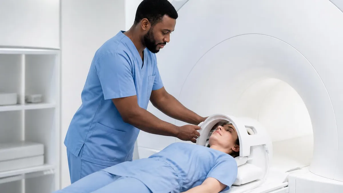
What Is an MRI Test? How Magnetic Resonance Imaging Scans Diagnose Disease in 2026
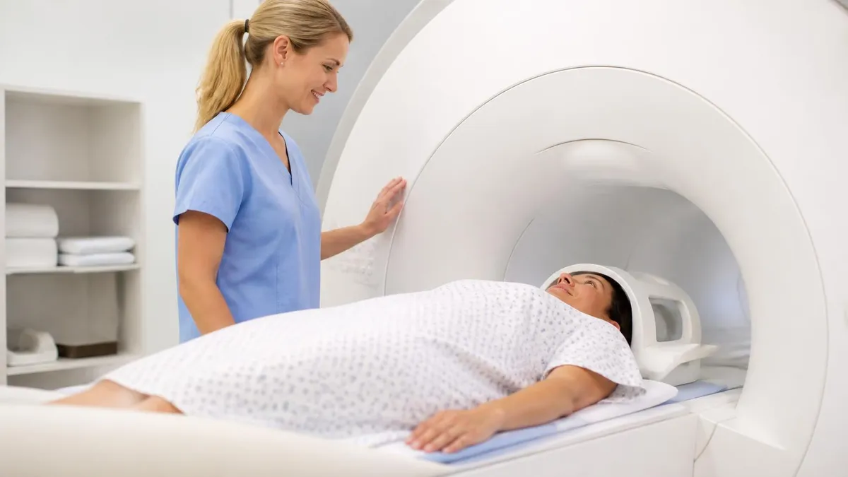
MRI Medical Abbreviation: What MRI Stands For and Why It Matters
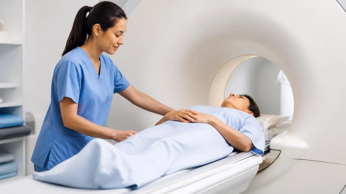
The History of MRI: From Discovery to Modern Medicine

Knee MRI Images: A Complete Guide to Reading, Understanding, and Interpreting Knee Scans
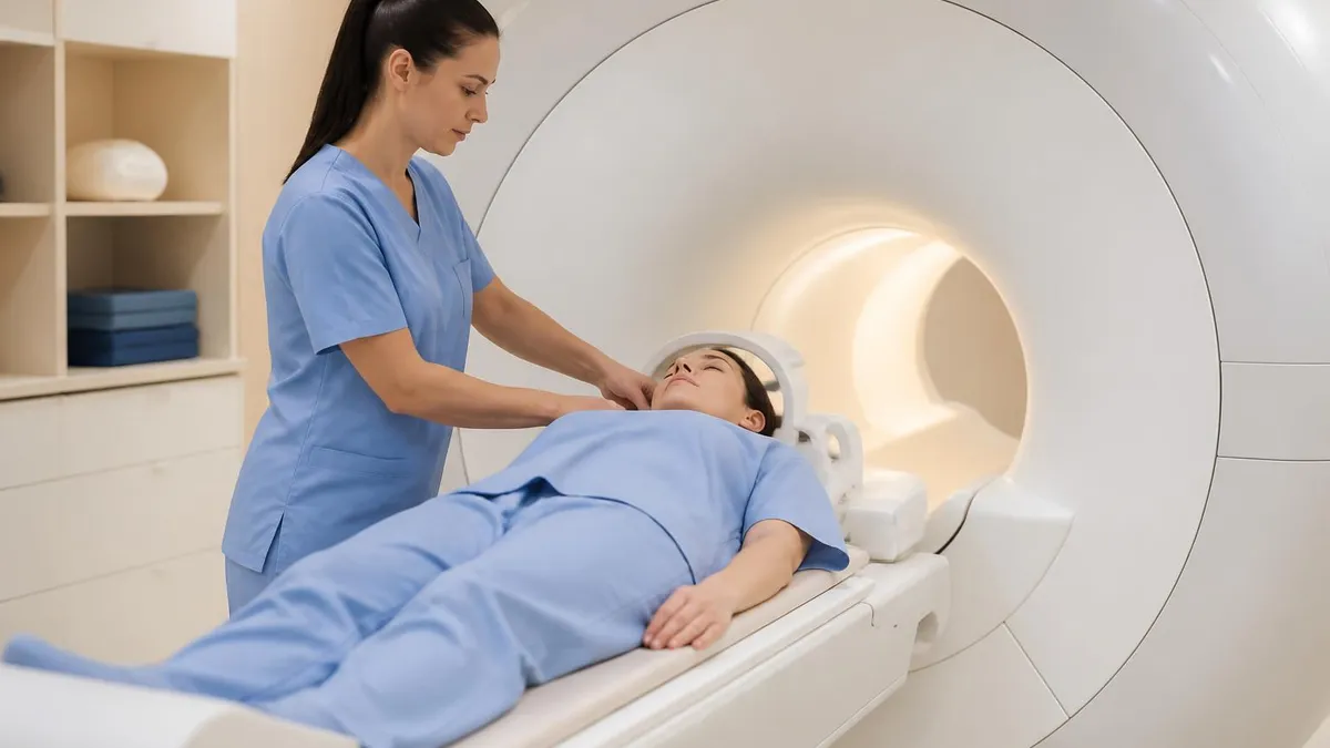
Noise of MRI Machine: Why MRI Scanners Are So Loud and What to Expect
Join the Discussion
Connect with other students preparing for this exam. Share tips, ask questions, and get advice from people who have been there.
View discussion (8 replies)