Is MRI Radiology? Understanding How MRI Fits Into the Field
Is MRI radiology? Learn how MRI fits within the radiology field, who reads MRI scans, and how it compares to X-ray, CT, and ultrasound imaging.
Is MRI radiology? The short answer is yes — magnetic resonance imaging is a core subspecialty within the broader field of radiology, and the physicians who interpret MRI scans are board-certified radiologists. However, MRI differs significantly from traditional radiology in how it produces images. Instead of using ionizing radiation like X-rays or computed tomography, MRI relies on powerful magnetic fields and radiofrequency pulses to generate detailed images of soft tissues, organs, and structures throughout the human body.
The confusion around whether MRI counts as radiology stems from the word itself. Radiology historically referred to imaging using radiation, and MRI uses no radiation whatsoever. Yet the field of radiology has expanded over the past four decades to encompass every major form of medical imaging, including ultrasound, nuclear medicine, and MRI. Hospitals organize all of these modalities under one radiology department, and radiologists complete cross-modality training during residency to read studies from each imaging technique.
Understanding where MRI fits within radiology matters for patients, students, and aspiring imaging professionals. Patients want to know who is qualified to interpret their results. Students choosing a healthcare career path benefit from understanding how MRI technologists differ from radiologic technologists. Hospital administrators rely on this organizational clarity to staff departments and credential physicians. Even insurance billing codes are organized around the radiology umbrella, with MRI procedures classified under diagnostic radiology categories.
The American College of Radiology, the Radiological Society of North America, and the American Board of Radiology all recognize MRI as part of diagnostic radiology. MRI physicists, MRI safety officers, and MRI technologists work alongside CT technologists and X-ray technologists in the same imaging departments. Continuing education credits for radiologic technologists often include MRI content, and many cross-trained technologists hold certifications in multiple modalities. This professional integration confirms MRI's permanent place within radiology.
From a clinical perspective, radiologists who specialize in MRI often subspecialize further into neuroradiology, musculoskeletal imaging, abdominal imaging, breast imaging, or cardiac imaging. Each subspecialty requires fellowship training after residency, and MRI is a major component of every one of these fellowships. A neuroradiologist, for example, may interpret 30 to 50 brain MRI studies per day, while a musculoskeletal radiologist focuses on knee, shoulder, and spine MRI exams.
This article explains exactly how MRI fits into radiology, who is qualified to perform and interpret MRI scans, what makes MRI distinct from other imaging methods, and how the modality is regulated and taught. We will cover the educational pathways, the equipment differences, the clinical applications, and the future direction of MRI within the broader field of radiology in 2026 and beyond.
Whether you are a patient receiving your first scan, a student preparing for the ARRT MRI registry, or simply curious about how modern imaging works, this guide will give you the complete picture. By the end, you will understand why the answer to "is MRI radiology" is more nuanced than a simple yes or no — and why the relationship between these two fields shapes everything from clinical workflow to career opportunities.
MRI in Radiology by the Numbers
How MRI Fits Within Radiology
MRI sits within the diagnostic radiology specialty, alongside X-ray, CT, ultrasound, and nuclear medicine. All five modalities share interpretation responsibilities under a single board-certified radiologist who completes residency training.
Unlike traditional radiology methods, MRI uses magnetic fields and radio waves instead of X-rays. This makes MRI a unique subset of radiology — radiation-free, but still managed by the radiology department in every hospital nationwide.
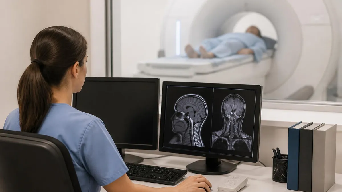
Every MRI scan in the United States is officially read and signed by a radiologist. Although MRI technologists operate the scanner, only a physician trained in diagnostic radiology can issue the final report and diagnosis.
The American College of Radiology accredits MRI facilities through its quality assurance program. This same body accredits CT, mammography, and ultrasound centers — confirming MRI's place within the formal radiology framework.
MRI is central to neuroradiology, musculoskeletal radiology, abdominal imaging, breast imaging, and cardiac radiology fellowships. Radiologists choose subspecialties largely based on the type of MRI work they want to focus on.
The distinction between MRI and traditional radiology becomes clear when you examine the underlying physics. Conventional radiology, dating back to Wilhelm Röntgen's discovery in 1895, uses ionizing radiation to create images. X-rays pass through the body and are absorbed differently by bone, soft tissue, and air, producing the familiar black-and-white pictures we associate with broken bones or chest exams. Computed tomography uses the same X-ray principle but in cross-sectional slices, building three-dimensional reconstructions from multiple angles.
MRI works on completely different principles. Inside the scanner, a powerful magnet — typically 1.5 Tesla or 3 Tesla in clinical use — aligns the hydrogen protons in your body's water molecules. Radiofrequency pulses then knock these protons out of alignment, and as they return to their original state, they emit signals that the scanner detects and converts into images. This process, called nuclear magnetic resonance, was first described in the 1940s but did not enter clinical medicine until the early 1980s when whole-body scanners became feasible.
For a deeper look at how the modality evolved from physics experiment to indispensable clinical tool, see The History of MRI: From Discovery to Modern Medicine. The transformation took roughly forty years and required breakthroughs in superconducting magnet technology, computational power, and clinical protocol development. Today's MRI scanners can image structures as small as one millimeter and produce hundreds of slices in a single exam.
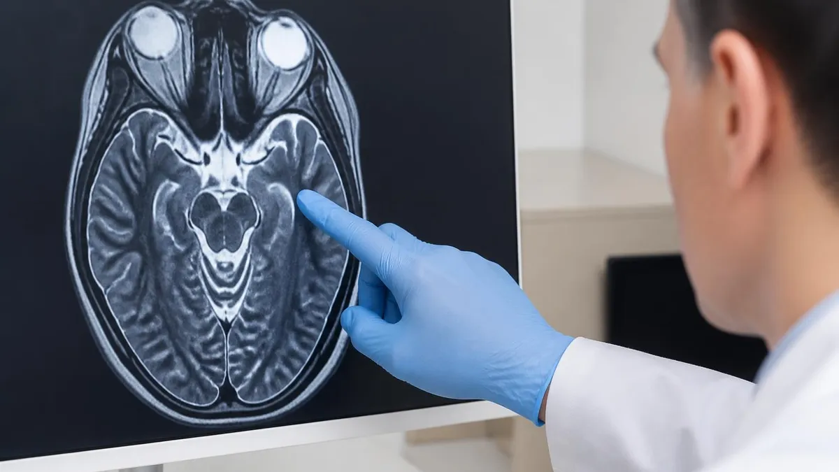
The clinical advantages of MRI over X-ray-based radiology are significant. MRI provides superior soft-tissue contrast, making it the gold standard for imaging the brain, spinal cord, joints, ligaments, and abdominal organs. A radiologist can distinguish gray matter from white matter in the brain, identify a small meniscus tear in the knee, or detect early multiple sclerosis lesions — all imaging tasks that would be impossible or unreliable with X-ray or CT alone.
However, MRI is not always the right choice. CT remains faster and more accessible for trauma cases, acute stroke evaluation, and lung imaging. X-rays are still the first-line modality for suspected fractures, chest infections, and dental imaging. Ultrasound is preferred for obstetric care, vascular studies, and many bedside applications because it is portable and uses no radiation. Each modality has its place, and radiologists are trained to recommend the right test for each clinical question.
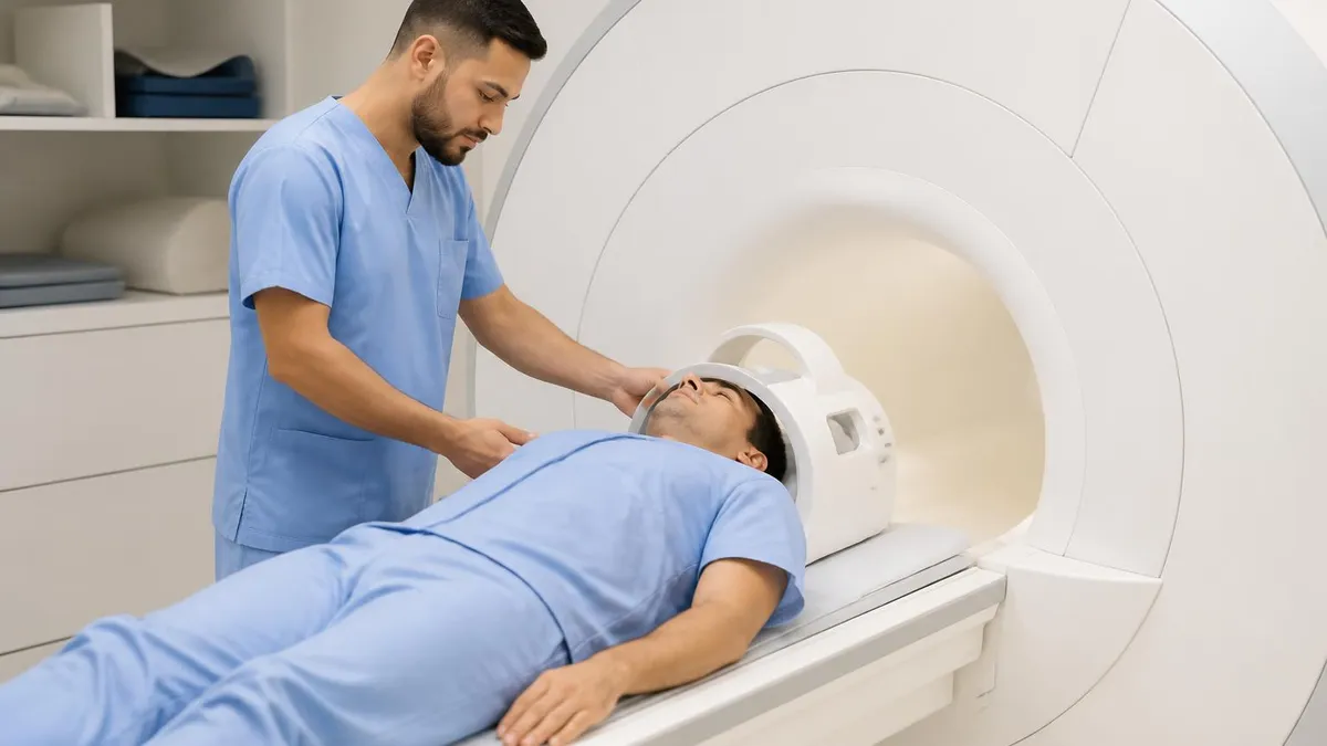
Another important difference involves the personnel who operate the equipment. Radiologic technologists, also called X-ray techs or rad techs, complete a two-year associate's degree and pass the ARRT primary certification exam. MRI technologists either start as rad techs and add MRI as a post-primary certification, or complete a primary MRI educational program. Both pathways require continuing education credits and registry maintenance, and both fall under the broader umbrella of radiologic sciences.
The infrastructure required for MRI also differs dramatically from traditional radiology. An X-ray suite needs only a lead-lined room and a small generator. A CT scanner needs power, cooling, and shielding. An MRI scanner requires a Faraday cage to block external radiofrequency interference, helium cooling for the superconducting magnet, and a strictly controlled five-zone safety perimeter to prevent metallic objects from becoming dangerous projectiles. These engineering challenges make MRI suites the most expensive and complex installations in any radiology department.
MRI Training and Credentials Within Radiology
Radiologists who interpret MRI scans complete four years of medical school followed by a one-year internship and a four-year diagnostic radiology residency. During residency, trainees rotate through every imaging modality including MRI, learning to interpret thousands of studies under supervision. After residency, they sit for the American Board of Radiology Core Exam and Certifying Exam to become board-certified diagnostic radiologists qualified to read MRI scans.
Many radiologists pursue additional fellowship training in MRI-heavy subspecialties such as neuroradiology, musculoskeletal imaging, or body imaging. These one-to-two-year fellowships provide deep expertise in MRI protocols, advanced sequences, and complex case interpretation. By the time a radiologist completes fellowship, they have personally interpreted between 15,000 and 30,000 MRI studies, building the pattern recognition essential for accurate diagnosis in everyday clinical practice.
Is MRI the Right Choice Compared to Other Radiology Methods?
- +Superior soft-tissue contrast for brain, spine, and joint imaging
- +No ionizing radiation exposure, making it safer for repeated scans
- +Can image in any anatomical plane without repositioning the patient
- +Excellent for detecting tumors, inflammation, and demyelinating disease
- +Functional MRI can map brain activity in real time
- +Contrast agents are generally safer than iodinated CT contrast
- +Useful for pediatric and obstetric patients when radiation must be avoided
- −Significantly longer scan times than X-ray or CT, often 30-90 minutes
- −Much higher cost per study compared to other imaging modalities
- −Strict contraindications for patients with pacemakers and metal implants
- −Loud scanner noise can require hearing protection during exams
- −Claustrophobic environment causes anxiety in roughly 5-10% of patients
- −Limited availability for emergency or after-hours imaging in rural areas
- −Image quality severely degraded by patient motion during acquisition
MRI Safety Checklist for Radiology Staff
- ✓Screen every patient for pacemakers, defibrillators, and neurostimulators before entry
- ✓Verify all metallic implants against MRI compatibility databases for the specific field strength
- ✓Remove all external metallic objects including jewelry, watches, and hearing aids
- ✓Confirm renal function before administering gadolinium-based contrast agents
- ✓Provide hearing protection — earplugs or headphones — for every patient and accompanying staff
- ✓Document patient claustrophobia history and offer sedation or open MRI when appropriate
- ✓Maintain Zone 4 access control to prevent unauthorized entry near the magnet
- ✓Train all staff on quench procedures and emergency magnet shutdown protocols
- ✓Verify pregnancy status in women of childbearing age before scanning
- ✓Use ferromagnetic detection systems at the entrance to the scanner room
MRI is radiology — but it is also more.
While MRI sits firmly within the diagnostic radiology specialty, it operates on physics principles entirely distinct from radiation-based imaging. This dual identity means MRI professionals must master both the radiology workflow and the unique safety, physics, and protocol challenges that come with strong magnetic fields. Understanding this distinction is essential for anyone working in modern imaging.
The clinical applications of MRI span virtually every organ system in the human body, and each application has shaped how radiologists practice within their subspecialties. Neuroradiology relies on MRI for nearly every brain and spine study, with the exception of acute trauma and stroke evaluation where CT remains faster. Brain MRI can detect tumors, multiple sclerosis plaques, dementia patterns, hemorrhage, and developmental abnormalities that no other modality can visualize as clearly.
Musculoskeletal radiology has been transformed by MRI. Before MRI became widely available in the 1990s, evaluating ligament tears, cartilage damage, and bone marrow disease required invasive procedures like arthrography or bone scans. Today, a knee MRI can show a torn ACL, meniscal injury, cartilage defects, and bone bruising in a single thirty-minute exam. Shoulder, hip, ankle, and spine MRI studies have similarly replaced older diagnostic approaches across orthopedic medicine.
In abdominal and pelvic imaging, MRI has become the second-line modality after ultrasound and CT for many indications. MRI excels at characterizing liver lesions, evaluating pancreatic disease, staging rectal cancer, and assessing the female pelvis for fibroids, endometriosis, and gynecologic malignancies. Prostate MRI has emerged as a standard tool for prostate cancer detection and biopsy guidance, with multiparametric protocols now recommended before any prostate biopsy.
Breast MRI plays a specific role in high-risk screening and complex cases. Women with BRCA mutations or strong family histories of breast cancer typically receive annual breast MRI in addition to mammography. MRI also helps stage newly diagnosed breast cancers, evaluate response to chemotherapy, and assess silicone implant integrity. Breast imaging fellowships now include extensive MRI training as a result.
Cardiac MRI represents one of the fastest-growing applications within radiology. This subspecialty examines heart muscle viability, congenital heart disease, cardiomyopathies, and inflammatory conditions like myocarditis. Many cardiac MRI studies are performed jointly by radiologists and cardiologists, with both specialties contributing to interpretation. The complexity of cardiac MRI protocols has created a niche subspecialty that often requires additional fellowship training beyond standard body imaging.
One area where the lines blur is interventional radiology with MRI guidance. While most interventional procedures use fluoroscopy or CT, an increasing number of biopsies, ablations, and targeted treatments now use MRI guidance because of its superior soft-tissue visualization. MR-guided focused ultrasound ablation of fibroids and bone metastases is a growing field, and MR-guided radiation therapy planning has become standard practice at many cancer centers.
The administrative side of MRI within radiology is also significant. MRI scans generate substantial revenue for hospitals and imaging centers, often more than X-ray or ultrasound. Insurance reimbursement, pre-authorization requirements, and appropriate use criteria all influence how MRI is ordered and performed. Radiology departments dedicate considerable resources to MRI scheduling, protocol optimization, and quality assurance to ensure efficient and accurate service delivery in 2026.
Despite the absence of ionizing radiation, MRI carries significant safety risks that traditional radiology does not. Ferromagnetic projectiles, implant heating, gadolinium contrast reactions, and acoustic noise injury can all occur if safety protocols are not strictly followed. Every patient must be screened thoroughly before entering the scanner room, regardless of how routine the exam appears to be.
The future of MRI within radiology is shaped by three converging forces: artificial intelligence, faster acquisition techniques, and new clinical applications. AI tools are already reshaping how radiologists interpret MRI studies, with FDA-approved algorithms now assisting with brain volumetry, prostate cancer detection, spine measurement, and stroke triage. By 2026, most large radiology practices are incorporating at least one AI tool into their MRI workflow.
Acceleration techniques are dramatically shortening scan times. Compressed sensing, parallel imaging, and deep learning reconstruction allow a brain MRI that took thirty minutes in 2015 to be completed in eight to ten minutes today, with equivalent or better image quality. This improves patient comfort, increases department throughput, and reduces costs across the system. For patients who struggle with claustrophobia, faster scans mean less time inside the magnet bore and a better overall experience.
For a practical look at how contrast factors into modern MRI exams, see MRI With and Without Contrast: How It Works, What to Expect. Contrast use continues to evolve, with newer macrocyclic gadolinium agents replacing older linear agents and protocols being refined to use less contrast overall. Some research is even exploring contrast-free MRI techniques for indications where contrast was previously considered essential.
New magnet technologies are also expanding access. Low-field MRI scanners at 0.55 Tesla and even 0.064 Tesla are entering the market, offering lower cost, easier siting, and the potential for point-of-care imaging in emergency departments and rural settings. Portable MRI scanners can now be wheeled to a patient's bedside in the intensive care unit, an unthinkable capability just five years ago. These developments are democratizing access to MRI imaging.
High-field research scanners at 7 Tesla are pushing the boundaries in the opposite direction. These ultra-high-field systems provide unprecedented resolution for brain imaging, musculoskeletal research, and metabolic studies. While 7T scanners remain primarily research tools, an increasing number of clinical 7T scanners are receiving FDA approval for specific indications such as epilepsy evaluation and brain tumor characterization in academic medical centers.
The workforce implications of these changes are significant. Radiologists must continue learning throughout their careers, incorporating new sequences, AI tools, and clinical indications into daily practice. MRI technologists need updated training as protocols evolve and new safety considerations emerge. Educational programs are reshaping curricula to prepare the next generation for an imaging environment that looks dramatically different from the one in which their instructors trained twenty years ago.
Looking forward, MRI's place within radiology will only become more central. As CT use is scrutinized for radiation dose and as ultrasound reaches its resolution limits, MRI fills the gap with safer, more detailed imaging. The fundamental answer to "is MRI radiology" will remain yes — but the technology, training, and applications behind that answer will continue evolving rapidly through 2026 and into the coming decade.
For patients preparing for an MRI scan, understanding that MRI is part of radiology can ease confusion when scheduling appointments and reviewing results. Your scan will likely be performed at a radiology center, an outpatient imaging facility, or a hospital radiology department. The technologist who operates the scanner is a registered MRI technologist, and the physician who interprets your images is a board-certified radiologist who may further specialize in the body region being imaged.
If you're considering an MRI career, the choice between becoming a radiologist and becoming an MRI technologist comes down to time, cost, and clinical role. Becoming a radiologist requires thirteen years of post-secondary education and training, but offers physician-level salary and decision-making authority. Becoming an MRI technologist requires two to four years of training and offers a strong middle-class salary with hands-on patient contact and direct involvement in image acquisition every day.
Students preparing for the ARRT MRI registry exam should focus on five core content areas: patient interactions and management, safety, image production, procedures, and equipment operation. Each section carries roughly equal weight on the exam, and the questions emphasize practical decision-making rather than rote memorization. Setting aside ten to twelve weeks of structured study, using practice tests weekly, and reviewing missed questions in detail is the most reliable preparation strategy in 2026.
For working radiology professionals, staying current with MRI involves more than continuing education credits. Subscribe to subspecialty journals, attend annual meetings such as RSNA or ISMRM, and participate in case-based learning platforms. The pace of innovation in MRI is accelerating, and what was considered cutting-edge five years ago may now be standard of care. Continuous learning is no longer optional in modern radiology departments.
Patients with metallic implants should always discuss MRI safety with their referring physician and the imaging facility before scheduling. Many older implants that were once contraindications are now considered MRI conditional, meaning they can be safely scanned under specific protocols. New databases and manufacturer documentation make this process easier, but it still requires careful verification before the patient enters the scanner room for any MRI exam.
Healthcare administrators considering capital investment in MRI should evaluate field strength, vendor service contracts, patient experience features, and AI capabilities. A 3 Tesla scanner offers higher resolution and faster scan times but costs more to purchase, install, and maintain than a 1.5 Tesla system. The right choice depends on the case mix the facility expects to handle, including pediatric volume, cardiac imaging plans, and research aspirations. Total cost of ownership matters more than sticker price.
Finally, the broader healthcare system benefits when MRI is used appropriately. Ordering an MRI for the right indication at the right time improves outcomes and reduces overall costs. Ordering it unnecessarily contributes to system inefficiency without improving patient care. The American College of Radiology publishes Appropriateness Criteria to guide referring physicians, and these criteria reflect the consensus of imaging experts about when MRI provides genuine value compared to other radiology options available today.
MRI Questions and Answers
About the Author
Attorney & Bar Exam Preparation Specialist
Yale Law SchoolJames R. Hargrove is a practicing attorney and legal educator with a Juris Doctor from Yale Law School and an LLM in Constitutional Law. With over a decade of experience coaching bar exam candidates across multiple jurisdictions, he specializes in MBE strategy, state-specific essay preparation, and multistate performance test techniques.