Hippocampus MRI: Complete Guide to Imaging, Anatomy, Volumetry, and Clinical Interpretation
Hippocampus MRI guide: dedicated protocols, coronal oblique planning, volumetry, atrophy scoring, sclerosis findings, and clinical interpretation tips.
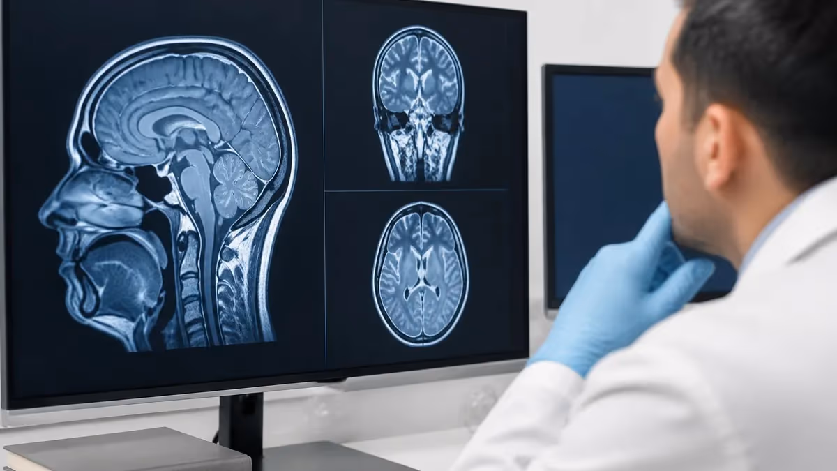
A hippocampus MRI is a dedicated magnetic resonance imaging examination focused on the medial temporal lobe, designed to evaluate the size, shape, signal characteristics, and internal architecture of the hippocampal formation. Unlike a standard brain MRI, a hippocampus MRI uses thin-slice coronal oblique sequences angled perpendicular to the long axis of the hippocampus, allowing radiologists to detect subtle atrophy, mesial temporal sclerosis, vascular insults, and developmental anomalies that routine scans often miss. This focused approach is the cornerstone of imaging in epilepsy, dementia, and amnestic disorders.
The hippocampus sits along the floor of the temporal horn of the lateral ventricle and forms part of the limbic system, the brain's memory and emotion network. Because the structure measures only about 4 to 4.5 cubic centimeters in healthy adults, even a 10 to 15 percent volume loss can carry profound clinical meaning. Dedicated MRI protocols, often combined with quantitative volumetry software, give clinicians a reproducible way to track these subtle changes across years, making MRI the gold standard for evaluating mesial temporal pathology.
Indications for a dedicated hippocampal study include suspected temporal lobe epilepsy, early Alzheimer disease, transient global amnesia, autoimmune limbic encephalitis, post-anoxic memory loss, and herpes simplex encephalitis. Neurologists increasingly order hippocampus-specific protocols when patients present with progressive memory complaints, especially when standard brain MRI is reported as unremarkable. The exam complements neuropsychological testing and biomarker studies such as cerebrospinal fluid amyloid-beta and tau, helping to localize the underlying lesion and guide treatment.
Modern hippocampal imaging typically requires a 3 Tesla scanner, a 32-channel head coil, and sequences that include high-resolution coronal T2, FLAIR, 3D T1 MPRAGE or BRAVO for volumetry, and diffusion-weighted imaging. Contrast is reserved for suspected encephalitis, neoplasm, or vascular pathology. Acquisition usually takes 25 to 40 minutes, and the radiologist will review images side by side, comparing left and right hippocampi for symmetry, signal, and morphology along the head, body, and tail.
The clinical impact of a high-quality hippocampus MRI cannot be overstated. In drug-resistant temporal lobe epilepsy, identification of unilateral hippocampal sclerosis can transform a patient's prognosis, since surgical resection produces seizure freedom in roughly 65 to 80 percent of selected candidates. In suspected Alzheimer disease, hippocampal atrophy ratings using the Scheltens medial temporal atrophy scale support earlier diagnosis and entry into disease-modifying therapy trials, including anti-amyloid antibody treatments that have recently entered clinical practice.
This guide walks through everything you need to understand a hippocampus MRI, from anatomy and protocol design to volumetry, common pathologies, and reporting language. Whether you are a radiology technologist preparing to scan a memory-clinic patient, a registry candidate studying limbic anatomy, or a patient trying to make sense of your own report, the sections below break the topic into manageable pieces with concrete examples and review checklists you can use immediately.
Because hippocampal imaging blends anatomy, physics, and clinical neurology, it appears frequently on advanced MRI examinations. If you are studying for boards, the same principles that govern coronal oblique planning also show up in pituitary, internal auditory canal, and cranial nerve imaging. Consider reviewing the MRI medical abbreviation reference alongside this article to lock in the terminology you will see repeatedly on reports and exam questions.
Hippocampus MRI by the Numbers
Hippocampal Anatomy and Subfields You Must Identify
The anterior, bulbous portion shows characteristic digitations on coronal T2 images. Loss of these undulating contours is one of the earliest signs of hippocampal sclerosis and should be specifically commented on in every report.
The midsection runs along the temporal horn floor and contains the CA1 through CA4 subfields plus the dentate gyrus. The body is the primary location for volume measurement and atrophy scoring on dedicated protocols.
The posterior portion curves medially around the brainstem and merges with the splenium region. Pathology here is easy to miss because slices become oblique relative to anatomy, making careful planning critical.
These transitional zones connect the hippocampus proper to the parahippocampal gyrus. Entorhinal atrophy is one of the earliest detectable changes in Alzheimer disease, often preceding hippocampal volume loss by years.
White matter tracts carrying hippocampal output toward the mammillary bodies. Thinning of the fornix on sagittal images is a useful secondary marker of hippocampal pathology and overall limbic system integrity.
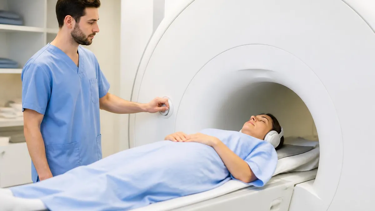
Building a dedicated hippocampus MRI protocol begins with patient positioning and coil selection. The patient lies supine with the head firmly secured inside a 32-channel head coil, which provides the signal-to-noise ratio necessary for sub-millimeter resolution. A pillow under the knees reduces lumbar strain on patients who must stay motionless for nearly 40 minutes. Technologists should explain breathing instructions, demonstrate the squeeze ball, and emphasize that even minor head motion during the high-resolution coronal sequences will blur the hippocampal digitations beyond recognition.
The localizer is acquired in three planes, and from there the technologist plans a sagittal T2 sequence that clearly displays the long axis of the hippocampus from head to tail. This sagittal image becomes the planning reference for every subsequent coronal acquisition. Slices are then prescribed perpendicular to this long axis, producing the classic coronal oblique orientation that displays both hippocampi symmetrically in cross section. Without this oblique angulation, asymmetric volumes and signal abnormalities cannot be reliably compared between sides.
The core sequence package includes coronal T2 fast spin echo, coronal FLAIR, coronal T1 inversion recovery, axial diffusion-weighted imaging, and a volumetric 3D T1 sequence such as MPRAGE or BRAVO with isotropic 1 mm voxels. The coronal T2 is the workhorse for detecting hippocampal sclerosis because it shows increased signal in the sclerotic hippocampus along with volume loss. FLAIR helps suppress cerebrospinal fluid signal and accentuates pathological hyperintensity, while the 3D T1 feeds automated volumetry software like NeuroQuant, Icometrix, or FreeSurfer.
Diffusion-weighted imaging is essential whenever clinical history suggests acute pathology. Herpes simplex encephalitis classically shows restricted diffusion in the medial temporal cortex within the first 48 hours, often before T2 or FLAIR changes are obvious. Similarly, status epilepticus can produce transient diffusion restriction in the hippocampus and pulvinar that mimics infarction. Including DWI on every hippocampus protocol takes only a few minutes and frequently changes management when these acute conditions are unsuspected by the referring clinician.
Contrast administration is reserved for specific indications: suspected encephalitis, mass lesion, vascular malformation, or post-treatment surveillance. Routine memory-loss imaging does not require gadolinium because the differential diagnosis is dominated by atrophic and degenerative processes that do not enhance. When contrast is used, post-gadolinium 3D T1 BRAVO or VIBE sequences in axial and coronal planes provide thin-section coverage of the entire mesial temporal region and adjacent meninges. This is especially helpful in autoimmune limbic encephalitis surveillance.
Field strength matters more for hippocampal imaging than for almost any other brain examination. While 1.5 Tesla scanners can identify gross hippocampal sclerosis, the subtle internal architecture changes, small cavernomas, focal cortical dysplasias, and early atrophy patterns are far better resolved at 3 Tesla. Many epilepsy centers now use 7 Tesla research scanners that resolve individual CA subfields, although these remain investigational. For most US clinical practices, 3T with a high-channel head coil represents the modern standard of care for evaluating mesial temporal structures.
Quality control begins before the patient leaves the scanner. The technologist should review every coronal image for motion, ghosting, and wraparound artifact, then verify that the prescription truly covers the entire hippocampus from head to tail. If any sequence is degraded, repeating it on the spot is far better than reporting a non-diagnostic study. For comparison and historical context on imaging evolution, the history of MRI shows how hippocampal protocols developed alongside advances in coil design and gradient performance.
Hippocampus MRI Volumetry, Atrophy Scoring, and Quantitative Analysis
Manual volumetry remains the reference standard against which all automated methods are validated. A trained reader segments the hippocampus slice by slice on coronal T1 or T2 images, traces the boundary along the alveus superiorly and the parahippocampal white matter inferiorly, and multiplies the summed pixel area by slice thickness. Total volumes are then normalized to total intracranial volume to control for head size differences between patients.
The technique is precise but time-consuming, requiring 20 to 40 minutes per hippocampus and significant anatomical expertise. Inter-rater variability typically runs 3 to 7 percent at experienced centers. Despite its labor cost, manual volumetry remains essential in research, in pre-surgical epilepsy workup at academic centers, and as the ground truth for training and validating automated segmentation algorithms used in everyday clinical practice today.
Dedicated Hippocampus MRI: Strengths and Limitations
- +Detects subtle mesial temporal sclerosis missed on standard brain MRI
- +Provides reproducible quantitative volumes for longitudinal tracking
- +Identifies surgical candidates for temporal lobectomy with high accuracy
- +Supports early Alzheimer diagnosis years before clinical dementia
- +No ionizing radiation, safe for repeated longitudinal studies
- +Reveals incidental findings such as cavernomas and dysplasias
- −Long scan times (25–40 min) can be difficult for restless patients
- −Motion artifact severely degrades high-resolution coronal slices
- −Requires 3T and high-channel coils for optimal subfield detail
- −Volumetry software adds cost and requires integration with PACS
- −Limited utility in patients with severe claustrophobia or metallic implants
- −Subtle bilateral sclerosis can be overlooked without volumetry
Hippocampus MRI Reading Checklist
- ✓Confirm coronal slices are perpendicular to the long axis of the hippocampus
- ✓Compare left and right hippocampal volumes at matched anatomic levels
- ✓Evaluate hippocampal digitations on the head for loss or blurring
- ✓Assess T2 and FLAIR signal for hyperintensity suggesting sclerosis
- ✓Measure the width of the choroid fissure and temporal horn for MTA scoring
- ✓Inspect the fornix and mammillary bodies for atrophy or signal change
- ✓Review DWI for acute restriction suggesting encephalitis or status
- ✓Check for incidental cavernomas, cysts, or focal cortical dysplasia
- ✓Cross-reference automated volumetry with age-matched normative data
- ✓Correlate imaging findings with clinical history and prior studies
The Triad of Mesial Temporal Sclerosis
Three findings together define MRI-positive hippocampal sclerosis: volume loss, increased T2 and FLAIR signal, and disturbed internal architecture with loss of digitations. When all three are present unilaterally and concordant with EEG lateralization, surgical resection produces seizure freedom in roughly 70 to 80 percent of carefully selected patients. Missing any one element should prompt a careful repeat review at 3T before excluding the diagnosis or recommending invasive monitoring.
Mesial temporal sclerosis is the most common pathology identified on dedicated hippocampus MRI and the leading cause of drug-resistant temporal lobe epilepsy. Imaging features include unilateral hippocampal atrophy, increased T2 and FLAIR signal, loss of internal digitations, and ipsilateral dilation of the temporal horn. Secondary findings such as atrophy of the ipsilateral fornix, mammillary body, and entorhinal cortex strengthen confidence in the diagnosis. Bilateral involvement occurs in roughly 10 to 15 percent of cases and complicates surgical planning considerably.
Alzheimer disease produces a characteristic pattern of bilateral, relatively symmetric hippocampal and entorhinal atrophy that progresses over years. Early in the disease, volume loss is greatest in the hippocampal head and the transentorhinal region, mirroring the Braak staging of neurofibrillary tangle pathology. Quantitative volumetry can detect changes years before clinical dementia, supporting earlier diagnosis. With recent approval of anti-amyloid antibody therapies, baseline hippocampal volume and longitudinal change have become important biomarkers for treatment monitoring.
Herpes simplex encephalitis is a neurological emergency where dedicated hippocampus MRI plays a critical role. The classic appearance is asymmetric T2 and FLAIR hyperintensity involving the medial temporal lobe, insula, and inferior frontal lobe, often with cortical restricted diffusion and patchy enhancement. The basal ganglia are characteristically spared. Recognition within hours can be life-saving because empiric acyclovir dramatically reduces mortality and morbidity. Any febrile patient with new memory loss or temporal lobe seizures warrants urgent imaging and lumbar puncture.
Autoimmune limbic encephalitis, often associated with antibodies against LGI1, CASPR2, or NMDA receptors, presents with subacute memory loss, seizures, and psychiatric symptoms. Imaging shows bilateral medial temporal T2 and FLAIR hyperintensity without significant restricted diffusion or hemorrhage. Volume initially appears preserved or even mildly increased due to edema, but progressive atrophy develops over months. Early recognition matters because immunotherapy with steroids, IVIG, or plasmapheresis can produce substantial recovery if started before irreversible neuronal loss occurs.
Transient global amnesia produces tiny punctate foci of restricted diffusion in the lateral hippocampus, usually CA1, that resolve within several days. These dot-like lesions are best detected on high-resolution DWI performed 24 to 72 hours after symptom onset. The clinical syndrome of sudden anterograde amnesia lasting under 24 hours, combined with these characteristic imaging findings, confirms the diagnosis and reassures patients about the benign nature and low recurrence rate of the condition.
Developmental and structural abnormalities seen on hippocampus MRI include incomplete hippocampal inversion, hippocampal malrotation, focal cortical dysplasia of the temporal pole, cavernous malformations, and low-grade tumors such as gangliogliomas and DNETs. These lesions account for a substantial fraction of MRI-negative epilepsy cases when only standard brain protocols are performed. Dedicated thin-section coronal imaging at 3T with surface coil techniques significantly improves detection rates and should be the default for any pharmacoresistant focal epilepsy workup at a comprehensive epilepsy center.
Post-hypoxic and post-status epilepticus injury produce bilateral hippocampal swelling and restricted diffusion acutely, followed by atrophy and gliosis over weeks to months. The hippocampus has high metabolic demand and abundant NMDA receptors, making it exquisitely vulnerable to global ischemia, hypoglycemia, and prolonged seizure activity. Recognizing these patterns helps clinicians counsel families about prognosis after cardiac arrest, near-drowning, severe hypoglycemia, or refractory status epilepticus, since persistent hippocampal injury strongly predicts long-term memory deficits and overall functional recovery.
Any patient with fever, altered mental status, and new memory or behavior changes requires urgent MRI and lumbar puncture. Empiric IV acyclovir should be started immediately without waiting for confirmation, because each hour of delay increases mortality. Restricted diffusion in the medial temporal lobe within 48 hours of symptom onset is the most sensitive early imaging finding and must trigger immediate clinical action.
Reporting a hippocampus MRI follows a structured template that ensures completeness and consistency. Begin with a clear statement of the indication and protocol, including field strength, sequences performed, and whether contrast was used. Describe the hippocampi by region — head, body, and tail — addressing volume, signal, and internal architecture on each side. Compare with prior studies whenever available, since longitudinal change is often more meaningful than any single measurement. Conclude with quantitative volumetry results when performed and a clear impression linking findings to the clinical question.
Volumetric results should always be reported with reference to age- and sex-matched normative data. A hippocampal volume in the 5th percentile carries different weight in an 80-year-old than in a 55-year-old. Most automated tools include color-coded percentile charts that translate raw volumes into clinically actionable categories. Asymmetry indices greater than 8 to 10 percent are considered significant and should prompt detailed visual review for sclerosis, even when individual volumes fall within the normal range for both hippocampi compared to controls.
For epilepsy patients, the report must address several specific questions: Is there imaging evidence of mesial temporal sclerosis? Is it unilateral or bilateral? Are there secondary findings such as fornix atrophy? Are there dual pathologies such as cortical dysplasia, cavernoma, or tumor? Concordance between imaging, EEG, and semiology determines surgical candidacy, so the radiologist's clear localization directly impacts whether a patient becomes seizure-free or requires invasive intracranial monitoring before a resection can safely proceed.
For dementia workup, the report should include an MTA score for each hippocampus, comments on entorhinal cortex and parahippocampal gyrus thickness, and a Koedam score for parietal atrophy. The pattern of atrophy helps differentiate Alzheimer disease from frontotemporal dementia, Lewy body dementia, and normal aging. Vascular contributions should be quantified using the Fazekas scale for white matter hyperintensities and a count of lacunes and microbleeds, since mixed pathology is common in older adults and significantly affects prognosis and treatment.
Communication with referring clinicians is essential, particularly for time-critical findings. Restricted diffusion suggesting encephalitis, new hemorrhage, or a mass lesion warrants immediate phone or secure-message notification, not just a routine report. Many institutions now use automated critical-results notification systems that document the communication and acknowledgment. A well-written report that is never read in time helps no one, so building reliable communication pathways is as important as the interpretation itself for high-quality patient care delivery.
Patient-facing communication deserves attention too. Patients receiving hippocampal MRI reports are often anxious about memory loss and may search online for terms they do not understand. Plain-language summaries, when offered, dramatically reduce anxiety and improve compliance with follow-up. Many practices now include a one-paragraph patient-friendly summary that explains the key findings in non-technical language, what they mean, and what next steps the clinical team recommends based on the imaging and overall clinical picture being managed.
Finally, remember that imaging is one component of a comprehensive evaluation. Hippocampus MRI findings must always be integrated with neuropsychological testing, EEG, cerebrospinal fluid biomarkers, and clinical assessment. A normal MRI does not exclude epilepsy or early Alzheimer disease, and conversely, age-related hippocampal atrophy does not by itself diagnose dementia. For patients curious about how MRI compares to other modalities in their workup, the MRI alternatives guide explains when CT, PET, or ultrasound add value beyond what MRI alone can provide.
If you are a technologist preparing to perform your first dedicated hippocampus MRI, practical preparation makes the difference between a brilliant study and a non-diagnostic one. Spend extra time with the patient before the scan, explaining the length and the importance of holding still. Offer earplugs and headphones, demonstrate the squeeze ball, and reassure them that the table will not move during most of the exam. Patients with claustrophobia or significant anxiety may benefit from premedication, and you should coordinate with the ordering provider well in advance whenever possible.
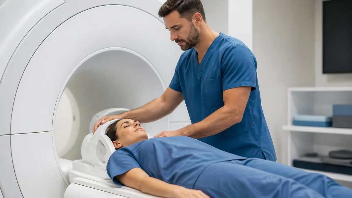
Coil positioning deserves careful attention. The head should sit deep in the coil so that the temporal lobes are positioned at the center of the magnetic field, where homogeneity and signal are highest. Tape and foam pads keep the head from rotating during the scan, while a small pillow under the knees reduces lower back strain. Verify that the patient is comfortable before starting, because shifting position even once during a 7-minute coronal acquisition will ruin the sequence and require a complete repeat that adds significant time.
Planning the coronal oblique slices is the single most important technical step. Display the sagittal T2 image showing the long axis of the hippocampus, then prescribe coronal slices perpendicular to that axis. Coverage must extend from the temporal pole anteriorly to the splenium posteriorly to capture the entire hippocampal length. Slice thickness of 2 mm with no gap is standard, and the field of view should be tightened to the temporal lobes to maximize in-plane resolution without aliasing into the rest of the head.
Sequence parameters at 3T typically use a TR of 4000 to 6000 ms and a TE of 90 to 110 ms for the coronal T2, with a matrix of 384 by 384 or higher. FLAIR uses similar TR but TE around 120 ms and an inversion time around 2200 to 2500 ms. The 3D T1 MPRAGE volume uses 1 mm isotropic voxels with TR around 2000 ms and TE near 3 ms. Always check vendor-specific recommendations because optimal parameters vary between Siemens, GE, Philips, and Canon platforms.
Motion management is the technologist's continuous job. Watch the patient on the in-room camera and pause between sequences to check in. If you see motion on the just-acquired series, repeat it before moving on rather than after the patient leaves. Modern scanners offer prospective motion correction such as PROMO on GE or scout-based correction on Siemens, and using these tools routinely on hippocampal protocols substantially improves diagnostic quality. The few extra minutes are far cheaper than calling the patient back the next week.
For studying purposes, hippocampus MRI represents one of the highest-yield topics on advanced certification exams. ARRT MRI registry questions frequently address coronal oblique planning, mesial temporal sclerosis findings, and protocol design. Boards examinations for radiology trainees include detailed anatomy of the hippocampal subfields and clinical correlations with epilepsy and dementia. Building a mental library of normal and abnormal coronal T2 appearances pays dividends not only on tests but in every neuroimaging shift you will work throughout your career as a practicing imaging professional.
Lastly, never underestimate the value of repetition. Reviewing twenty normal hippocampus MRIs gives you a visual baseline that makes subtle pathology jump out. Pair this with structured reading of one or two cases per week from teaching files such as Radiopaedia, the ASNR case of the week, or your institution's own archive. Within six months of focused study, most trainees develop confident pattern recognition for hippocampal sclerosis, Alzheimer-pattern atrophy, encephalitis, and the common developmental abnormalities that define mesial temporal imaging.
MRI Questions and Answers
About the Author
Medical Laboratory Scientist & Clinical Certification Expert
Johns Hopkins UniversityDr. Sandra Kim holds a PhD in Clinical Laboratory Science from Johns Hopkins University and is certified as a Medical Technologist (MT) and Medical Laboratory Scientist (MLS) through ASCP. With 16 years of clinical laboratory experience spanning hematology, microbiology, and molecular diagnostics, she prepares candidates for ASCP board exams, MLT, MLS, and specialist certification tests.