Hip MRI Images: A Complete Guide to Reading, Understanding, and Interpreting Hip Scans
Learn to read hip effusion MRI images, interpret labral tears, AVN, and bursitis on T1, T2, and PD sequences with clinical examples.
A hip effusion MRI is one of the most informative musculoskeletal exams a radiologist or technologist can review, because the joint capsule, labrum, articular cartilage, marrow, and surrounding soft tissues all appear within a single field of view. When fluid accumulates inside the joint capsule, fluid-sensitive sequences reveal high signal that wraps around the femoral head and neck, immediately raising suspicion for synovitis, occult fracture, septic arthritis, or transient osteoporosis. Knowing how each of these conditions appears on hip MRI images allows clinicians to make confident decisions about aspiration, surgery, or watchful waiting.
Reading a hip MRI begins with orientation. The standard protocol acquires coronal, axial, and sagittal planes with at least one T1-weighted sequence and one fluid-sensitive sequence such as fat-saturated T2 or proton density. Coronal images give the cleanest view of the labrum, articular cartilage, and the relationship between the femoral head and the acetabulum, while axial images highlight the anterior and posterior capsule, the iliopsoas tendon, and the hamstring origin at the ischial tuberosity.
Sagittal images, although often overlooked, are critical for evaluating anterior labral pathology and the rectus femoris origin. Most academic centers add a coronal STIR or PD fat-sat sequence to suppress marrow fat and emphasize edema, which is particularly useful for detecting stress fractures, transient bone marrow edema, and early avascular necrosis. A 3 Tesla magnet improves cartilage detail considerably, but a well-tuned 1.5 Tesla scan with surface coil placement remains diagnostic for most clinical questions.
The femoral head and acetabulum form a deep ball-and-socket joint stabilized by the fibrocartilaginous labrum, a structure that mirrors the meniscus in function but is much more vulnerable to tears at its anterosuperior margin. On normal MRI, the labrum appears as a uniformly dark triangular structure between the femoral head and the bony acetabulum. Any linear high signal extending to the labral surface should prompt suspicion for a tear, especially in patients reporting groin pain, clicking, or limited internal rotation.
Joint fluid is normally minimal, with a thin rim no thicker than four millimeters in the dependent recess. When fluid distends the capsule, the volume often exceeds ten milliliters and may extend along the iliopsoas bursa, mimicking an isolated bursitis. Distinguishing simple effusion from complex septic fluid requires assessing signal heterogeneity, synovial enhancement on post-contrast images, and surrounding soft tissue edema. Septic effusions typically show thick, irregular synovial enhancement and may communicate with adjacent soft tissue abscesses requiring urgent drainage.
Comparing imaging modalities is part of every clinical workup. Some hip questions, such as cortical fracture mapping or hardware position, are still better answered with CT, which is why understanding MRI alternatives remains essential for ordering physicians. MRI excels at marrow, cartilage, and labral evaluation, while CT remains superior for cortical bone and metallic implants. Choosing the right modality based on clinical question and contraindications optimizes diagnostic accuracy and reduces unnecessary follow-up imaging studies.
This guide walks through hip anatomy as it appears on MRI, the specific signal patterns of effusion and the most common hip pathologies, the protocols used in clinical practice, and the practical tips that help students and new technologists feel confident in front of a hip exam. Whether you are preparing for boards, training in MR, or simply curious about what your radiologist sees, the goal is to make hip imaging readable, predictable, and clinically useful for every patient encounter.
Hip MRI Imaging by the Numbers
Hip MRI Protocol and Sequences
Coronal T1 highlights marrow and fat planes for evaluating AVN and stress reactions, while coronal PD with fat saturation reveals labral tears, cartilage defects, and joint fluid distribution clearly.
Axial fat-suppressed T2 shows iliopsoas bursitis, anterior and posterior capsule distension, hamstring tendinopathy, and surrounding soft tissue edema that points to inflammation or infection.
Sagittal imaging best demonstrates anterior labral pathology, rectus femoris origin tears, and the trochanteric bursa, often missed on coronal and axial planes alone in athletes.
Direct injection of dilute gadolinium into the joint capsule distends the labrum, increasing sensitivity for subtle tears, paralabral cysts, and chondral delamination near the acetabular rim.
Intravenous gadolinium with subtracted post-contrast sequences highlights synovial enhancement in septic arthritis, pigmented villonodular synovitis, and inflammatory arthropathies for treatment planning.
Recognizing normal hip anatomy on MRI is the foundation that makes pathology obvious. On a well-positioned coronal image, the femoral head sits perfectly centered within the acetabular cup, with a uniformly thin layer of articular cartilage visible as intermediate signal on proton density sequences. The fovea capitis, where the ligamentum teres attaches, appears as a small notch in the medial femoral head and should not be mistaken for a focal cartilage defect or an osteochondral lesion in patients with hip pain.
The labrum is the next structure to scrutinize. In a healthy young adult, it forms a sharp, uniformly dark triangle hugging the acetabular rim, most reliably evaluated on radial reformats or thin coronal slices. The anterosuperior quadrant is where ninety percent of tears occur, partly because of repetitive impingement against the femoral neck and partly because of relative hypovascularity. A thin rim of intermediate signal at the chondrolabral junction is normal and should not be over-called as a tear by inexperienced readers.
Femoral neck morphology deserves careful attention because cam-type femoroacetabular impingement directly causes labral tears and chondral damage over time. Measure the alpha angle on an oblique axial image through the center of the femoral neck. Values above 55 degrees suggest cam deformity, while a deep acetabulum with a center-edge angle above 40 degrees suggests pincer-type impingement. Both morphologies frequently coexist and produce the classic anterosuperior labral tear with adjacent chondral delamination.
The marrow signal of the proximal femur changes predictably with age. In children and adolescents, hematopoietic red marrow appears intermediate to low on T1 and may persist into the femoral neck and intertrochanteric region in adults, particularly in smokers, athletes, and patients with anemia. Pathologic marrow replacement appears darker than adjacent skeletal muscle on T1, a useful rule that distinguishes benign red marrow from metastatic disease, lymphoma, or chronic infection without needing additional sequences in most cases.
The iliopsoas tendon courses anterior to the joint capsule and inserts on the lesser trochanter. An iliopsoas bursa sits between the tendon and the anterior capsule and may communicate with the joint in up to fifteen percent of adults, allowing joint fluid to track into the bursa and present as a pelvic cystic mass. The hamstring origin at the ischial tuberosity, the abductor insertions on the greater trochanter, and the rectus femoris origin on the anterior inferior iliac spine are common sites of tendinopathy and avulsion injuries.
Surrounding soft tissues should be reviewed systematically. Look at the gluteus medius and minimus tendons for tendinosis or tears, the trochanteric bursa for fluid, the sciatic nerve for piriformis syndrome, and the inguinal region for sports hernia signs in athletes. Comparison with the contralateral side is invaluable on coronal images, because subtle asymmetry in muscle bulk, fat infiltration, or fluid distribution often points to chronic disease that would otherwise be overlooked in isolation by even experienced readers.
Understanding the language of MRI reporting also helps. Knowing what every MRI medical abbreviation means, from FS to STIR to PD, helps technologists communicate with radiologists and helps students grasp protocols faster. When you can name the sequence, predict its appearance, and connect it to a specific clinical question, hip imaging stops feeling like memorization and starts feeling like applied anatomy with consistent, predictable, learnable rules across patients.
Hip Effusion MRI Signal Characteristics
Simple joint effusion appears as homogeneous high signal on T2 and PD fat-saturated images, with corresponding low signal on T1. Fluid distends the anterior and posterior capsular recesses uniformly, and the femoral head appears to float within a halo of bright signal on coronal sequences. There should be no internal debris, no septations, and no synovial thickening.
Common causes include transient synovitis in children, early osteoarthritis, and post-traumatic hemarthrosis that has resorbed. Volume estimation matters because effusions greater than fifteen milliliters often indicate active intraarticular pathology. Aspiration may be considered when clinical signs suggest infection, but uncomplicated reactive effusions usually resolve with rest, anti-inflammatory medication, and treatment of the underlying cause within several weeks of conservative management overall.
MRI vs Other Imaging for Hip Pathology
- +Superior visualization of labral tears, cartilage, and bone marrow edema not seen on radiographs
- +No ionizing radiation, making it safe for repeated imaging in young patients and athletes
- +Multiplanar imaging in coronal, axial, and sagittal planes without repositioning the patient
- +Detects early avascular necrosis weeks before changes appear on X-ray or CT
- +Evaluates surrounding soft tissues, tendons, and bursae in a single examination
- +MR arthrography improves sensitivity to over 90% for subtle labral and chondral lesions
- +Differentiates septic from sterile effusion with synovial enhancement patterns
- −Long scan times of 30-45 minutes can be difficult for patients in significant pain
- −Claustrophobia and noise limit tolerance in some patients without sedation
- −Metallic hardware, pacemakers, and certain implants may be contraindicated
- −Higher cost compared to ultrasound or radiographs for initial screening evaluations
- −Limited availability in rural areas with longer wait times for non-urgent indications
- −Cortical bone detail is inferior to CT for complex fracture characterization
- −Patient body habitus and motion artifacts can degrade image quality significantly
Hip MRI Reading Checklist
- ✓Verify patient positioning, coil placement, and field of view covers both hips for comparison
- ✓Check for joint effusion volume and signal characteristics on fluid-sensitive sequences
- ✓Evaluate the labrum systematically from anterior to posterior for tears or paralabral cysts
- ✓Assess articular cartilage thickness and signal on PD fat-saturated coronal images
- ✓Review femoral head and neck marrow for edema, AVN, or occult fracture lines on T1
- ✓Measure alpha angle and center-edge angle to identify femoroacetabular impingement morphology
- ✓Inspect iliopsoas, gluteus medius, and hamstring tendons for tendinosis or partial tears
- ✓Look for bursal fluid in iliopsoas, trochanteric, and ischiogluteal locations
- ✓Compare contralateral hip for subtle asymmetry in muscle bulk or signal intensity
- ✓Correlate imaging findings with patient symptoms, age, activity level, and clinical history
Always check T1 marrow signal first
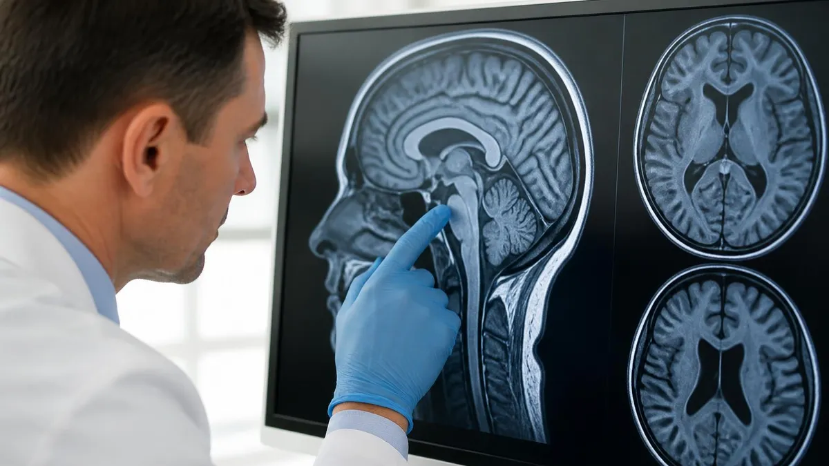
The single most important sequence on a hip MRI is the coronal T1. Normal marrow should be brighter than skeletal muscle. If any portion of the femoral head or neck appears darker than muscle, suspect AVN, infection, fracture, or marrow replacing process and investigate further with fluid-sensitive and post-contrast sequences before concluding the study is normal.
Several hip pathologies dominate clinical practice and every one of them has a characteristic MRI signature worth memorizing. Avascular necrosis of the femoral head, also known as osteonecrosis, classically presents with the double-line sign on T2 imaging, where a bright inner line of granulation tissue parallels a dark outer line of reactive sclerosis surrounding a necrotic segment. Early changes appear only as subtle marrow edema on STIR, which is why MRI is far more sensitive than radiographs for detecting AVN within the first weeks of symptoms.
Femoroacetabular impingement is now recognized as a leading cause of early hip osteoarthritis in young adults. Cam impingement produces an aspherical femoral head-neck junction that grinds against the anterosuperior acetabulum during flexion, tearing the labrum and delaminating the adjacent cartilage. Pincer impingement, where the acetabulum is overcoverage, crushes the labrum directly against the femoral neck. Both patterns produce the characteristic anterosuperior labral tear with a paralabral cyst extending into the surrounding soft tissues.
Stress fractures of the femoral neck are emergencies because completion of a tension-side fracture can require total hip replacement. On MRI, look for a linear low signal line surrounded by extensive marrow edema on the superior aspect of the femoral neck. Compression-side stress fractures on the inferior cortex are less urgent but still warrant protected weight bearing. Athletes, military recruits, and patients with metabolic bone disease are at highest risk and benefit from early MRI when clinical suspicion is elevated.
Transient bone marrow edema syndrome presents with sudden hip pain and dramatic marrow edema throughout the femoral head and neck without any discrete fracture line or necrotic segment. It typically affects middle-aged men and pregnant women and resolves spontaneously over six to nine months. The challenge is distinguishing it from early AVN, which has a focal subchondral abnormality, and from occult fracture, which shows a discrete low-signal fracture line on T1 sequences with appropriate clinical history.
Greater trochanteric pain syndrome encompasses gluteus medius and minimus tendinopathy, partial-thickness tears, and trochanteric bursitis. MRI shows tendon signal abnormality, fluid in the trochanteric bursa, and sometimes fatty atrophy of the abductor muscles in chronic cases. Distinguishing reparable partial tears from full-thickness retracted tears guides decisions between physical therapy, corticosteroid injection, platelet-rich plasma therapy, and surgical repair for these increasingly recognized causes of lateral hip pain in active middle-aged adults.
Hip osteoarthritis on MRI shows joint space narrowing, subchondral cysts, marrow edema in load-bearing regions, osteophytes, and reactive synovitis with joint effusion. Cartilage loss begins at the superior weight-bearing surface and progresses circumferentially. MRI cartilage mapping with T2 or T1 rho techniques can detect early biochemical changes before morphologic loss, useful for research and clinical trials of cartilage-preserving interventions in young patients with early osteoarthritis from prior trauma or developmental conditions.
Less common but important diagnoses include pigmented villonodular synovitis with characteristic low-signal hemosiderin deposits on all sequences, synovial chondromatosis with multiple intraarticular bodies, septic arthritis as already discussed, and metastatic disease to the proximal femur. Knowing the typical age, location, and signal pattern of each diagnosis transforms a long differential into a focused, evidence-based interpretation that directly improves patient outcomes through appropriate, timely referral and treatment decisions in real clinical practice.
In any elderly patient presenting with hip pain after a fall and negative radiographs, MRI is the gold standard for ruling out occult femoral neck fracture. Missing this diagnosis can lead to displacement, avascular necrosis, and the need for hip replacement instead of simple pin fixation. Always include a coronal T1 sequence and look carefully for any low-signal line traversing the femoral neck trabeculae.
Practical clinical tips separate competent hip MRI readers from confident ones. Always begin with the clinical history, because the same finding has different significance in a 25-year-old runner, a 55-year-old construction worker, and an 85-year-old fall victim. Read the request thoughtfully, look at any prior imaging including radiographs and CT, and form a focused differential before opening the MRI. This top-down approach prevents premature closure on the first abnormality and ensures comprehensive evaluation of all relevant anatomy in every case.
Window and level adjustments matter more than students realize. Cartilage assessment requires narrow windowing on PD fat-saturated images to differentiate normal from delaminated cartilage. Marrow evaluation requires wider T1 windowing to compare femoral head signal against muscle reliably. Bone edema on STIR is best appreciated with mid-range windowing. Spending a few seconds optimizing display before interpretation pays off in diagnostic accuracy and reduces the chance of overcalling or missing subtle abnormalities consistently across challenging cases.
Compare every finding to the contralateral side. Hip MRI typically includes both joints in the coronal plane, providing an internal control that highlights subtle asymmetries in marrow signal, cartilage thickness, muscle bulk, or fluid distribution. A finding that looks borderline in isolation often becomes definitively abnormal when compared with the opposite side. This rule is especially valuable in evaluating early AVN, stress reactions, transient osteoporosis, and gluteus tendinopathy, where bilateral comparison frequently reveals important asymmetries.
Be cautious about magic-angle artifact in tendons. The gluteus medius and minimus tendons, the iliopsoas tendon, and the hamstring origin can show artificial high signal on short echo-time sequences when oriented at 55 degrees to the main magnetic field. Always confirm any tendon abnormality on long echo-time T2 or fluid-sensitive sequences before reporting a tear. This single pitfall accounts for many false-positive tendon diagnoses in hip MRI interpretation by trainees and experienced radiologists alike.
Recognize normal variants that mimic pathology. The sublabral sulcus is a normal cleft between the posteroinferior labrum and the acetabular cartilage and should not be called a tear. The pulvinar fat pad in the cotyloid fossa is a normal structure that can simulate a mass on coronal images. Os acetabuli, accessory bones around the acetabular rim, can simulate fractures. Knowing these variants prevents unnecessary follow-up imaging, anxiety, and inappropriate referrals to orthopedic specialists for surgical consultation.
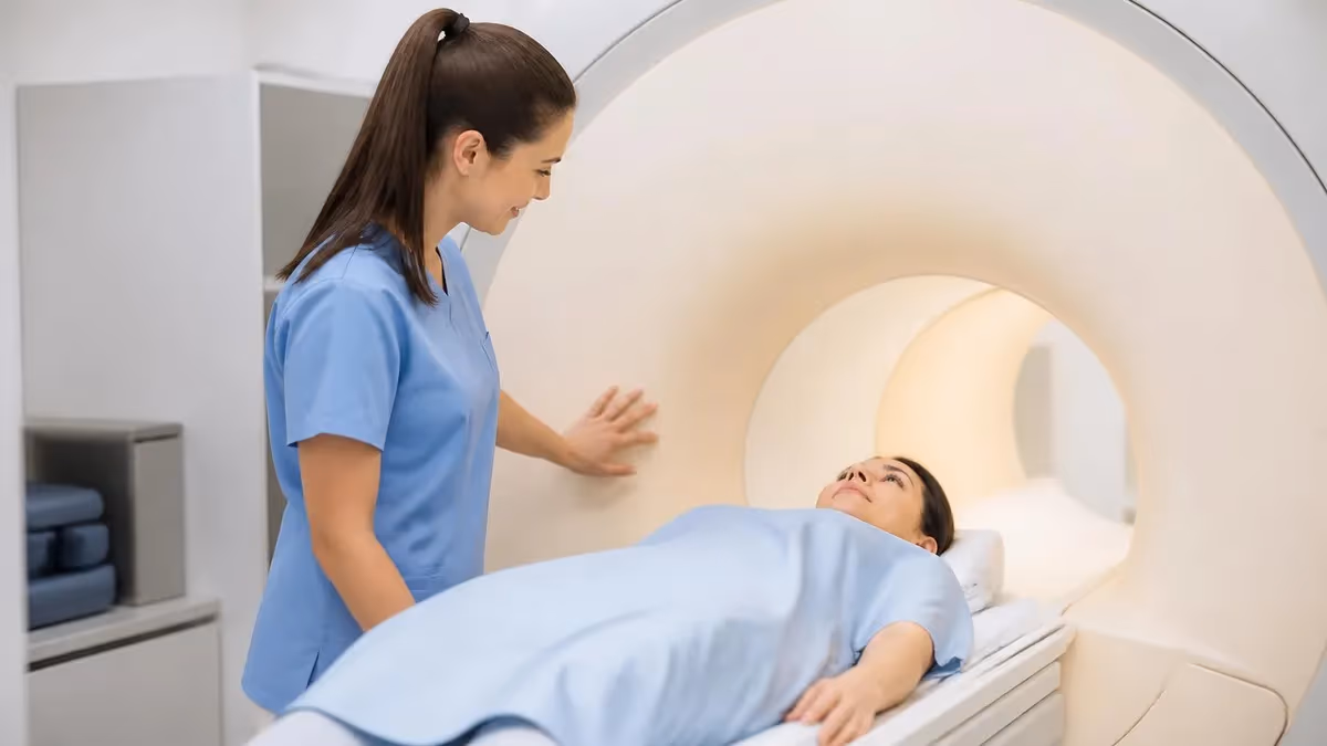
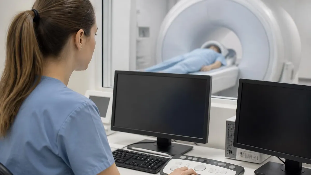
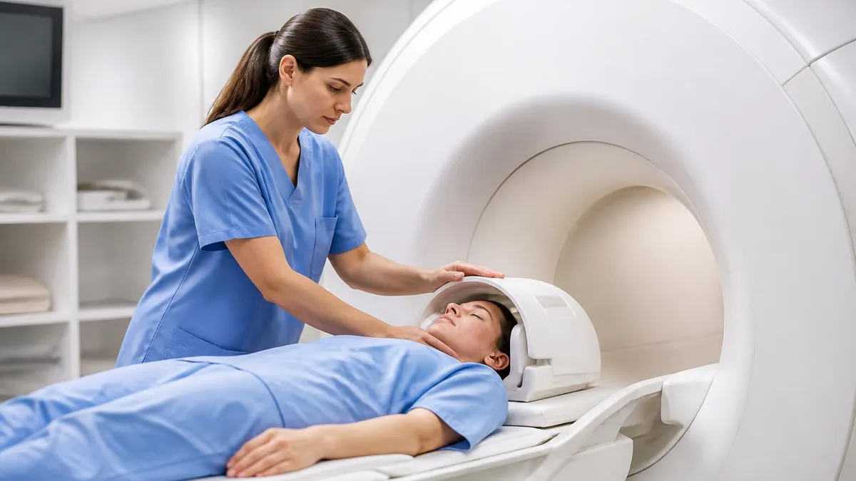
Patient experience matters in MRI. The hip exam takes 30-45 minutes, requires the patient to lie still while the magnet produces loud knocking and clicking sounds. Understanding the noise of an MRI machine and explaining it to patients in advance reduces anxiety and motion artifacts, which directly improves image quality. Offering earplugs, headphones, music, and a clear understanding of scan duration transforms patient cooperation and yields more diagnostic studies on the first attempt without repeats.
Finally, write structured reports that answer the clinical question directly. Lead with the relevant finding, describe its location, signal characteristics, and severity, and offer a focused differential when appropriate. Compare to prior studies when available. Recommend specific next steps such as aspiration, orthopedic referral, or follow-up imaging timeline. Clinicians read your report under time pressure, so clarity and actionability transform a competent interpretation into a clinically valuable consultation that drives appropriate, timely patient care decisions.
Becoming proficient at hip MRI interpretation is a marathon, not a sprint. Most radiology residents and MRI technologists need at least 100 supervised reads before they feel comfortable with the full range of hip pathology. The fastest learners use a structured approach: review the protocol, read in a consistent order, dictate using templates, and immediately look up any finding they cannot name with confidence. Building this habit early prevents bad shortcuts from calcifying into permanent gaps in your knowledge base over time.
Use teaching files relentlessly. Cases from radiopaedia.org, the Society of Skeletal Radiology, the Radiological Society of North America, and your institutional archive provide an endless supply of confirmed diagnoses with explanatory captions. Focus on classic appearances first, then progressively work through atypical presentations and mimics. Quizzing yourself on findings before reading the discussion forces active recall, the single most powerful learning technique supported by cognitive science research and successful radiology training programs worldwide for decades now.
Build a personal differential generator for the most common clinical scenarios. Acute hip pain in a child has a different differential than acute hip pain in an elderly woman, which differs again from chronic groin pain in a young athlete. Memorize the three to five most likely diagnoses for each scenario, then refine your differential based on imaging findings. This pattern recognition approach mirrors how experienced radiologists actually think and dramatically accelerates accurate diagnosis in clinical practice every single day.
Cross-train with related modalities. Reviewing CT and ultrasound for the same patient provides invaluable correlation that strengthens MRI interpretation. Understand what each modality can and cannot show, when MR arthrography adds value over conventional MRI, and when nuclear medicine bone scan or PET-CT is needed. Multimodality literacy makes you a far more valuable consultant to orthopedic surgeons and primary care physicians who order your studies and depend on integrated interpretations to plan patient management.
Practice dictating concise, clinically focused reports. Avoid the temptation to list every minor finding without context. Instead, organize your report by relevance to the clinical question, then anatomic region. Address the labrum, cartilage, marrow, soft tissues, and joint fluid in a consistent order. Save the impression for actionable conclusions: confirm or refute the suspected diagnosis, suggest next steps, and offer differential diagnoses when appropriate. Clinicians remember and trust readers who report clearly under time pressure consistently.
Stay current with evolving techniques. Three-dimensional isotropic sequences allow reformats in any plane from a single acquisition, reducing scan time and improving labral evaluation. T2 mapping and dGEMRIC provide quantitative cartilage assessment. Synthetic MRI generates multiple contrasts from a single sequence. Knowing what is on the horizon helps you adapt as protocols evolve in your department and gives you talking points when discussing scan optimization with MRI technologists and physicists during quality improvement initiatives every quarter.
Finally, never stop reading the literature. Subscribe to AJR, Radiology, Skeletal Radiology, and the European Journal of Radiology to stay current on emerging diagnoses, imaging biomarkers, and management changes. Attend MSK conferences whenever possible, and participate in tumor boards or hip preservation conferences if your institution offers them. Lifelong learning is the hallmark of every excellent radiologist and technologist, and few subspecialties evolve as rapidly or rewardingly as musculoskeletal MRI of the hip and pelvis.
MRI Questions and Answers
About the Author
Medical Laboratory Scientist & Clinical Certification Expert
Johns Hopkins UniversityDr. Sandra Kim holds a PhD in Clinical Laboratory Science from Johns Hopkins University and is certified as a Medical Technologist (MT) and Medical Laboratory Scientist (MLS) through ASCP. With 16 years of clinical laboratory experience spanning hematology, microbiology, and molecular diagnostics, she prepares candidates for ASCP board exams, MLT, MLS, and specialist certification tests.