
Brain CT vs MRI: Complete Guide to Choosing the Right Brain Scan 2026 September
Brain CT vs MRI explained: speed, cost, radiation, and when doctors choose each scan. ✅ Clear comparison for patients and students.
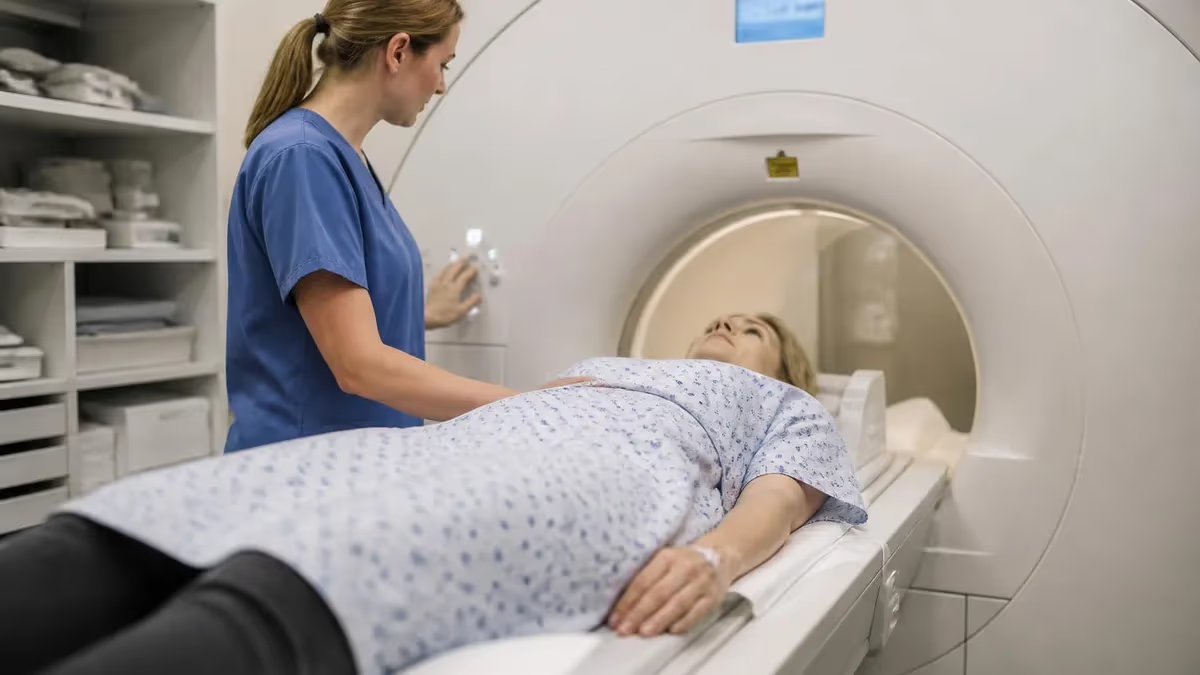
When a physician orders a brain imaging study, the choice between brain CT vs MRI is one of the most consequential decisions in diagnostic medicine. Computed tomography and magnetic resonance imaging both produce detailed pictures of the brain's internal structures, but they rely on completely different physical principles, excel in different clinical situations, and carry different risk profiles for patients. Understanding these differences helps patients ask informed questions and helps radiology students master a concept that appears frequently on registry examinations.
CT scanning uses ionizing X-ray radiation rotated around the patient to produce cross-sectional images in a matter of seconds. The speed advantage makes CT the default choice in emergency departments across the United States when a physician suspects acute stroke, traumatic brain injury, or intracranial hemorrhage. A non-contrast head CT can be completed and interpreted in under ten minutes, which can be the difference between life and permanent disability when brain tissue is actively dying.
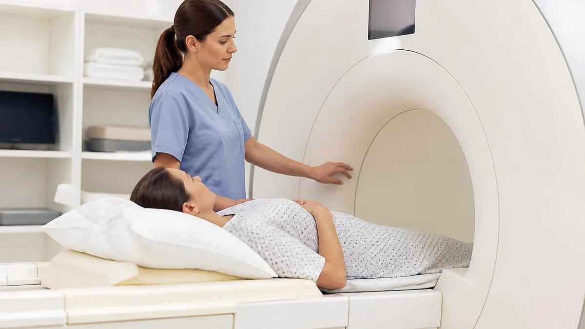
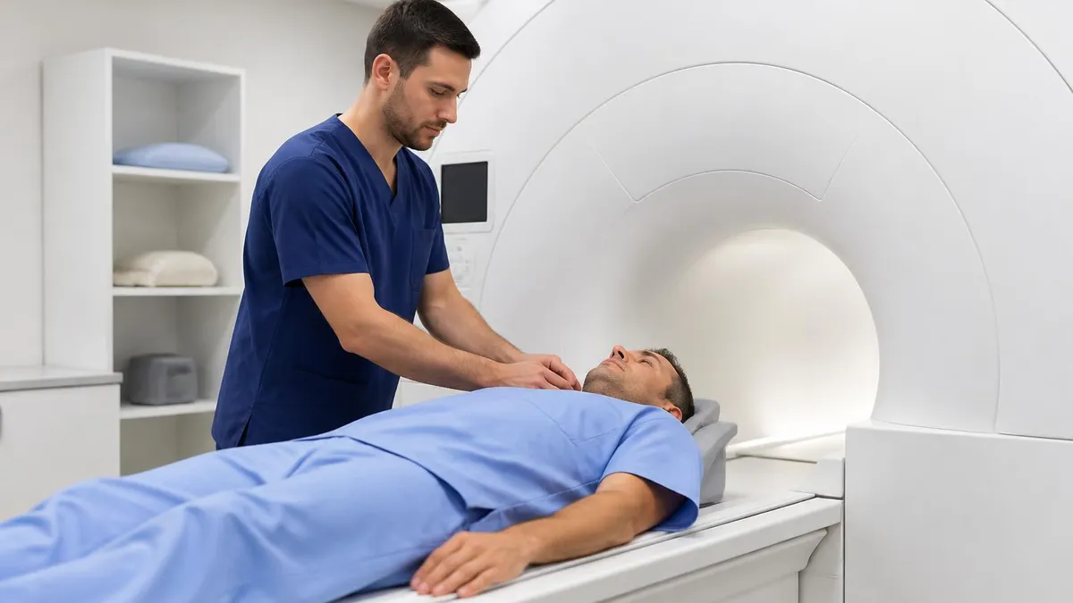
MRI, by contrast, uses powerful magnetic fields and radiofrequency pulses to excite hydrogen protons in tissue water, generating images with superior soft-tissue contrast. A standard brain MRI protocol takes 30 to 60 minutes, making it impractical for unstable emergency patients but ideal for evaluating tumors, multiple sclerosis plaques, subtle cortical abnormalities, and posterior fossa pathology that CT often misses entirely due to beam-hardening artifact from the surrounding skull bone.
The concept of brain ct vs mri extends beyond simple speed vs. quality tradeoffs. Each modality has unique contraindications, preparation requirements, and insurance coverage considerations that affect real-world utilization. Patients with cardiac pacemakers, cochlear implants, or certain aneurysm clips may be excluded from MRI but can safely undergo CT. Conversely, pregnant patients and children who require repeated imaging benefit significantly from MRI's lack of ionizing radiation.
Cost is another decisive factor in clinical and patient decision-making. A head CT without contrast typically costs between $200 and $900 in the United States depending on facility type and geographic region. A brain MRI without contrast ranges from $400 to $3,500 at the same facilities. Insurance coverage varies, but most major carriers will approve the modality a physician deems medically necessary. For uninsured patients or those with high deductibles, these cost differences are highly significant.
From an educational standpoint, radiology technologists and medical imaging students must understand both modalities deeply. Registry examinations test not only technical parameters such as magnetic field strength, slice thickness, and window settings but also clinical indications, patient preparation protocols, and safety screening procedures. The distinction between CT and MRI brain imaging is foundational content for the ARRT MRI registry examination and frequently appears in multiple-choice question sets across every major practice test platform.
This article provides a comprehensive, clinically grounded comparison of brain CT and MRI. You will learn how each technology works, when physicians choose one over the other, what patients experience during each procedure, how radiologists interpret the images, and what emerging technologies are changing the landscape of brain imaging in 2026. Whether you are a patient preparing for a scan or a student preparing for board examinations, this guide will give you the depth and clarity you need.
Brain CT vs MRI by the Numbers
Key Differences Between Brain CT and MRI
CT uses ionizing X-ray radiation and reconstructs images via computer algorithms. MRI uses a strong magnetic field plus radiofrequency pulses to detect hydrogen proton signals in tissue, producing images with no radiation exposure whatsoever.
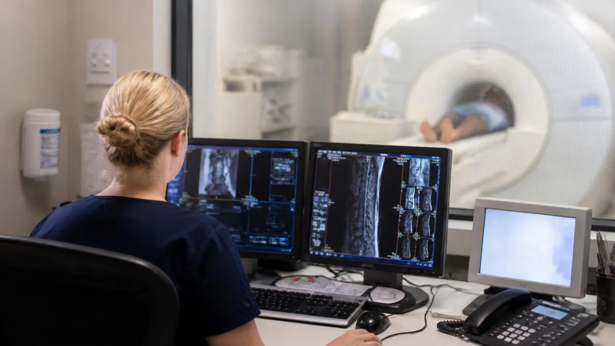
CT scanners are faster (2–5 minutes per study), more widely available in community hospitals and urgent care settings, and are operational 24/7 in most emergency departments. MRI requires longer scan times and specialized facilities with controlled magnetic environments.
MRI provides dramatically superior soft-tissue contrast, making it the gold standard for brain tumors, demyelinating diseases, and posterior fossa pathology. CT excels at detecting acute blood, calcifications, and bony skull fractures with high spatial resolution.
CT exposes patients to ionizing radiation — a concern for children and pregnant patients. MRI carries no radiation risk but is contraindicated for patients with ferromagnetic implants, certain pacemakers, and some cochlear devices. Claustrophobia is a practical barrier for some MRI patients.
Head CT is significantly less expensive ($200–$900) than brain MRI ($400–$3,500). Both are generally covered by Medicare, Medicaid, and private insurance when medically indicated, but prior authorization requirements for MRI are more common than for CT.
To truly understand how to choose between brain CT and MRI, it helps to understand the underlying physics of each modality. CT scanning, introduced clinically in the 1970s, works by rotating an X-ray tube and detector array around the patient's head. The detector measures how much radiation is attenuated by different tissue types — dense bone absorbs more X-rays than soft brain tissue, which absorbs more than air-filled sinuses. A computer algorithm called filtered back projection reconstructs these attenuation values into a cross-sectional image, with each pixel assigned a Hounsfield unit value.
On a CT image, fresh blood appears bright white (hyperdense) because hemoglobin has high X-ray attenuation. This is why CT is the first-line test for suspected intracranial hemorrhage — a subarachnoid bleed shows up as brilliant white contrast against the gray brain parenchyma, and radiologists can identify it within seconds of viewing the images. Bone appears even brighter, while cerebrospinal fluid and edema appear darker than normal brain tissue. CT's sensitivity for acute hemorrhage within the first 24 hours exceeds 98 percent, making it essentially the definitive test in this time window.
MRI physics is considerably more complex. In simple terms, the scanner's powerful magnet aligns hydrogen protons in the patient's body water along the magnetic field direction. A radiofrequency pulse then tips these protons out of alignment. As the protons relax back to their equilibrium state, they emit a radiofrequency signal that the scanner detects via receiver coils placed around the patient's head. The timing of when this signal is measured — controlled by the pulse sequence parameters TR (repetition time) and TE (echo time) — determines whether the image emphasizes T1 relaxation, T2 relaxation, or other tissue properties.
T1-weighted images make fat and subacute blood appear bright, making them ideal for anatomy and contrast-enhancement studies. T2-weighted images make water appear bright, which is valuable for detecting edema, tumors, and demyelinating lesions that have increased water content compared to normal white matter.
FLAIR (fluid-attenuated inversion recovery) sequences suppress the bright CSF signal while preserving the bright signal of periventricular lesions, making MS plaques far more conspicuous. DWI (diffusion-weighted imaging) detects the restricted movement of water molecules inside acutely infarcted brain cells, making it the most sensitive sequence for early ischemic stroke detection — often showing abnormality within minutes of symptom onset.
The posterior fossa — the region of the brain containing the cerebellum, brainstem, and cranial nerve nuclei — is notoriously difficult to evaluate with CT due to beam-hardening artifact from the thick surrounding skull bone. This artifact creates streaking and obscures subtle pathology. MRI is completely immune to this artifact, providing crisp, artifact-free images of the cerebellum and brainstem. For patients with vertigo, ataxia, or cranial nerve deficits, MRI is nearly always the preferred study because the posterior fossa is the most likely anatomical region of interest.
Gadolinium-based contrast agents are used with MRI to identify areas of blood-brain barrier breakdown, which occurs in tumors, abscesses, active demyelinating plaques, and meningitis. Unlike iodinated CT contrast agents, gadolinium is generally considered safer in patients with mild-to-moderate renal impairment, although nephrogenic systemic fibrosis remains a concern in patients with severe renal failure. Both modalities offer contrast-enhanced options, but MRI's contrast resolution is superior, detecting lesions as small as 2 to 3 millimeters that CT would miss entirely.
Functional MRI (fMRI) and MR spectroscopy extend the capabilities of brain MRI far beyond structural anatomy. fMRI maps brain activation patterns by detecting BOLD (blood oxygen level-dependent) signal changes during cognitive tasks, and is used pre-surgically to identify eloquent cortex — the speech, motor, and vision areas that surgeons must avoid. MR spectroscopy measures metabolite concentrations within a voxel of brain tissue, distinguishing tumor recurrence from treatment-related necrosis and characterizing brain tumors by their metabolic profile. These advanced applications have no equivalent in CT imaging, underscoring MRI's central role in modern neuroimaging.
Clinical Indications: When Doctors Choose CT vs MRI for Brain Imaging
In emergency medicine, CT is almost always the first brain imaging study ordered. When a patient arrives with sudden severe headache, altered mental status, focal neurological deficit, or head trauma, a non-contrast head CT can be performed and read within 10 minutes. This speed is critical for ruling out hemorrhage before administering tPA (tissue plasminogen activator) for ischemic stroke, since giving thrombolytics to a hemorrhagic stroke patient can be fatal. CT is also the study of choice for evaluating skull fractures, epidural and subdural hematomas, and midline shift from mass effect.
MRI with DWI sequences is actually more sensitive than CT for detecting early ischemic stroke — within the first 6 hours, CT may be completely normal while DWI shows the infarcted territory clearly. Many comprehensive stroke centers now perform both CT (for immediate hemorrhage exclusion) and MRI-DWI (for ischemia characterization) within the same emergency evaluation. For penetrating head trauma with metallic fragments, CT is mandatory since MRI is absolutely contraindicated if ferromagnetic foreign bodies may be present near critical brain structures.
Brain CT vs MRI: Advantages and Disadvantages
- +CT completes in 2–5 minutes — critical for emergency diagnosis of hemorrhage and trauma
- +CT is available at virtually every hospital in the US, including small community facilities
- +CT clearly shows acute blood, calcifications, and bone fractures with high accuracy
- +CT costs significantly less than MRI — important for uninsured or underinsured patients
- +CT has no absolute contraindications related to implanted metallic devices or pacemakers
- +MRI provides superior soft-tissue contrast for tumors, MS, dementia, and posterior fossa lesions
- −CT uses ionizing radiation — approximately 2 mSv per head study, a concern in children and pregnant patients
- −CT cannot detect early ischemic stroke within the first 6 hours in most cases
- −CT beam-hardening artifact from skull bone severely limits posterior fossa evaluation
- −MRI takes 30–60 minutes — impractical for uncooperative, claustrophobic, or critically ill patients
- −MRI is contraindicated for patients with ferromagnetic implants, certain pacemakers, and some cochlear devices
- −MRI is significantly more expensive and less available than CT in rural or resource-limited settings
Brain Scan Preparation Checklist: CT and MRI
- ✓Notify your ordering physician and the imaging facility of all implanted devices, including pacemakers, stents, joint replacements, and cochlear implants.
- ✓Inform the technologist if you are pregnant or may be pregnant before any CT examination involving radiation exposure.
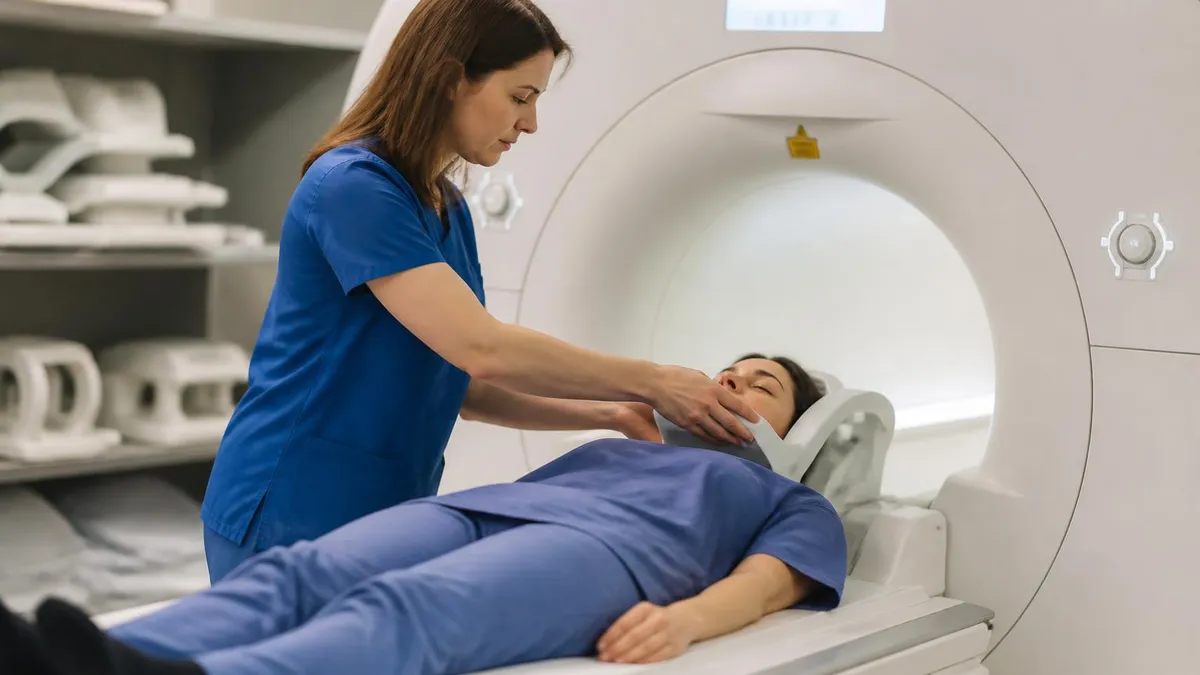
- ✓Remove all metallic jewelry, hairpins, piercings, and hearing aids before entering the MRI scan room.
- ✓Disclose any history of claustrophobia to the scheduling staff so anxiolytic medication can be prescribed before the MRI appointment if needed.
- ✓Report all kidney disease, diabetes managed with metformin, or prior contrast reactions before receiving iodinated CT contrast or gadolinium MRI contrast.
- ✓Follow facility-specific fasting instructions — typically 4 hours before contrast-enhanced studies — unless told otherwise by your physician.
- ✓Bring a list of all current medications, including over-the-counter drugs and supplements, to review for potential contrast interaction risks.
- ✓Arrive 15–30 minutes early to complete safety screening questionnaires that identify MRI contraindications before you enter the magnet room.
- ✓Wear comfortable, loose-fitting clothing without metallic fasteners or zippers if possible, to reduce the need to change into a gown.
- ✓Arrange transportation if you are receiving sedation or anxiolytic medication, as driving afterward is unsafe and prohibited by most facilities.
The 6-Hour Window Changes Everything in Stroke Care
Within the first 6 hours of ischemic stroke onset, a non-contrast head CT may appear completely normal — yet MRI with diffusion-weighted imaging (DWI) can already show the affected brain territory with high sensitivity. This is why comprehensive stroke centers perform CT first to exclude hemorrhage and then immediately follow with MRI-DWI to map ischemia, enabling the most targeted treatment decisions and often dramatically improving patient outcomes.
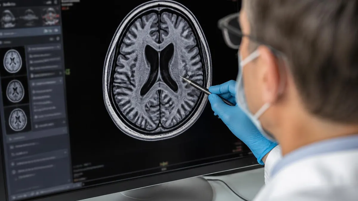
Interpreting brain CT and MRI images requires systematic training and pattern recognition that takes radiologists years to develop, but radiology technologists and medical imaging students must develop a working understanding of normal versus abnormal appearances on both modalities. On a non-contrast head CT, the radiologist or radiographer evaluates the study in a specific order: checking for hemorrhage using brain windows, assessing bone integrity using bone windows, evaluating the ventricles for hydrocephalus, examining the gray-white matter differentiation, and assessing sulcal effacement that might indicate elevated intracranial pressure.
Normal brain parenchyma on CT appears as a uniform gray density with a Hounsfield unit value of approximately 20 to 40 HU. Gray matter is slightly denser than white matter, creating visible differentiation on careful inspection. Loss of this gray-white differentiation in the middle cerebral artery territory is an early CT sign of ischemic stroke, sometimes visible as early as 3 to 6 hours after onset. The dense MCA sign — a hyperdense linear appearance of the middle cerebral artery itself — indicates intraluminal thrombus and carries prognostic significance for infarct size and functional outcome.
On brain MRI, the radiologist evaluates multiple sequences simultaneously rather than relying on a single image set. The T1 sequence provides anatomical reference — gray matter appears darker than white matter on T1, the opposite of CT. T2 and FLAIR sequences identify areas of increased water content, such as edema, gliosis, and demyelinating lesions. Post-contrast T1 sequences identify blood-brain barrier breakdown. DWI and the corresponding ADC (apparent diffusion coefficient) map identify restricted diffusion that indicates acute ischemia or highly cellular tumors such as lymphoma.
Brain tumor characterization on MRI involves evaluating several features: the lesion's location (intra-axial versus extra-axial), signal characteristics on T1 and T2, degree and pattern of contrast enhancement, presence of necrosis or hemorrhage within the lesion, surrounding edema extent, and mass effect on adjacent structures. Meningiomas are classically extra-axial, isointense on T1, intensely and homogeneously enhancing, with a characteristic dural tail sign. High-grade gliomas are intra-axial, heterogeneous, ring-enhancing, surrounded by extensive vasogenic edema that displaces but does not destroy white matter tracts.
White matter disease is a common finding on brain MRI that requires careful interpretation in clinical context. Periventricular and subcortical white matter hyperintensities on T2/FLAIR sequences can represent normal aging, chronic small vessel ischemic disease, demyelination from MS, or other conditions. The Fazekas scale grades white matter hyperintensity burden from 0 (none) to 3 (confluent), providing reproducible quantification that correlates with vascular risk factors and cognitive function. MS lesions follow specific distribution patterns described by the McDonald criteria — periventricular lesions perpendicular to the ventricles (Dawson fingers), juxtacortical lesions, and infratentorial lesions are particularly characteristic.
Advanced MRI techniques add functional and metabolic dimensions to structural brain imaging. Magnetic resonance angiography (MRA) visualizes intracranial arteries without radiation, detecting aneurysms, arteriovenous malformations, and stenoses. MR venography evaluates the dural venous sinuses for thrombosis — a condition that can cause stroke in young patients and is beautifully demonstrated on MRI while being poorly visible on CT. Susceptibility-weighted imaging (SWI) detects microhemorrhages and iron deposition invisible to other sequences, providing important information about cerebral amyloid angiopathy and traumatic diffuse axonal injury.
For students preparing for the ARRT MRI registry examination, understanding image interpretation concepts — not just technical parameters — is essential. Questions about artifact recognition, sequence selection, and pathology appearance are common on board examinations. The ability to explain why a specific sequence is chosen for a specific clinical indication demonstrates the integrative knowledge that distinguishes a competent technologist from one who simply presses buttons. Practice identifying normal anatomy on both CT and MRI, and study classic pathological findings with radiological images until recognition becomes automatic and confident.
Never assume an implanted device is MRI-safe without verified documentation. Cardiac pacemakers older than 2015, cochlear implants, intracranial ferromagnetic aneurysm clips, and retained metallic orbital foreign bodies are absolute contraindications to MRI. A ferromagnetic object inside the MRI bore can torque, heat, or migrate due to the magnetic field, causing serious injury or death. Always complete the MRI safety screening questionnaire thoroughly and honestly, and always verify implant safety with the manufacturer's conditional MRI documentation before the scan.
For radiology students and medical imaging professionals preparing for registry examinations, the brain CT vs MRI comparison is not merely academic — it is a foundational competency that underpins clinical decision-making throughout an imaging career. The ARRT MRI examination tests candidates across six content categories: patient care, safety, image production, procedures, and physical principles of image formation. Questions about when MRI is appropriate, which sequences to use, and how to screen patients for safety constitute a significant portion of the registry examination content specification.
Understanding pulse sequence physics is essential for registry success. Students must know that T1-weighted sequences use short TR (repetition time less than 600 ms) and short TE (echo time less than 30 ms), producing images where fat appears bright and free water appears dark. T2-weighted sequences use long TR (greater than 2000 ms) and long TE (greater than 60 ms), producing images where free water appears bright. These fundamental relationships govern contrast behavior across all MRI sequences and must be memorized and understood conceptually, not just as isolated facts.
Gradient echo sequences differ from spin echo sequences in that they use a radiofrequency flip angle less than 90 degrees and a gradient to refocus the echo rather than a 180-degree refocusing pulse. This makes gradient echo sequences faster — suitable for breath-hold abdominal imaging and cardiac MRI — but also more sensitive to magnetic susceptibility effects from metal, air-tissue interfaces, and hemorrhage products. Understanding this susceptibility sensitivity is why SWI (susceptibility-weighted imaging), a gradient echo-based sequence, is so valuable for detecting cerebral microhemorrhages in traumatic brain injury patients.
Safety content on the registry examination covers the FDA's classification of MRI conditions: MR-safe (poses no known hazards), MR-conditional (safe within specific conditions of use), and MR-unsafe (poses unacceptable risks). The 2026 ARRT content specifications require candidates to demonstrate knowledge of radiofrequency energy deposition (measured as SAR — specific absorption rate), the biological effects of gradient switching (peripheral nerve stimulation), and acoustic noise management. These safety principles protect both patients and staff and have real clinical consequences when violated.
The ACR (American College of Radiology) publishes MRI safety guidelines that are the industry standard in the United States. The ACR divides the MRI environment into four zones with progressive levels of restriction: Zone I is unrestricted public access, Zone II is the reception and screening area, Zone III is the controlled area adjacent to the magnet room, and Zone IV is the magnet room itself.
The ferromagnetic detection system — a screening arch installed at Zone III entry — identifies metallic objects that patients or staff might inadvertently bring near the magnet. Understanding this zone model is tested on registry examinations and is mandatory knowledge for safe clinical practice.
Students often ask whether they should focus more on MRI or CT content for registry preparation. The answer depends on which registry they are pursuing. The ARRT MRI registry is entirely focused on MRI physics, procedures, and safety — CT appears only for comparison and context. The ARRT Radiography registry covers CT more extensively since radiographers rotate through CT suites.
For students pursuing dual certification in both modalities, building parallel competency in CT and MRI physics creates a comprehensive skill set that is increasingly valued in the modern imaging department, where multimodality imaging pathways are standard of care for complex neurological conditions.
Preparation resources for the registry examination should include a combination of content review textbooks, physics problem sets, and timed practice examinations. Reviewing normal brain anatomy systematically — identifying the thalamus, basal ganglia, internal capsule, corpus callosum, hippocampus, and brainstem structures by name and location — builds the anatomical foundation that makes pathology recognition intuitive. When you know exactly what normal looks like, abnormal findings become conspicuous rather than confusing. Use every practice test opportunity to reinforce both technical knowledge and clinical reasoning simultaneously.
Looking ahead to the future of brain imaging, both CT and MRI technology are advancing rapidly, blurring some of the traditional boundaries between these modalities. Photon-counting CT detectors, now entering clinical use at major academic centers, provide dramatically improved spatial resolution and spectral imaging capabilities without increasing radiation dose. Spectral CT can differentiate iodine from calcium at arterial phase imaging, improving stroke characterization and potentially detecting early ischemic changes that conventional CT cannot resolve. These advances may reduce the gap between CT and MRI for certain neurological applications.
On the MRI side, ultra-high-field scanners operating at 7 Tesla field strength are producing brain images with sub-millimeter spatial resolution that reveals cortical layers, small perforating arteries, and iron deposition patterns invisible at standard clinical field strengths. While 7T MRI is currently restricted to research and select clinical applications due to cost, safety considerations at ultra-high field strength, and specific absorption rate limitations, the technology is gradually transitioning toward broader clinical availability. At 7T, conditions like focal cortical dysplasia causing drug-resistant epilepsy can be identified and precisely localized for surgical planning with far greater success than at 1.5T or 3T.
Artificial intelligence and machine learning are transforming both CT and MRI brain imaging workflows. Deep learning algorithms can now detect intracranial hemorrhage on CT with sensitivity and specificity comparable to board-certified radiologists, and these systems are deployed in dozens of hospital systems to flag critical findings for immediate radiologist review — reducing time to treatment for hemorrhagic stroke patients. On the MRI side, AI-powered image reconstruction algorithms can produce diagnostic-quality images from significantly undersampled k-space data, reducing scan time by 50 to 75 percent without meaningful loss of diagnostic accuracy.
Hybrid imaging modalities represent another frontier in brain investigation. PET-MRI scanners acquire simultaneous positron emission tomography metabolic data and MRI structural data in a single session, providing co-registered functional and anatomical brain maps with no additional patient burden or time. Amyloid PET combined with MRI is increasingly used in the early detection and monitoring of Alzheimer's disease, particularly as FDA-approved anti-amyloid therapies require accurate patient selection based on amyloid burden. Understanding the complementary roles of structural MRI and nuclear medicine brain imaging is becoming important for technologists in all modalities.
Teleradiology and cloud-based AI diagnostic platforms are changing how brain imaging results reach patients and clinicians. Stroke alert systems now transmit CT and MRI images directly from the scanner to a radiologist's smartphone within 30 seconds of acquisition, enabling remote expert interpretation for community hospitals without an on-site neuroradiologist. These systems have documented time savings of 20 to 40 minutes in stroke diagnosis workflows — directly translating to preserved brain tissue and improved patient outcomes. For imaging professionals, understanding the technical and clinical workflow implications of these systems is increasingly a required competency.
For patients navigating their own care, the most practical advice is to ask your physician a straightforward question when a brain scan is ordered: why this modality for my specific situation? A good physician should be able to explain whether speed, radiation avoidance, soft-tissue detail, or another factor drove the choice. If you have concerns about radiation exposure from CT, ask about MRI alternatives.
If you have implanted devices, confirm your safety with the imaging facility before your appointment — never assume compatibility. If cost is a concern, ask whether a less expensive initial study would be clinically appropriate, or ask the facility's financial counselors about assistance programs before the scan date.
Whether you are a patient trying to understand your upcoming brain scan or a radiology student building the knowledge base for your career, the fundamental principle is the same: no single imaging modality is superior in all situations. CT and MRI are complementary tools, each uniquely suited to specific clinical problems.
The skilled clinician, radiologist, and technologist knows not just how to operate each tool but when and why to choose it — and that nuanced, evidence-based judgment is what separates excellent patient care from routine image acquisition. Mastering this distinction is both a clinical imperative and a career-defining competency in the field of medical imaging.
MRI Questions and Answers
DWI MRI: Diffusion-Weighted Imaging Explained (2026 Guide)
Side Effects of Brain MRI: Understanding Risks, Safety, and What Every Patient Should Know
MRI Lumbar Spine Without Contrast: Complete Guide to the Scan, What It Shows, and How to Prepare
MRI History: From Nuclear Resonance to Modern Scanners

MRI of Cervical Spine: Complete Guide to Imaging, Anatomy, Pathology, and Patient Preparation
About the Author

Medical Laboratory Scientist & Clinical Certification Expert
Johns Hopkins UniversityDr. Sandra Kim holds a PhD in Clinical Laboratory Science from Johns Hopkins University and is certified as a Medical Technologist (MT) and Medical Laboratory Scientist (MLS) through ASCP. With 16 years of clinical laboratory experience spanning hematology, microbiology, and molecular diagnostics, she prepares candidates for ASCP board exams, MLT, MLS, and specialist certification tests.
Join the Discussion
Connect with other students preparing for this exam. Share tips, ask questions, and get advice from people who have been there.
View discussion (8 replies)