Anatomy and Physiology of the Respiratory System: Complete Guide 2026 June
Master the anatomy and physiology of the respiratory system 🎯 — structures, functions, gas exchange, and exam-ready study tips.
Understanding the anatomy and physiology of the respiratory system is one of the most essential foundations in any health science curriculum. From nursing students to pre-med candidates, mastering how the lungs, airways, and gas-exchange mechanisms work together determines success not just on exams, but in clinical practice. The respiratory system is responsible for delivering oxygen to every living cell in your body and expelling carbon dioxide, making it one of the most life-critical systems you will ever study.
The respiratory system is conventionally divided into the upper and lower respiratory tracts. The upper tract includes the nose, nasal cavity, pharynx, and larynx, while the lower tract encompasses the trachea, bronchi, bronchioles, and the alveoli deep within the lungs. Each segment plays a specific, non-negotiable role in conditioning, conducting, and exchanging air. Failing to understand one component often leads to confusion about downstream pathophysiology, such as why COPD impairs gas exchange or why croup causes the distinctive stridor heard in pediatric emergencies.
Breathing itself is not a passive event driven solely by the lungs. It is an active mechanical process orchestrated by a network of skeletal muscles — primarily the diaphragm and the intercostal muscles — regulated by chemoreceptors that detect blood oxygen and carbon dioxide concentrations in real time. The brainstem's respiratory control centers, including the medullary rhythmicity center, continuously adjust breathing rate and depth in response to metabolic demands, exercise, altitude, and disease states.
Gas exchange occurs at the alveolar-capillary membrane, a structure so thin — roughly 0.5 micrometers — that oxygen and carbon dioxide can diffuse across it in milliseconds. About 300 million alveoli in each healthy adult lung create a total surface area of approximately 70 square meters, roughly the size of half a tennis court. This enormous surface area is what allows the respiratory system to meet the oxygen demands of a body at rest, during moderate exercise, and even during high-intensity athletic performance.
The physiology of ventilation depends on pressure gradients governed by Boyle's Law: as lung volume increases during inhalation, intrapulmonary pressure drops below atmospheric pressure, drawing air inward. During exhalation, the reverse occurs. Lung compliance — how easily the lungs stretch — and airway resistance — how much friction air faces moving through the bronchial tree — are the two chief mechanical variables that clinicians assess when evaluating respiratory function, particularly in patients on mechanical ventilation or those with obstructive versus restrictive lung disease.
Pulmonary volumes and capacities are standardized measurements that describe how much air moves in and out of the lungs under various conditions. Tidal volume (about 500 mL at rest), inspiratory reserve volume, expiratory reserve volume, and residual volume combine to form the four primary lung volumes. Understanding how spirometry measures forced vital capacity (FVC) and forced expiratory volume in one second (FEV1) is critical for identifying obstructive conditions like asthma and COPD versus restrictive conditions like pulmonary fibrosis.
Whether you are preparing for the HESI, TEAS, NCLEX, or a comprehensive anatomy and physiology course exam, a deep grasp of respiratory system anatomy and physiology will give you a decisive advantage. This guide walks you through every major structure, function, and physiological process — with the clinical context, real numbers, and conceptual clarity that will help your knowledge stick long after the exam is over.
Respiratory System by the Numbers

Upper and Lower Respiratory Tract: Key Structures
Filters, warms, and humidifies incoming air using mucous membranes and cilia. The nasal turbinates increase surface area dramatically. Paranasal sinuses lighten the skull and contribute to voice resonance, and their drainage pathways are clinically significant in sinusitis.
The pharynx serves as a shared passageway for air and food, divided into nasopharynx, oropharynx, and laryngopharynx. The larynx houses the vocal cords and the epiglottis, which flips down during swallowing to prevent aspiration of food into the airway.
The trachea is a 10–12 cm tube reinforced by C-shaped cartilage rings. It bifurcates at the carina into the right and left primary bronchi, which branch progressively into secondary, tertiary bronchi, and then into bronchioles — all the way to the terminal bronchioles.
Alveoli are microscopic grape-like air sacs where gas exchange actually occurs. Type I pneumocytes form the thin walls; Type II pneumocytes secrete surfactant, which reduces surface tension and prevents alveolar collapse. The respiratory membrane is only 0.5 µm thick.
Each lung is enclosed in a double-layered serous membrane called the pleura. The visceral pleura lines the lung surface; the parietal pleura lines the thoracic wall. Between them, a thin film of pleural fluid reduces friction during breathing and creates the pressure gradient that keeps lungs inflated.
The mechanics of breathing involve two distinct phases: inspiration and expiration. During inspiration, the diaphragm contracts and flattens, moving downward by about 1.5 centimeters during quiet breathing and up to 10 centimeters during deep inhalation. Simultaneously, the external intercostal muscles elevate the rib cage, expanding the thoracic cavity in all three dimensions — vertical, lateral, and anterior-posterior. This expansion drops the intrapulmonary pressure approximately 1–3 mmHg below atmospheric pressure, and air rushes in to equalize the gradient.
Quiet expiration, unlike inspiration, is largely a passive process. When inspiratory muscles relax, the elastic recoil of the lung tissue and the chest wall naturally reduces thoracic volume, raising intrapulmonary pressure above atmospheric pressure and pushing air out. Forced expiration — as occurs during exercise, coughing, or spirometry testing — recruits the internal intercostal muscles and the abdominal muscles to compress the thoracic cavity more rapidly and forcefully, generating the high airflow rates needed for effective airway clearance.
Lung compliance is the measure of how easily lung tissue stretches in response to pressure changes. Healthy lungs have high compliance, meaning they expand easily with relatively small pressure changes. In pulmonary fibrosis, scar tissue makes lungs stiff and non-compliant, dramatically increasing the work of breathing. Conversely, in emphysema, destruction of alveolar walls and loss of elastic tissue gives lungs extremely high (abnormal) compliance, so they inflate easily but cannot recoil adequately to push air out — leading to air trapping and the characteristic barrel chest appearance.
Surface tension within the alveoli is a critical physical force that, without countermeasure, would cause alveolar collapse between each breath. Pulmonary surfactant — a complex mixture of phospholipids (primarily dipalmitoylphosphatidylcholine, or DPPC) and proteins secreted by Type II alveolar cells — reduces this surface tension by roughly 70%. In premature infants born before 28–32 weeks gestation, surfactant production is insufficient, resulting in neonatal respiratory distress syndrome (NRDS), treated with exogenous surfactant therapy administered directly into the trachea immediately after birth.
Airway resistance is the opposition that air encounters as it flows through the bronchial tree. The bronchioles are the primary site of resistance regulation because they lack cartilaginous support and depend entirely on smooth muscle tone, which is controlled by the autonomic nervous system. Sympathetic stimulation triggers bronchodilation — the mechanism exploited by albuterol and other beta-2 agonist medications — while parasympathetic activity causes bronchoconstriction. In asthma, chronic inflammation and smooth muscle hyperresponsiveness lead to episodic, reversible increases in airway resistance that manifest as wheezing, chest tightness, and dyspnea.
Ventilation-perfusion (V/Q) matching is one of the most clinically important concepts in respiratory physiology. For efficient gas exchange, airflow (ventilation) to each lung region must match blood flow (perfusion). The lungs employ hypoxic pulmonary vasoconstriction — a local reflex that diverts blood away from poorly ventilated, low-oxygen regions — to optimize V/Q matching. When this mechanism fails, as in pneumonia or pulmonary embolism, hypoxemia results. Understanding V/Q mismatch explains why supplemental oxygen corrects the hypoxemia of pneumonia but not that of a massive pulmonary embolism.
Dead space is another key ventilation concept. Anatomical dead space refers to the roughly 150 mL of air that fills the conducting airways — the trachea, bronchi, and bronchioles — and never reaches the alveoli to participate in gas exchange. Physiological dead space adds any alveoli that are ventilated but not perfused. In a healthy person, anatomical and physiological dead space are nearly equal. In pulmonary embolism, however, large regions of alveoli receive airflow but no blood, massively increasing physiological dead space and making each breath progressively less effective at maintaining normal blood gas levels.
Gas Exchange, Transport, and Control of Breathing
Gas exchange at the alveolar-capillary membrane is driven entirely by partial pressure gradients. Oxygen moves from the alveoli (PO₂ ≈ 104 mmHg) into the pulmonary capillary blood (PO₂ ≈ 40 mmHg on arrival), while carbon dioxide diffuses in the opposite direction from capillary blood (PCO₂ ≈ 45 mmHg) into the alveoli (PCO₂ ≈ 40 mmHg). Diffusion is so rapid that equilibration is nearly complete within the first third of the time a red blood cell spends in the pulmonary capillary.
Fick's Law of Diffusion governs the rate of gas exchange and states that diffusion rate is proportional to the surface area of the membrane, the partial pressure gradient, and the diffusion coefficient of the gas — and inversely proportional to membrane thickness. Carbon dioxide diffuses about 20 times faster than oxygen because of its much greater solubility in biological membranes, which is why CO₂ retention (hypercapnia) tends to appear later than low oxygen (hypoxemia) in diseases that thicken the respiratory membrane, such as pulmonary fibrosis or pulmonary edema.

Studying Respiratory System Anatomy: Structured Course vs. Self-Study
- +Structured courses provide a clear sequence, moving logically from upper airway anatomy to alveolar gas exchange
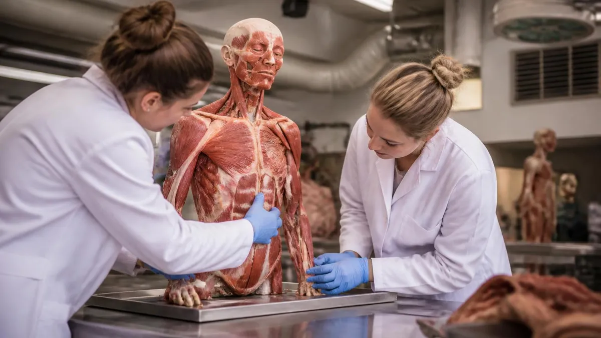
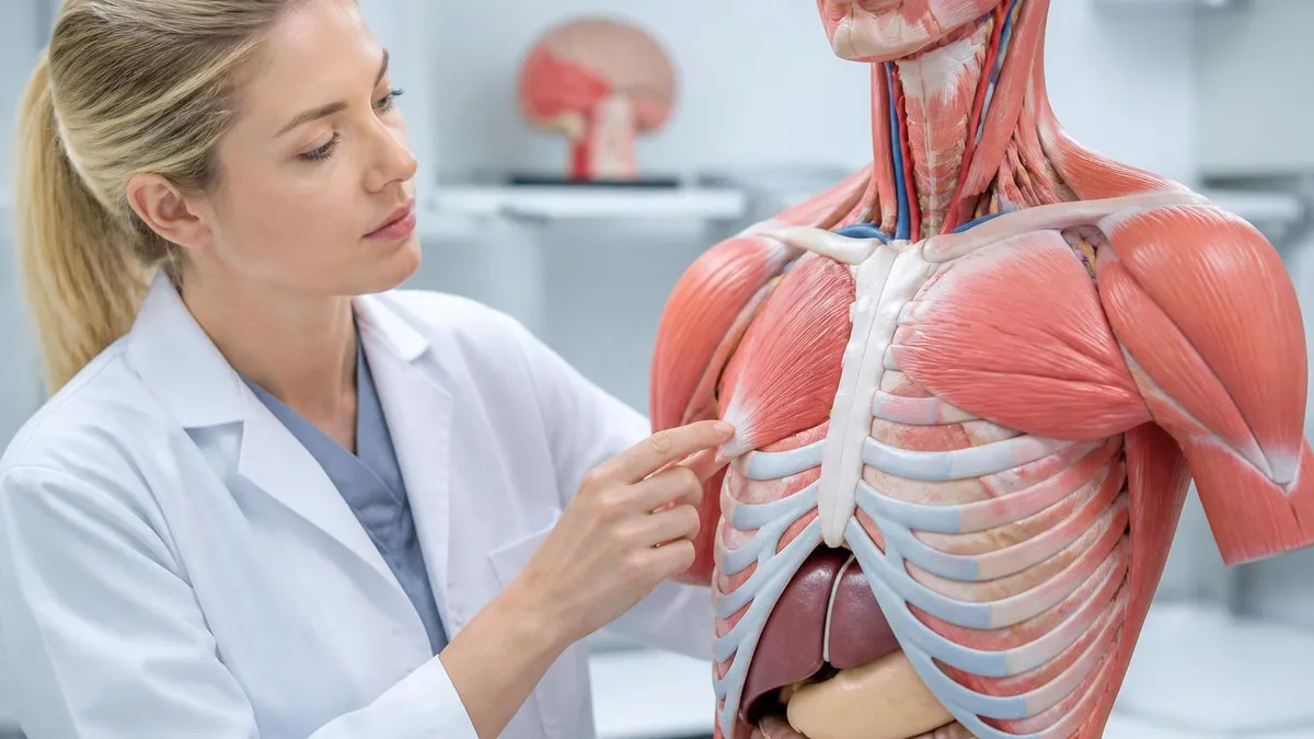
- +Instructor-led labs with cadavers or plastinated specimens make three-dimensional anatomy viscerally memorable
- +Scheduled exams in formal courses create accountability checkpoints that combat procrastination
- +Peer study groups in formal settings expose you to different mnemonics and explanations that reinforce your own understanding
- +Formal courses often integrate clinical correlations, such as linking surfactant deficiency directly to NRDS case studies
- +Transcripts and grades from accredited programs satisfy prerequisites for nursing, PA, and medical school admissions
- −Formal courses move at a fixed pace, which may be too fast for complex topics like V/Q matching or hemoglobin dissociation curves
- −Tuition and lab fees for accredited anatomy and physiology courses can exceed $2,000 per semester at community colleges
- −Lecture-heavy formats may underemphasize the functional physiology needed to understand clinical respiratory conditions
- −Self-directed learners may struggle to identify which respiratory subtopics carry the most exam weight without a syllabus
- −Online self-study lacks hands-on laboratory experiences that reinforce three-dimensional spatial understanding of lung anatomy
- −Without external deadlines, self-study students risk spending too much time on familiar topics and under-preparing on weaker areas
Respiratory System Study Checklist: What You Must Know Before Your Exam
- ✓Label all structures of the upper respiratory tract from the nasal cavity through the larynx on a blank diagram.
- ✓Describe the three functions of the nasal cavity: filtration, warming, and humidification of inspired air.
- ✓Trace airflow from the trachea through the bronchial tree down to the terminal bronchioles and alveoli.
- ✓Explain the role of Type I and Type II alveolar cells, including surfactant production and its clinical significance in NRDS.
- ✓Describe the pressure changes that drive inspiration and quiet expiration using Boyle's Law as your framework.
- ✓Distinguish between lung compliance and airway resistance, and name one disease that alters each parameter.
- ✓Draw and interpret the oxygen-hemoglobin dissociation curve, including the Bohr effect and rightward versus leftward shifts.
- ✓Explain the partial pressure gradients that drive O₂ and CO₂ diffusion across the alveolar-capillary membrane.
- ✓Define tidal volume, inspiratory reserve volume, expiratory reserve volume, residual volume, vital capacity, and total lung capacity.
- ✓Describe how central and peripheral chemoreceptors regulate minute ventilation in response to changes in PCO₂ and PO₂.
Why the Respiratory Membrane Thickness Matters Clinically
The alveolar-capillary membrane is only 0.5 micrometers thick — about 140 times thinner than a human hair. Any disease that thickens this membrane (pulmonary edema, fibrosis, or ARDS) impairs oxygen diffusion first, because CO₂ diffuses 20× faster. This is why hypoxemia appears before hypercapnia in early interstitial lung disease, and why supplemental oxygen can temporarily compensate until the underlying pathology worsens significantly.
Respiratory control is a sophisticated, multi-layered system that integrates chemical, neural, and mechanical feedback to maintain blood gas homeostasis within remarkably tight margins. The medullary respiratory centers — the dorsal and ventral respiratory groups — generate the baseline rhythm of breathing through a process of automatic, rhythmic neural discharge. This automatic rhythm continues even without conscious input, which is why breathing persists during sleep, anesthesia, and unconsciousness, and why voluntary breath-holding can only be sustained until rising arterial CO₂ overwhelms the voluntary suppression of the medullary drive.
Central chemoreceptors located on the ventral surface of the medulla oblongata are the most sensitive monitors of blood carbon dioxide levels. They respond not to CO₂ directly but to the pH changes caused when CO₂ diffuses across the blood-brain barrier and reacts with water to form carbonic acid, which dissociates into bicarbonate and hydrogen ions. Rising H⁺ concentration stimulates the central chemoreceptors, increasing ventilation to blow off CO₂ and restore pH. This system is so sensitive that a rise in arterial PCO₂ of just 1 mmHg increases minute ventilation by approximately 2–3 liters per minute.
Peripheral chemoreceptors in the carotid bodies (at the bifurcation of the common carotid artery) and aortic bodies (near the aortic arch) respond to three stimuli: a drop in arterial PO₂ below 60 mmHg, a rise in PCO₂, and a fall in arterial pH. The carotid bodies are far more important clinically than the aortic bodies.
Their response to hypoxia is critical at high altitude, during acute respiratory failure, and in patients with chronic lung disease who have adapted to chronically elevated CO₂ and now rely on low PO₂ as their primary ventilatory stimulus — the so-called hypoxic drive that every nursing student learns to respect when administering oxygen therapy.
Beyond chemical control, pulmonary stretch receptors — known as slowly adapting receptors — detect over-inflation of the lungs and send signals via the vagus nerve to the pontine respiratory group to shorten inspiration. This protective mechanism, the Hering-Breuer reflex, prevents the lungs from being dangerously over-distended. Rapidly adapting receptors (irritant receptors) respond to noxious chemicals, dust, and smoke, triggering coughing, bronchoconstriction, and increased breathing rate. J-receptors in the alveolar walls respond to increased capillary pressure and interstitial fluid accumulation, producing the sensation of dyspnea experienced in pulmonary edema and heart failure.
Voluntary control of breathing originates in the cerebral cortex, which can temporarily override the automatic medullary rhythm through direct projections to respiratory motor neurons in the spinal cord. This is what allows singers to sustain long phrases, free divers to hold their breath for several minutes, and speakers to control the rhythm of their speech. However, voluntary suppression always fails when CO₂ rises to critical levels, demonstrating the absolute priority of the chemical drive over conscious control in preserving homeostasis.
Temperature has a direct and significant effect on respiratory rate. During fever, increased metabolic rate accelerates CO₂ production, which stimulates the medullary chemoreceptors and drives compensatory hyperventilation. During vigorous exercise, ventilation can increase from a resting 6 L/min to over 100 L/min in trained athletes — a 15-fold increase driven primarily by rising CO₂ from muscle metabolism, lactic acidosis, increased body temperature, and direct neural input from exercising limbs via mechanoreceptors and proprioceptors in muscles and joints.
Understanding the interplay between neural and chemical control of ventilation is not only academically important — it has direct clinical applications. Opioid medications, for example, suppress the medullary respiratory centers, reducing both rate and depth of breathing, which can cause life-threatening respiratory depression and hypercapnia. Benzodiazepines potentiate this effect when combined with opioids, explaining why the combination is far more dangerous than either drug alone. Recognizing these mechanisms helps clinicians anticipate and prevent respiratory complications in pain management, post-operative care, and ICU settings.

Administering high-flow oxygen to patients with chronic hypercapnic COPD can suppress their hypoxic ventilatory drive, leading to worsening CO₂ retention and respiratory acidosis. On exams, this scenario is frequently tested — always identify the patient's baseline blood gas status before selecting the appropriate oxygen delivery device and target saturation (typically 88–92% SpO₂ in chronic CO₂ retainers, not the standard 94–98%).
Clinical applications of respiratory anatomy and physiology span virtually every area of medicine and nursing practice. The most common respiratory disorders — asthma, COPD, pneumonia, pulmonary embolism, and acute respiratory distress syndrome (ARDS) — each represent a specific disruption of a normal anatomical or physiological mechanism that students and clinicians must be able to identify and explain. When you understand the underlying mechanism, clinical presentations, diagnostic findings, and treatments follow a logical, predictable pattern rather than seeming like a random list of facts to memorize.
Asthma is fundamentally a disease of airway inflammation and bronchospasm. Exposure to triggers such as allergens, cold air, exercise, or respiratory infections activates mast cells and eosinophils in the bronchial mucosa, releasing inflammatory mediators including histamine, leukotrienes, and prostaglandins. These mediators cause smooth muscle contraction, mucus hypersecretion, and mucosal edema — all of which dramatically increase airway resistance and produce the classic triad of wheezing, dyspnea, and cough. Spirometry reveals a decreased FEV1/FVC ratio that improves with bronchodilator therapy, confirming the reversible obstructive pattern.
COPD, by contrast, encompasses two related but distinct entities: chronic bronchitis and emphysema, though most patients have features of both. Chronic bronchitis is defined clinically as a productive cough for at least three months per year for two consecutive years, caused by chronic irritant exposure (most commonly cigarette smoke) that triggers goblet cell hyperplasia and mucus gland enlargement. Emphysema involves irreversible destruction of alveolar walls by inflammatory enzymes (particularly elastase from neutrophils), reducing surface area for gas exchange and eliminating the elastic tissue recoil that normally aids expiration — resulting in air trapping and hyperinflation.
Pneumonia illustrates the vulnerability of the respiratory system to infection when host defenses are overwhelmed. Bacteria, viruses, or fungi that reach the alveoli trigger an intense inflammatory response, flooding the alveolar spaces with exudate (consolidation) and dramatically reducing the surface area available for gas exchange. Chest X-ray reveals lobar or diffuse infiltrates, and the hypoxemia results primarily from intrapulmonary shunting — blood flowing through consolidated, non-ventilated alveoli — rather than diffusion impairment, which is why supplemental oxygen (which increases FiO₂ to ventilated alveoli) partially corrects the hypoxemia.
Pulmonary embolism occurs when a blood clot — typically originating in the deep veins of the lower extremities — lodges in the pulmonary arterial circulation, obstructing blood flow to lung regions that continue to be ventilated. This creates high V/Q mismatch (dead space physiology). The affected alveoli receive air but no blood, so they cannot participate in gas exchange.
Compensatory hyperventilation attempts to maintain oxygenation through remaining functional alveoli but cannot overcome the physiological dead space created by the embolized regions. The result is hypoxemia, hypocapnia (from hyperventilation), and respiratory alkalosis — a combination that is highly suggestive of PE on arterial blood gas analysis.
ARDS represents one of the most severe forms of acute respiratory failure, characterized by diffuse alveolar damage, non-cardiogenic pulmonary edema, bilateral infiltrates on chest imaging, and a PaO₂/FiO₂ ratio below 300 mmHg. The pathophysiology involves a massive inflammatory response — triggered by sepsis, trauma, aspiration, or severe pneumonia — that disrupts the alveolar-capillary membrane, allowing protein-rich fluid to flood the alveolar spaces. Type II pneumocyte damage reduces surfactant production, causing widespread alveolar collapse (atelectasis) and dramatically reducing lung compliance. Treatment requires protective mechanical ventilation with low tidal volumes (6 mL/kg ideal body weight) to prevent ventilator-induced lung injury.
Respiratory failure is classified as either hypoxemic (Type 1) or hypercapnic (Type 2). Type 1 failure involves a PaO₂ below 60 mmHg with a normal or low PaCO₂ — seen in conditions with V/Q mismatch or shunting like pneumonia, ARDS, and PE. Type 2 failure involves a PaCO₂ above 45 mmHg with hypoxemia — seen when the respiratory pump fails due to COPD exacerbation, neuromuscular disease, opioid overdose, or chest wall deformity. Distinguishing these two types on arterial blood gas is one of the most high-yield clinical skills tested on boards and licensing exams across every health profession.
Effective exam preparation for respiratory system content requires more than passive reading — it demands active recall, clinical application, and repeated self-testing across all cognitive levels from simple identification to complex analysis. Students who consistently outperform their peers on anatomy and physiology exams share a common approach: they build their study strategy around the way exam questions are actually written, not just around the way textbooks are organized. This means anticipating scenario-based questions that require you to apply physiological principles to patient situations, not just recall definitions.
Start your preparation by building a solid anatomical framework before moving to physiology. Use a systematic approach: trace the path of an air molecule from the external nares through every structure to the alveolus, labeling the function of each stop along the way. Then reverse the journey and trace an oxygen molecule from the alveolus into a red blood cell, through the pulmonary veins, through the heart, and out to a peripheral tissue. This bidirectional mental walkthrough prevents the common mistake of memorizing structures in isolation without understanding how they connect functionally.
Active recall through practice questions is the single most effective study strategy supported by cognitive science research. After reading each major topic section — upper airway anatomy, lung mechanics, gas exchange, oxygen transport, or respiratory control — close the book and write down everything you can recall from memory. Compare your notes to the source material, then immediately practice 10–20 questions on the same topic. This retrieval practice consolidates memory far more effectively than re-reading, and spaced repetition — returning to the same material at increasing intervals — prevents the forgetting curve from erasing what you have learned.
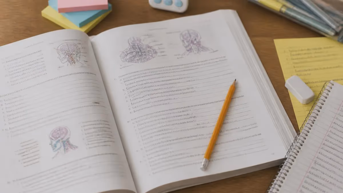
Diagrams and drawings are particularly powerful tools for respiratory anatomy because so much of the system is three-dimensional and spatially complex. Drawing the bronchial tree from memory, labeling the lobes of each lung (right: upper, middle, lower; left: upper, lower), sketching the pleural layers, and recreating the oxygen-hemoglobin dissociation curve by hand all force your brain to actively reconstruct the information rather than passively recognize it on a page. Color-coding different structures — airways in blue, vasculature in red, nerves in yellow — further enhances retention by adding a visual dimension to memory encoding.
Clinical context makes physiological concepts dramatically more memorable and exam-ready. For every major physiological mechanism you study, pair it with a clinical disorder that disrupts it. Surfactant physiology becomes unforgettable when linked to NRDS in premature infants. Pulmonary compliance makes intuitive sense when you picture a patient with pulmonary fibrosis laboring to draw each breath against stiff, scarred lung tissue. V/Q mismatch becomes concrete when you consider why a patient with a saddle pulmonary embolism becomes acutely short of breath despite normal airway anatomy. Clinical pairing transforms abstract physiology into applicable, testable knowledge.
Group study can accelerate learning if structured correctly. Rather than passively reviewing notes together, conduct peer teaching sessions where each student takes responsibility for explaining one topic — gas exchange, lung volumes, or respiratory chemoreceptors — to the others. The act of explaining forces you to identify gaps in your own understanding and reorganize information into a teachable narrative. Peer questions will reveal nuances and edge cases that solo study misses, and discussing disagreements over mechanisms deepens conceptual understanding far more than agreeing on answers from an answer key.
Time management in the weeks leading up to a respiratory system exam should follow a structured schedule. In the first week, focus on anatomy: master the structural components, their locations, and their immediate functions. In the second week, shift to mechanics and physiology: breathing mechanics, lung volumes, gas exchange, and oxygen transport.
In the third week, integrate clinical applications and practice exclusively with scenario-based questions. In the final days before the exam, focus on weak areas identified through practice testing, review the highest-yield facts (alveolar surface area, normal values for blood gases, V/Q concepts), and avoid cramming new material that has not been processed through active recall and repetition.
Anatomy Physiology Questions and Answers

How Hard Is Anatomy and Physiology? The Complete Honest Guide for Students
Human Anatomy and Physiology Book: The Complete Guide to Choosing, Using, and Mastering the Best Textbooks for 2026

Eye Anatomy and Physiology: The Complete Guide to Understanding How Your Eyes Work
Anatomy and Physiology PDF: Complete Free Download Guide for 2026 Students

Best Anatomy and Physiology Book: The Complete Guide to Top Textbooks for 2026
About the Author
Educational Psychologist & Academic Test Preparation Expert
Columbia University Teachers CollegeDr. Lisa Patel holds a Doctorate in Education from Columbia University Teachers College and has spent 17 years researching standardized test design and academic assessment. She has developed preparation programs for SAT, ACT, GRE, LSAT, UCAT, and numerous professional licensing exams, helping students of all backgrounds achieve their target scores.