ACLS Rhythms: The Complete Guide to Recognizing Cardiac Rhythms for ACLS Certification
Master ACLS rhythms with this complete guide to recognizing VF, 🔎 VT, asystole, PEA, bradycardia and tachycardia for ACLS certification and exam prep.

Learning the core ACLS rhythms is the single most important skill that separates a confident provider from one who freezes at the bedside. Advanced Cardiovascular Life Support is built around fast, accurate rhythm recognition, because the rhythm on the monitor dictates every decision that follows: whether you shock, whether you push epinephrine, whether you pace, or whether you simply continue high-quality CPR. Before you memorize a single drug dose, you must be able to look at a six-second strip and name what you see within a few seconds.
The American Heart Association organizes ACLS around a handful of rhythm families, and almost every cardiac emergency you will ever encounter falls into one of them. There are the two shockable arrest rhythms, ventricular fibrillation and pulseless ventricular tachycardia. There are the two non-shockable arrest rhythms, asystole and pulseless electrical activity. Then there are the peri-arrest rhythms: symptomatic bradycardias such as sinus bradycardia and high-grade heart blocks, and the tachycardias, which split into narrow-complex and wide-complex categories.
Why does this matter so much? Because the treatment pathways diverge sharply depending on the category. A patient in ventricular fibrillation needs immediate defibrillation, while a patient in asystole gains nothing from a shock and instead needs compressions and epinephrine. Misclassify the rhythm and you waste precious seconds, deliver the wrong therapy, or both. Speed and accuracy together are what the AHA is testing, and what real patients depend on.
This guide walks through each rhythm family in plain language, gives you the distinguishing ECG features, and ties every rhythm back to the algorithm it triggers. You will learn to ask the three questions that resolve nearly every strip: Is there a pulse? Is the QRS narrow or wide? Is the rhythm regular or irregular? Those three questions, asked in order, route you to the correct branch of the correct algorithm almost every time.
We will also cover the practical realities that textbooks gloss over, such as how artifact can mimic a lethal rhythm, why a flat line on one lead does not always mean asystole, and how to tell coarse VF from fine VF when seconds count. These are the nuances that trip up candidates on the megacode station and clinicians during real codes, so we give them the attention they deserve throughout this study hub.
Finally, this article is designed as a certification prep hub. Each section links to free practice questions and ECG interpretation drills so you can test yourself as you read. Rhythm recognition is a perishable skill that improves dramatically with repetition, so plan to read actively, sketch the waveforms, and quiz yourself often. By the end you should be able to glance at any ACLS strip and confidently route it to the right treatment in seconds.
ACLS Rhythms by the Numbers
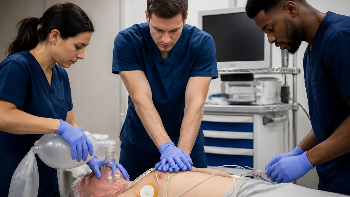
The Six Core ACLS Rhythm Families
A chaotic, disorganized quivering of the ventricles with no identifiable QRS complexes. There is no pulse and no effective cardiac output. VF is shockable and demands immediate defibrillation followed by CPR.
A rapid, wide-complex, regular rhythm originating in the ventricles with no palpable pulse. Like VF, it is treated with immediate defibrillation, compressions, and epinephrine every three to five minutes.
A complete absence of electrical activity producing a flat or nearly flat line. It is non-shockable. Treatment centers on high-quality CPR, epinephrine, and a focused search for reversible causes.
Any organized rhythm on the monitor without a detectable pulse. Non-shockable. Management mirrors asystole and emphasizes identifying and reversing the underlying Hs and Ts.
The peri-arrest rhythms in patients with a pulse. Symptomatic bradycardia may need atropine or pacing, while unstable tachycardia may require synchronized cardioversion or adenosine.
The most useful first cut you can make in any cardiac arrest is whether the rhythm is shockable or non-shockable, because that single distinction determines whether a defibrillator earns its place in the next ten seconds. The shockable rhythms are ventricular fibrillation and pulseless ventricular tachycardia. Both reflect disorganized or excessively rapid ventricular electrical activity that prevents coordinated contraction, and both respond to a well-timed, high-energy shock that can reset the heart toward an organized rhythm.
Ventricular fibrillation appears as a wandering, irregular baseline with no discernible P waves, QRS complexes, or T waves. Clinicians describe it as coarse when the deflections are large and fine when they are small. Coarse VF generally indicates a more recent arrest and a better chance of successful defibrillation, while fine VF can resemble asystole and may reflect a heart that has been fibrillating for several minutes. Either way, the treatment is the same: shock, then immediately resume compressions without waiting to recheck the pulse.
Pulseless ventricular tachycardia presents very differently on the strip. You will see a regular, rapid, wide-complex rhythm, often a monotonous sawtooth pattern, yet the patient has no pulse. The absence of a pulse is what makes it an arrest rhythm rather than a peri-arrest tachycardia. Because the underlying problem is again a ventricular rhythm incompatible with output, the response is identical to VF: defibrillate, perform two minutes of CPR, then reassess. Many candidates review the full sequence in an ACLS study guide before drilling it.
The non-shockable rhythms are asystole and pulseless electrical activity, and here a defibrillator offers no benefit. Asystole is the absence of meaningful electrical activity. Before you call it, confirm the leads are connected, the gain is turned up, and you have checked a second lead, because a disconnected electrode or a fine VF viewed in one plane can masquerade as a flat line. Treating true asystole with a shock wastes time and interrupts the compressions that actually matter.
Pulseless electrical activity is the great imitator. The monitor shows an organized rhythm that looks like it should be producing a pulse, perhaps a normal-appearing sinus rhythm or a slow idioventricular pattern, yet the patient has no detectable circulation. PEA is fundamentally a problem of something interfering with the heart's ability to pump, so the algorithm pushes you to hunt aggressively for reversible causes while delivering compressions and epinephrine.
For both non-shockable rhythms, the cornerstone of care is uninterrupted, high-quality CPR and epinephrine every three to five minutes, paired with a disciplined search through the Hs and Ts: hypovolemia, hypoxia, hydrogen ion acidosis, hypo- and hyperkalemia, hypothermia, tension pneumothorax, tamponade, toxins, and thrombosis of the lungs or heart. Correcting the underlying cause is often the only path to return of spontaneous circulation in these patients.
Mastering the shockable versus non-shockable split early gives you a mental fork that simplifies the entire cardiac arrest algorithm. Once you can reliably place a strip into one of these two buckets, the rest of the decision tree, including drug timing and energy selection, follows in a predictable, rehearsable sequence that holds up under the pressure of a real code.
ACLS Practice Test Questions
Prepare for the ACLS Advanced Cardiovascular Life Support Practice exam with our free practice test modules. Each quiz covers key topics to help you pass on your first try.
ACLS Cardiac Rhythms & ECG Interpretation
ACLS Exam Questions covering ACLS Cardiac Rhythms & ECG Interpretation. Master ACLS Test concepts for certification prep.
ACLS Pharmacology & Medications
Free ACLS Practice Test featuring ACLS Pharmacology & Medications. Improve your ACLS Exam score with mock test prep.
ACLS Best Practices & Methodologies
ACLS Mock Exam on Best Practices & Methodologies. ACLS Study Guide questions to pass on your first try.
ACLS Communication & Stakeholder Relations
ACLS Test Prep for Communication & Stakeholder Relations. Practice ACLS Quiz questions and boost your score.
ACLS Core Concepts & Fundamentals
ACLS Questions and Answers on Core Concepts & Fundamentals. Free ACLS practice for exam readiness.
ACLS Data Analysis & Reporting
ACLS Mock Test covering Data Analysis & Reporting. Online ACLS Test practice with instant feedback.
ACLS Documentation & Record Keeping
Free ACLS Quiz on Documentation & Record Keeping. ACLS Exam prep questions with detailed explanations.
ACLS Industry Regulations & Compliance
ACLS Practice Questions for Industry Regulations & Compliance. Build confidence for your ACLS certification exam.
ACLS Leadership & Team Management
ACLS Test Online for Leadership & Team Management. Free practice with instant results and feedback.
ACLS Professional Standards & Ethics
ACLS Study Material on Professional Standards & Ethics. Prepare effectively with real exam-style questions.
ACLS Quality Assurance & Improvement
Free ACLS Test covering Quality Assurance & Improvement. Practice and track your ACLS exam readiness.
ACLS Risk Management & Assessment
ACLS Exam Questions covering Risk Management & Assessment. Master ACLS Test concepts for certification prep.
Recognizing Cardiac Rhythms on the ECG Strip
Start by estimating the heart rate, because it instantly narrows the possibilities. The classic shortcut is to count large boxes between two QRS complexes and divide 300 by that number, giving you 300, 150, 100, 75, 60, and 50 for one through six boxes. A rate above 100 is a tachycardia and a rate below 60 is a bradycardia, though ACLS usually intervenes only when the rate is below 50 with symptoms.
For irregular rhythms, the box method fails, so count the QRS complexes in a six-second strip and multiply by ten. This six-second method works for atrial fibrillation and other irregular patterns where beat-to-beat spacing varies. Always pair the rate with the clinical picture, since the same rate can be benign in one patient and dangerous in another depending on symptoms and stability.

Manual ECG Interpretation vs Relying on the Monitor's Automated Read
- +Manual interpretation builds a durable skill that works on any monitor or defibrillator
- +You can catch artifact that fools automated algorithms into false alarms
- +Distinguishing fine VF from asystole is a human judgment the machine often gets wrong
- +You retain control during pauses, deciding whether to shock based on the actual waveform
- +Manual reading reinforces the rate, regularity, and QRS-width habit that ACLS rewards
- +It prepares you for the megacode station where you must verbalize your rhythm call
- −Manual interpretation requires practice and can feel slow for new providers
- −Stress during a real code can degrade pattern recognition without rehearsal
- −Subtle distinctions like coarse versus fine VF take repetition to master
- −Lead placement errors can mislead even an experienced human interpreter
- −Fatigue late in a long resuscitation increases the chance of misreading a strip
- −Without regular drilling, rhythm recognition skills decay between certifications
ACLS Rhythm Recognition Checklist
- ✓Confirm whether the patient has a pulse before interpreting the rhythm.
- ✓Check that all ECG leads are connected and the gain is set appropriately.
- ✓Estimate the heart rate using the box method or six-second count.
- ✓Determine whether the rhythm is regular or irregular.
- ✓Measure the QRS width to classify narrow versus wide complex.
- ✓Look for P waves and assess their relationship to the QRS.
- ✓Decide immediately if the rhythm is shockable or non-shockable.
- ✓View a second lead before declaring asystole to rule out fine VF.
- ✓Match the rhythm to its correct ACLS algorithm branch.
- ✓Reassess the rhythm during the next pulse and rhythm check.
Pulse, Width, Regularity — In That Order
When a strip appears, ask three questions in sequence: Is there a pulse? Is the QRS narrow or wide? Is the rhythm regular or irregular? Those three answers route almost every ACLS rhythm to the correct algorithm branch. Drilling this order until it becomes automatic is the fastest way to build calm, accurate decision-making under pressure.
Bradycardias and heart blocks make up the slow end of the peri-arrest spectrum, and they matter in ACLS only when the patient is symptomatic. A heart rate below 50 beats per minute becomes a clinical problem when it produces hypotension, altered mental status, signs of shock, ischemic chest discomfort, or acute heart failure. A well-conditioned athlete with a resting rate of 45 and no symptoms needs no intervention, while a patient with the same rate who is confused and hypotensive needs urgent treatment. The rhythm alone never decides; the rhythm plus symptoms decides.
Sinus bradycardia is the simplest slow rhythm: a normal P wave precedes every QRS, the PR interval is constant, and the rate is simply slow. It often reflects increased vagal tone, medications such as beta-blockers, or inferior wall ischemia. When symptomatic, the first-line drug is atropine, and if atropine fails, the algorithm escalates to transcutaneous pacing or a dopamine or epinephrine infusion to support the rate and perfusion until a definitive solution is arranged.
Heart blocks describe failures of conduction between the atria and ventricles, and they range from benign to immediately life-threatening. First-degree block is a uniformly prolonged PR interval with every P wave still conducting to a QRS; it rarely needs treatment. Second-degree block comes in two types, and distinguishing them is a common exam point because they behave very differently and carry different risks of deteriorating into complete block.
In second-degree type I, also called Mobitz I or Wenckebach, the PR interval lengthens progressively until a QRS is dropped, then the cycle resets. It is usually a problem at the AV node, often responds to atropine, and tends to be relatively stable. In second-degree type II, or Mobitz II, the PR interval stays constant but P waves intermittently fail to conduct without warning. Mobitz II is more dangerous because it signals disease below the AV node and frequently progresses to complete heart block, so it often warrants pacing rather than relying on atropine.
Third-degree, or complete heart block, is the most dangerous of the group. Here the atria and ventricles beat completely independently, a phenomenon called AV dissociation. The P waves march out at their own rate while the QRS complexes appear at a separate, usually slower rate driven by an escape pacemaker. Because atropine often fails when the escape rhythm originates low in the conduction system, the AHA emphasizes early transcutaneous pacing and prompt expert consultation for a transvenous pacemaker.
A practical pearl ties these together: when you see a slow rhythm, immediately ask whether each P wave is connected to a QRS in a consistent way. Consistent connection points toward sinus bradycardia or first-degree block, progressive lengthening points to Wenckebach, intermittent dropped beats with a fixed PR suggest Mobitz II, and complete independence of P waves and QRS complexes means third-degree block. That single observation about the P-to-QRS relationship reliably sorts the bradycardias into their correct categories and tells you how aggressive your treatment must be.
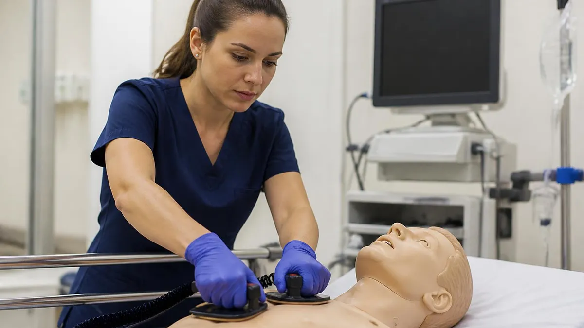
In high-grade blocks such as Mobitz II and third-degree block, atropine may be ineffective and can paradoxically worsen the situation by increasing the atrial rate without improving ventricular conduction. The AHA recommends moving quickly to transcutaneous pacing in these patients. Do not waste time cycling through atropine doses when pacing is indicated.
Tachycardias are the fast end of the peri-arrest spectrum, and the ACLS approach hinges on two questions answered almost simultaneously: Is the patient stable or unstable, and is the QRS narrow or wide? Stability is the first gate. An unstable tachycardia, defined by hypotension, altered mental status, signs of shock, ischemic chest pain, or acute heart failure that is caused by the rapid rate, calls for immediate synchronized cardioversion regardless of the precise rhythm. Stability, not the exact diagnosis, drives that first decision under the algorithm.
If the patient is stable, you gain time to classify the rhythm precisely, and the QRS width becomes the key fork. Narrow-complex tachycardias are supraventricular, meaning the impulse uses the normal conduction system. The most common are sinus tachycardia, supraventricular tachycardia, atrial fibrillation, and atrial flutter. Sinus tachycardia is a response to an underlying stressor such as fever, pain, hypovolemia, or anxiety, so the correct treatment is to find and fix the cause rather than slow the heart directly with drugs.
Supraventricular tachycardia is a regular, rapid, narrow-complex rhythm, often between 150 and 250 beats per minute, in which P waves are frequently buried in the preceding T wave. For stable SVT, the algorithm starts with vagal maneuvers such as the modified Valsalva, then moves to adenosine, a fast-acting drug that briefly blocks the AV node and can break the reentry circuit. If adenosine fails, rate-controlling agents such as diltiazem or beta-blockers are considered next, always watching the blood pressure.
Atrial fibrillation and atrial flutter are the irregular and sawtooth members of the narrow-complex family. Atrial fibrillation shows an irregularly irregular rhythm with no organized P waves and a chaotic baseline, while atrial flutter produces classic flutter waves with a sawtooth appearance, often conducting in a fixed ratio such as 2:1. Both are managed with rate control or, in selected cases, cardioversion, with careful attention to the duration of the arrhythmia and the risk of dislodging an atrial clot.
Wide-complex tachycardias are the higher-stakes category, and the safest rule in ACLS is to treat a wide, regular, monomorphic tachycardia as ventricular tachycardia until proven otherwise. Stable monomorphic VT may be treated with antiarrhythmics such as amiodarone, procainamide, or sotalol, while any sign of instability sends you straight to synchronized cardioversion. Reviewing the ACLS drugs guide helps you match each antiarrhythmic to its indication, dose, and cautions before the exam.
Polymorphic ventricular tachycardia deserves special mention because its QRS complexes vary in shape and amplitude, producing a twisting pattern. When associated with a prolonged QT interval, it is called torsades de pointes, and the treatment includes intravenous magnesium and correcting the underlying cause, such as electrolyte abnormalities or offending drugs. Polymorphic VT in a pulseless patient is treated like VF with defibrillation, illustrating again how the presence or absence of a pulse continually reshapes the management pathway.
Bringing the tachycardias together, your mental flow should run from stability to QRS width to regularity. Unstable means cardiovert now. Stable and narrow points toward vagal maneuvers, adenosine, or rate control. Stable and wide points toward antiarrhythmics with VT assumed. Memorizing this flow, rather than a long list of individual rhythms, gives you a reliable framework that performs under the time pressure of a real resuscitation or a megacode evaluation.
With the rhythm families understood, the final task is turning knowledge into the kind of automatic recognition that holds up during a stressful code or a timed megacode station. The most effective study method is deliberate, repetitive exposure to strips. Set aside short daily sessions in which you view a strip, force yourself to commit to a rhythm call within five seconds, then check the answer. Spaced repetition over several weeks produces far better retention than one long cram session the night before your certification course.
Practice verbalizing your interpretation out loud, exactly as you would during a megacode. Say the rate, the regularity, the QRS width, and your conclusion, then state the treatment. For example: rate about 180, regular, narrow QRS, no visible P waves, this is SVT, and since the patient is stable I will start with vagal maneuvers and prepare adenosine. This scripted narration builds a groove that your brain can fall back on when adrenaline makes complex reasoning difficult.
Use a consistent algorithm-first mindset rather than trying to memorize every rhythm in isolation. If you anchor on the three questions of pulse, QRS width, and regularity, you can reconstruct the correct pathway even for an unfamiliar strip. Candidates who memorize isolated facts tend to freeze when a rhythm looks slightly different from the textbook example, while those who reason through the framework adapt smoothly to variation in real ECGs.
Pay deliberate attention to the high-yield distinctions that examiners and real patients punish most. Practice telling fine VF from asystole by always checking a second lead. Drill the difference between Mobitz I and Mobitz II by tracking the PR interval across several beats. Rehearse recognizing when a wide-complex tachycardia should be assumed to be VT. These specific decision points appear repeatedly on exams and carry the greatest consequences when missed at the bedside.
Integrate rhythm practice with pharmacology so the two reinforce each other. Every time you identify a rhythm, immediately recall the first-line drug, its dose, and its timing. Pair VF and pulseless VT with defibrillation and epinephrine every three to five minutes, symptomatic bradycardia with atropine and pacing, and stable SVT with adenosine. Linking the picture to the action turns two separate study tasks into one efficient, durable memory that mirrors how you will actually function during a resuscitation.
Finally, simulate the real environment as closely as you can before test day. Use timed quizzes, practice with background distractions, and review your missed questions until you understand why each answer is correct. Track which rhythm families give you trouble and weight your practice toward those weak spots. Approaching your certification with this structured, feedback-driven plan converts the intimidating wall of ACLS rhythms into a small, manageable set of patterns you can recognize confidently and treat correctly every time.
ACLS Questions and Answers
ACLS Study Guide: Complete 2026 Certification Prep with Algorithms, Drugs & Practice Tests

ACLS Guidelines 2026: Complete AHA Update on Algorithms, Drugs, CPR Quality & Post-Arrest Care
ACLS Algorithm PDF: Complete 2026 Download Guide with All AHA Cardiac Arrest, Bradycardia, Tachycardia & Post-Arrest Algorithms

Online ACLS Renewal: Complete 2026 Guide to Recertifying Your Advanced Cardiovascular Life Support Credential Fast