
ACLS Lethal Rhythms: Complete Training Guide for Recognition & Treatment 2026
Master ACLS lethal rhythms with this complete guide. Learn to identify and treat VF, VT, PEA & asystole. ✅ Includes algorithms, drugs & practice tests.
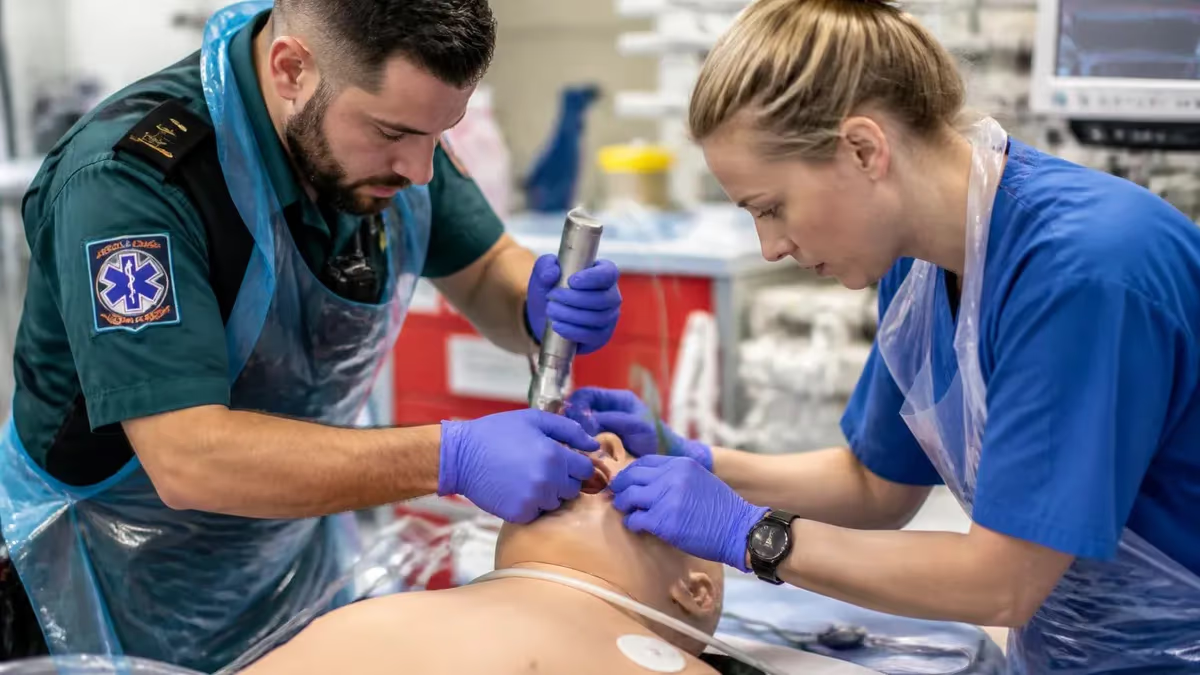
Understanding ACLS lethal rhythms is the most critical skill any advanced cardiovascular life support provider can master. These rhythms — ventricular fibrillation, pulseless ventricular tachycardia, pulseless electrical activity, and asystole — are directly responsible for the majority of sudden cardiac arrest deaths in the United States each year. The American Heart Association estimates that nearly 350,000 out-of-hospital cardiac arrests occur annually, and a provider's ability to rapidly identify and correctly treat the underlying rhythm determines whether a patient survives neurologically intact.
The four lethal rhythms recognized in ACLS protocols are divided into two major treatment pathways: shockable rhythms and non-shockable rhythms. Ventricular fibrillation (VF) and pulseless ventricular tachycardia (pVT) are shockable rhythms, meaning defibrillation is the primary intervention. Pulseless electrical activity (PEA) and asystole are non-shockable rhythms that require CPR, epinephrine, and aggressive identification of reversible causes using the Hs and Ts framework. Knowing which pathway to follow — and executing it without hesitation — is what the ACLS certification exam tests most heavily.
Healthcare providers frequently underestimate just how fast recognition must happen in a real resuscitation. From the moment a monitor displays a shockable rhythm, the AHA recommends that defibrillation occur within two minutes, and ideally within seconds if a defibrillator is already attached. Every 10-second delay in defibrillation for ventricular fibrillation reduces survival rates by approximately 7–10 percent. This is why rhythm recognition drills, scenario-based practice, and repeated exposure to 12-lead and monitor strips are built into every legitimate ACLS training curriculum.
Beyond initial recognition, ACLS providers must also understand the pharmacological management that accompanies each rhythm. Epinephrine 1 mg IV/IO is given every 3–5 minutes for all cardiac arrest rhythms, while amiodarone 300 mg (with a second dose of 150 mg) is reserved for refractory VF and pVT after the second shock. Lidocaine 1–1.5 mg/kg IV is an acceptable alternative when amiodarone is unavailable. Understanding the timing, dosage, and rationale behind each drug distinguishes a competent ACLS provider from one who is simply following a checklist.
Preparation for the ACLS written exam requires more than memorizing algorithms. Candidates must be able to look at a rhythm strip and within seconds identify rate, regularity, P-wave morphology, PR interval, QRS duration, and any aberrant features. The test presents rhythm strips in isolation, without clinical context, which means pattern recognition skills must be sharp and automatic. Providers who struggle with ECG interpretation are the most likely to fail the written component of their certification or recertification exam on the first attempt.
This guide covers everything you need to know about ACLS lethal rhythms, from the electrocardiographic features that define each rhythm to the step-by-step treatment algorithms you must execute during a simulated or real resuscitation. Each section includes clinical pearls, common exam pitfalls, and practical mnemonics that help the material stick under pressure. Whether you are preparing for your initial ACLS certification or your biennial renewal, working through this material systematically will build the confidence and competence needed to pass the exam and perform effectively at the bedside.
If you want to see how your rhythm recognition skills stack up right now, review the core concepts around acls lethal rhythms and acute presentations before diving into timed practice tests. The sections below break down each shockable and non-shockable rhythm in detail, walk through the algorithms, and explain the drug dosing sequences that the AHA expects every certified provider to know cold.
ACLS Lethal Rhythms by the Numbers
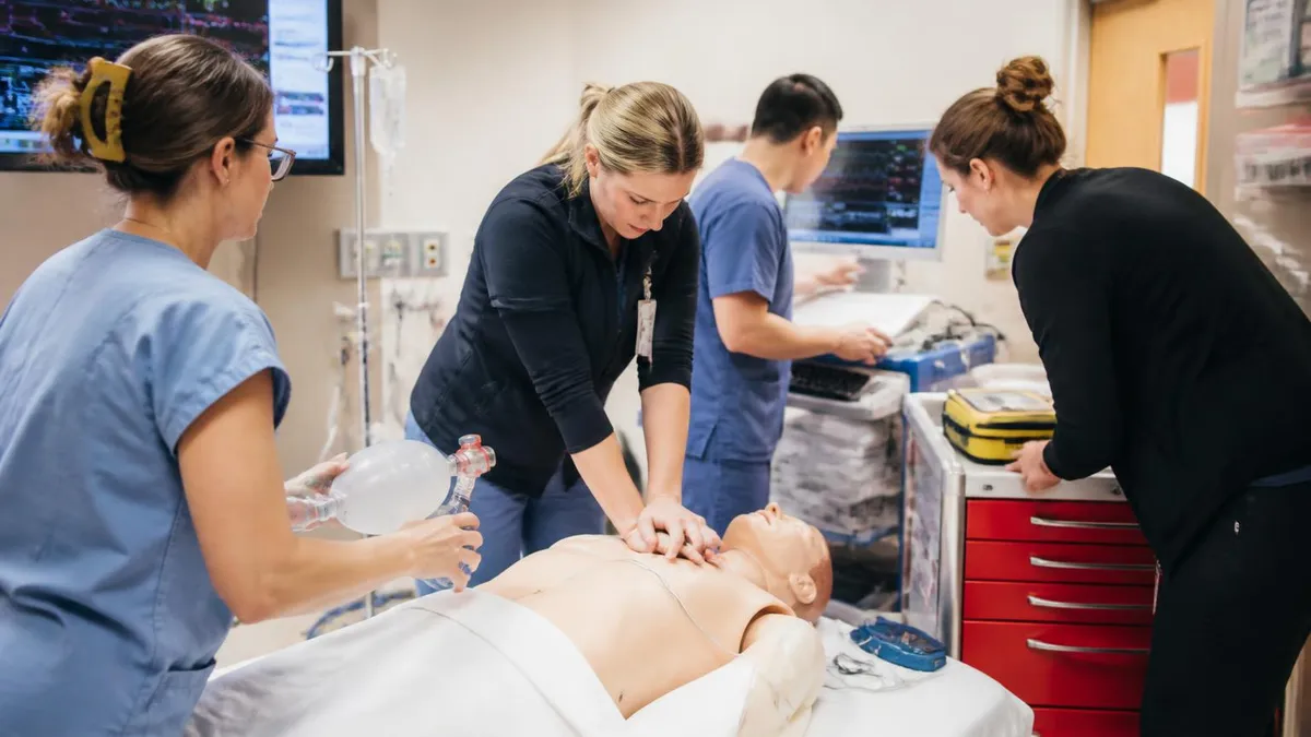
The Four ACLS Lethal Rhythms at a Glance
Chaotic, disorganized electrical activity with no discernible QRS complexes. The ventricles quiver instead of contracting. This is the most common initial rhythm in sudden cardiac arrest and the most treatable when defibrillation is delivered immediately.
Wide, regular QRS complexes at a rate >100 bpm with no palpable pulse. Often precedes VF and carries the same shockable treatment pathway. Monomorphic pVT has uniform QRS morphology; polymorphic pVT (Torsades) has twisting QRS amplitude.
Organized electrical activity on the monitor without a palpable pulse. PEA is a clinical diagnosis requiring immediate CPR and aggressive search for reversible causes using the Hs and Ts. Rate and rhythm on the monitor can appear almost normal.
Complete absence of electrical and mechanical cardiac activity, appearing as a flat or nearly flat line on the monitor. Confirm in two leads before diagnosing. Asystole carries the worst prognosis and requires CPR plus epinephrine without defibrillation.
Ventricular fibrillation is characterized on the ECG by rapid, chaotic electrical deflections that vary in amplitude and morphology with no recognizable QRS complex, P wave, or T wave. The baseline appears to oscillate between coarse (high-amplitude) and fine (low-amplitude) fibrillatory waves. Coarse VF generally responds better to defibrillation than fine VF, which may require a dose of epinephrine before the next shock to restore amplitude and increase the likelihood of successful cardioversion. Rate in VF is indeterminate and meaningless; the only relevant finding is the absence of organized electrical activity.
Pulseless ventricular tachycardia presents very differently. The ECG shows a rapid, regular rhythm with wide, bizarre QRS complexes typically exceeding 0.12 seconds in duration. The rate is usually between 100 and 250 bpm. Monomorphic pVT maintains the same QRS shape throughout, while polymorphic pVT — including Torsades de Pointes — shows twisting of the QRS axis around the isoelectric line, classically described as a rotation or spindle appearance. Torsades is particularly important to recognize because it is associated with prolonged QT interval and may respond to magnesium sulfate 1–2 g IV in addition to the standard shockable rhythm protocol.
Pulseless electrical activity encompasses a wide variety of organized rhythms seen on the monitor in a pulseless patient. You may see what appears to be a normal sinus rhythm, a bradycardic rhythm, a junctional escape, or even a relatively narrow complex tachycardia — all without a pulse.
This is why the clinical check is critical: never assume a patient is perfusing based on what the monitor shows. PEA is almost always caused by an underlying reversible condition, and the Hs and Ts mnemonic (covered in a dedicated section below) is the framework for rapid systematic identification and correction of those causes during resuscitation.
Asystole must be confirmed carefully because motion artifact, a loose lead, or a single-lead view can mimic a flat line. The AHA specifically recommends verifying asystole in two orthogonal leads before confirming the diagnosis. Fine ventricular fibrillation can occasionally be mistaken for asystole, especially on older or low-gain monitors. If there is any doubt, treat the rhythm as VF and attempt defibrillation. Delivering a shock to true asystole carries essentially no additional risk, while failing to shock fine VF carries catastrophic consequences for the patient.
On the ACLS certification exam, ECG interpretation questions typically present a rhythm strip without clinical context and ask you to identify the rhythm or select the correct immediate intervention. Common traps include a pulseless patient in a rhythm that looks organized (PEA), a wide-complex tachycardia that could be either VT or SVT with aberrancy (treat as VT if the patient is pulseless), and a very fine VF that resembles artifact. The exam expects you to know the defining ECG features of each lethal rhythm and to be able to apply the correct algorithm branch within seconds of seeing the strip.
Rate regularity and QRS morphology are the two most important axes for classifying rhythms during a code. A regular, wide-complex rhythm without a pulse = pVT. An irregular, chaotic rhythm without recognizable complexes = VF. An organized rhythm without a pulse = PEA. A flat or nearly flat line = asystole. Building this four-branch decision tree into automatic mental recall is the single most important skill you can develop for both the written exam and real-world resuscitation performance. Timed rhythm strip drills with a study partner or online simulator accelerate this process significantly.
Understanding how rhythms transition from one to another also matters clinically and for exam questions. VT commonly degenerates into VF if untreated. Successful defibrillation of VF sometimes produces a brief period of asystole or PEA before the sinus node recovers. Post-resuscitation bradycardia and hypotension are expected and managed with targeted temperature management, vasopressors, and close hemodynamic monitoring in the ICU. The ACLS written test may ask about post-resuscitation care, including target temperature ranges (32–36°C for therapeutic hypothermia) and the importance of avoiding hypoxia and hypotension in the immediate post-arrest period.
ACLS Drug Protocols for Lethal Rhythms
For ventricular fibrillation and pulseless ventricular tachycardia, the treatment sequence begins immediately with high-quality CPR and defibrillation as soon as the defibrillator is available. The first shock is delivered at 200 joules (biphasic) or 360 joules (monophasic). After each shock, CPR resumes immediately for two minutes before the rhythm is rechecked. Epinephrine 1 mg IV/IO is administered every 3–5 minutes starting after the second shock. Amiodarone 300 mg IV/IO is given for VF or pVT that persists after the third shock, with a second dose of 150 mg if the rhythm remains refractory.
Vasopressin 40 units IV/IO was previously listed as an alternative to the first or second dose of epinephrine, but the 2020 AHA guidelines removed vasopressin from the cardiac arrest algorithm, simplifying the protocol to epinephrine alone. Lidocaine 1–1.5 mg/kg IV/IO is an acceptable antiarrhythmic alternative when amiodarone is unavailable, with a second dose of 0.5–0.75 mg/kg if needed. Sodium bicarbonate is not recommended routinely but may be considered for hyperkalemia or tricyclic antidepressant overdose. Magnesium sulfate 1–2 g IV is specifically indicated for Torsades de Pointes.
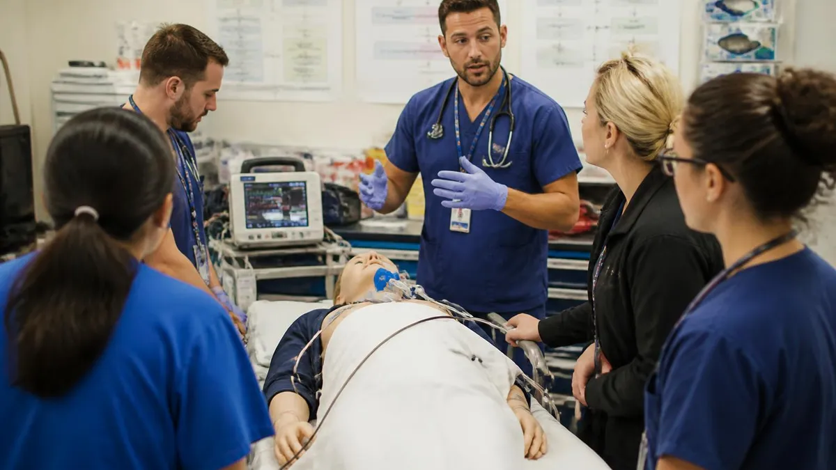
Early Defibrillation vs. Delayed Defibrillation: Key Trade-offs
- +Defibrillation within 3 minutes of VF onset yields survival rates of 40–60% in witnessed arrests
- +Automated external defibrillators (AEDs) allow rapid shock delivery before ACLS providers arrive
- +Each minute without defibrillation reduces VF survival by 7–10%, making speed the top priority
- +Modern biphasic defibrillators require lower energy (200 J) and cause less myocardial injury than older monophasic devices
- +Immediate defibrillation is preferred when the arrest is witnessed and a defibrillator is immediately available
- +Successful first shock terminates VF in over 90% of cases when delivered within the first few minutes
- −Defibrillation without prior CPR when arrest has been prolonged (>4–5 minutes) reduces shock success rates
- −Interrupting high-quality CPR for rhythm checks and shock delivery reduces perfusion pressure and coronary flow
- −Repeated shocks in refractory VF increase myocardial injury and reduce the chance of organized rhythm recovery
- −Fine VF that has not received epinephrine may not respond to defibrillation, wasting critical resuscitation time
- −Post-shock asystole or PEA is common and can be misinterpreted as resuscitation failure if the team is not prepared
- −Excessive interruptions for shock delivery without maintaining CPR compression fraction below 80% worsen outcomes
ACLS Lethal Rhythms Exam Preparation Checklist
- ✓Memorize the four lethal rhythms: VF, pVT, PEA, and asystole with their defining ECG features.
- ✓Practice identifying shockable vs. non-shockable rhythms on timed rhythm strips until automatic.
- ✓Know the cardiac arrest algorithm branches for shockable and non-shockable rhythms by memory.
- ✓Memorize epinephrine 1 mg IV/IO every 3–5 minutes for all pulseless rhythms.
- ✓Know amiodarone dosing: 300 mg first dose, 150 mg second dose for refractory VF/pVT.
- ✓Understand that defibrillation energy is 200 J biphasic (or 360 J monophasic) for VF and pVT.
- ✓List all 8 Hs and 8 Ts and be able to explain the intervention for each reversible cause.
- ✓Understand that vasopressin was removed from the 2020 AHA cardiac arrest algorithm.
- ✓Practice CPR quality metrics: rate 100–120/min, depth 2–2.4 inches, allow full chest recoil.
- ✓Review post-ROSC care priorities: SpO2 target, MAP target, TTM range, and 12-lead ECG timing.
Never Shock Asystole — Confirm in Two Leads First
The most dangerous error in cardiac arrest management is misidentifying fine VF as asystole. Always confirm a flat-line rhythm in at least two leads before diagnosing asystole. If there is any doubt about whether the rhythm is fine VF or true asystole, default to attempting defibrillation — a shock delivered to asystole causes no additional harm, but failing to shock VF is fatal.
The Hs and Ts framework is the standardized mnemonic the AHA uses to ensure that resuscitation teams systematically consider every potentially reversible cause of cardiac arrest during PEA and asystole. The eight Hs are: Hypovolemia, Hypoxia, Hydrogen ion (acidosis), Hypo/Hyperkalemia, Hypothermia, Hypoglycemia (added in later guideline revisions), and — depending on the version — occasionally Hypo/Hypermagnesemia. The eight Ts are: Tension pneumothorax, Tamponade (cardiac), Toxins, Thrombosis (pulmonary embolism), Thrombosis (coronary/ACS), and Trauma. Memorizing both lists and being able to rapidly cycle through them during a resuscitation is an explicit competency tested on the ACLS written exam.
Hypovolemia is the most common cause of PEA in trauma and post-surgical patients. The treatment is aggressive volume resuscitation with crystalloid or blood products, depending on the clinical context. Hypoxia — inadequate oxygenation before or during the arrest — is treated by ensuring a patent airway, appropriate ventilation, and supplemental oxygen. Hydrogen ion excess (severe metabolic acidosis) may respond to sodium bicarbonate, particularly in cases of prolonged arrest, tricyclic antidepressant overdose, or hyperkalemia. Electrolyte abnormalities, especially hyperkalemia, are treated with calcium chloride or calcium gluconate IV, sodium bicarbonate, glucose, and insulin.
Hypothermia is a particularly important reversible cause because patients in cardiac arrest due to severe hypothermia (core temperature below 30°C) can survive neurologically intact with aggressive rewarming — even after prolonged resuscitation. The adage in wilderness and emergency medicine is that a patient is not dead until they are warm and dead. Active internal rewarming strategies including warmed IV fluids, warmed humidified oxygen, bladder irrigation, peritoneal lavage, and in extreme cases extracorporeal membrane oxygenation (ECMO) are all appropriate interventions. Standard ACLS medications may be less effective until the core temperature rises above 30°C.
Tension pneumothorax presents during resuscitation with decreased or absent breath sounds on one side, tracheal deviation away from the affected side, and progressive hemodynamic deterioration. It is treated with immediate needle decompression at the second intercostal space, midclavicular line, followed by chest tube placement. Cardiac tamponade — blood accumulating in the pericardial space compressing the heart — causes PEA and is treated with pericardiocentesis or emergency pericardial window. The classic Beck's triad of hypotension, muffled heart sounds, and jugular venous distension may be present but is often absent during cardiac arrest.
Pulmonary embolism as a cause of PEA is suggested by the clinical context: sudden collapse in a patient with recent immobility, surgery, malignancy, or known hypercoagulable state. Massive PE can cause sudden pulseless arrest with a right-heart strain pattern on ECG (S1Q3T3, right bundle branch block, sinus tachycardia before arrest). Systemic thrombolysis with alteplase 50–100 mg IV is an option for confirmed or highly suspected PE during cardiac arrest, but CPR must continue for at least 60–90 minutes after administration to allow the drug to work. Emergency surgical or catheter-directed embolectomy are alternatives when thrombolytics fail or are contraindicated.
Toxin-induced cardiac arrest encompasses overdoses of beta-blockers, calcium channel blockers, digoxin, tricyclic antidepressants, cocaine, opioids, and many other substances. Each toxin has specific antidotes or treatment strategies that may need to be layered on top of standard ACLS. Opioid overdose causing respiratory arrest with secondary cardiac arrest should receive naloxone. Beta-blocker and calcium channel blocker toxicity may respond to high-dose insulin therapy, calcium, glucagon, and lipid emulsion therapy. These are tested on the ACLS exam as special circumstances that require modifications to standard algorithm management.
Understanding when to consider terminating resuscitation efforts is also part of ACLS provider competency. While there are no absolute time limits specified in AHA guidelines, factors associated with poor outcomes and reasonable resuscitation termination include unwitnessed asystole without a shockable rhythm at any point, prolonged resuscitation without return of spontaneous circulation, absence of reversible causes, and end-tidal CO2 consistently below 10 mmHg after 20 minutes of CPR. These decisions are always made by the team leader in context, and the ACLS exam may ask about prognostic indicators rather than specific time cutoffs.
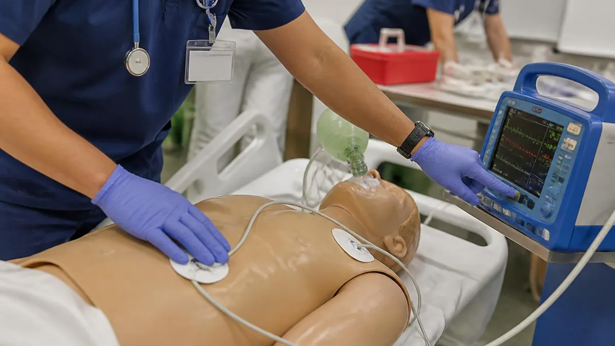
The 2020 AHA ACLS guidelines removed vasopressin from the cardiac arrest algorithm. Epinephrine 1 mg IV/IO every 3–5 minutes is now the sole vasopressor recommended for all pulseless rhythms. If your study materials or previous certification training included vasopressin as an option, update your knowledge base before your exam to reflect current guidelines — outdated information is a common source of errors on the written test.
High-performance resuscitation teams — the model promoted by both the AHA and resuscitation researchers — assign specific roles before and during every code to eliminate confusion, reduce errors, and maximize the quality of each intervention. The standard role assignments for a five-person ACLS team are: compressor, airway manager, IV/IO and medication administrator, monitor/defibrillator operator, and team leader.
The team leader does not perform hands-on interventions unless absolutely necessary; their role is to maintain situational awareness, direct the algorithm, call for rhythm checks, verify medication doses, communicate with the family and receiving unit, and keep accurate documentation of all interventions and their timing.
Closed-loop communication is one of the most consistently tested team dynamics concepts on the ACLS written exam. In closed-loop communication, the team leader issues a clear verbal directive, the team member receiving the directive acknowledges it by name and content, performs the task, and verbally confirms completion to the team leader. For example: team leader says, "Draw up epinephrine 1 mg IV now." The medication nurse responds, "Epinephrine 1 mg IV — drawing up now." After administration: "Epinephrine 1 mg IV given at 14:32." This three-step loop prevents errors caused by ambiguous communication in high-noise, high-stress environments.
CPR quality metrics are monitored in real time on modern monitors equipped with accelerometers and impedance sensors. Providers are expected to maintain a compression rate of 100–120 per minute, a compression depth of at least 2 inches (5 cm) but no more than 2.4 inches (6 cm), allow complete chest recoil between compressions, and minimize interruptions so that the chest compression fraction (CCF) remains at or above 60%, ideally above 80%. Hyperventilation — giving breaths too rapidly or with too much volume — increases intrathoracic pressure, reduces venous return, and lowers coronary perfusion pressure.
The ventilation rate target before advanced airway placement is 30:2 (30 compressions to 2 breaths).
Rotating compressors every two minutes is standard practice because fatigue-related compression depth degradation occurs rapidly, often without the compressor being aware of it. Research has shown that compression quality begins to decline within 60 to 90 seconds in many providers, particularly in scenarios requiring sustained effort. The two-minute rhythm check cycle coincides with the compressor rotation, allowing a seamless handoff that minimizes interruption time. The incoming compressor should be positioned and ready before the outgoing compressor stops, and the switch should occur in under five seconds.
Vascular access during cardiac arrest is obtained preferentially via large-bore peripheral IV (antecubital or external jugular) or intraosseous (IO) access. IO access — typically at the proximal humerus or proximal tibia — can be established in under 60 seconds with a powered drill device and delivers drugs and fluids as rapidly as central venous access in a collapsed vasculature.
Central venous access is not recommended during active chest compressions because of the procedural interruptions it requires and the risk of pneumothorax. Drug delivery via IO or peripheral IV should be followed by a 20 mL fluid bolus and limb elevation to accelerate central circulation of the drug.
The team leader is also responsible for recognizing when special circumstances apply and modifying the algorithm accordingly. Pregnancy, for instance, requires manual left uterine displacement throughout CPR to relieve aortocaval compression, and perimortem cesarean delivery should be initiated within four to five minutes of maternal cardiac arrest if ROSC has not been achieved. Opioid overdose causing isolated respiratory arrest without cardiac arrest should be treated with naloxone and rescue breathing before full ACLS arrest management. Hypothermic arrest, as discussed earlier, requires a longer resuscitation and different medication thresholds than normothermic arrest.
Providers who want to deepen their understanding of the teamwork and communication principles embedded in the ACLS curriculum should study the AHA's HeartCode ACLS learning system or attend a high-fidelity simulation course. The written exam tests cognitive knowledge, but the skills station tests behavioral performance — how fluidly you move through the algorithm, how clearly you communicate, and how quickly you adapt when a simulated patient's rhythm changes unexpectedly. Both components must be passed to earn certification, and preparation for the skills station requires practice in a team setting, not just solo study.
Many readers pair this guide with the aha acls algorithms to check their readiness before exam day.
Building a practical study plan for ACLS lethal rhythms requires more than reading a textbook from cover to cover. The most effective approach combines passive review of algorithms and drug dosing with active recall practice using rhythm strips, scenario-based questions, and timed simulations. Begin your preparation by creating a one-page summary of the cardiac arrest algorithm with both the shockable and non-shockable branches, all drug doses and intervals, and the full Hs and Ts list. This reference sheet becomes the foundation for spaced repetition review over the two to four weeks before your exam.
Rhythm strip practice is available through numerous free and paid online platforms, but the highest-yield approach is to work through strips that are presented in the same format as the ACLS written exam: a short strip of 6–10 seconds displayed without any clinical context, followed by a multiple-choice question about the rhythm or the correct intervention.
Aim to identify each of the four lethal rhythms within three seconds of viewing the strip. If you need more than five seconds, you are not yet at exam-ready speed. Use flashcards with rhythm strips on one side and the rhythm name plus treatment algorithm on the other for efficient repetition.
Pharmacology is the second most heavily tested domain after rhythm recognition on the ACLS written exam. Create a drug card for each cardiac arrest medication that includes the drug name, indication, dose, route, timing, and key contraindications or special considerations. Epinephrine, amiodarone, lidocaine, magnesium sulfate, sodium bicarbonate, calcium chloride, adenosine (for stable SVT, not cardiac arrest), atropine (for symptomatic bradycardia, not routinely for cardiac arrest), and vasopressors for post-ROSC hemodynamic support should all be on your list. Pay special attention to the distinction between drugs used during active cardiac arrest versus drugs used for peri-arrest rhythms.
Scenario-based practice is the most effective way to integrate rhythm recognition with algorithm execution and drug selection. Ask a colleague to present you with a verbal or written scenario — a patient collapses, the monitor shows a specific rhythm — and practice verbalizing your response in real time. Include the team role assignments, the first intervention, the drug sequence, the rhythm check timing, and the Hs and Ts you would consider. This type of active practice mimics the skills station component of the ACLS exam and builds the automatic decision-making pathways that are essential during an actual resuscitation.
One of the most common reasons candidates fail the ACLS written exam on the first attempt is failure to distinguish between peri-arrest rhythms (stable tachycardias, bradycardias, and rhythms with a pulse) and true cardiac arrest rhythms (pulseless). The exam frequently presents a rhythm strip or short clinical scenario and asks whether the patient is stable or unstable, and whether immediate synchronized cardioversion, unsynchronized defibrillation, medications, or CPR is the correct first step.
The critical distinction: synchronized cardioversion is used for organized rhythms with a pulse (unstable tachycardias); unsynchronized defibrillation (a shock) is used for VF and pVT (no pulse, no organized rhythm); and CPR plus epinephrine is used for PEA and asystole.
Post-exam preparation does not end with passing the written test. ACLS certification requires demonstrated competency in the megacode skills station, which tests your ability to manage a simulated cardiac arrest from initial rhythm recognition through ROSC or termination of resuscitation. The AHA instructor evaluating the megacode uses a standardized checklist that includes correct algorithm sequencing, appropriate drug dosing and timing, team communication quality, and CPR technique. Reviewing the megacode evaluation criteria in advance and practicing at least two or three full scenarios with a team will dramatically increase your confidence and performance on exam day.
Finally, candidates preparing for ACLS renewal after a two-year lapse should not underestimate how much the guidelines may have changed. The 2020 AHA updates included several significant algorithm modifications beyond the removal of vasopressin, including updated guidance on advanced airway timing, CPR quality monitoring, and post-resuscitation care bundles. Reading the AHA ACLS Provider Manual for the current guidelines cycle, supplementing with high-quality practice tests that reflect 2020 updates, and attending a refresher simulation session before your renewal exam are the three most effective strategies for maintaining and demonstrating competency in ACLS lethal rhythm management.
ACLS Questions and Answers

ACLS Acute Coronary Syndrome Algorithm: Complete Study Guide for Certification Prep
ACLS Renewal Class: Complete 2026 Guide to Recertification Requirements, Costs, Formats & Pass Strategies
ACLS Bradycardia Algorithm: Complete 2026 Guide to Recognition, Treatment Steps & Drug Doses

ACLS CPR Certification: Complete Training Guide for Healthcare Professionals
ACLS Study Guide: Complete 2026 Certification Prep with Algorithms, Drugs & Practice Tests
About the Author

Registered Nurse & Healthcare Educator
Johns Hopkins University School of NursingDr. Sarah Mitchell is a board-certified registered nurse with over 15 years of clinical and academic experience. She completed her PhD in Nursing Science at Johns Hopkins University and has taught NCLEX preparation and clinical skills courses for nursing students across the United States. Her research focuses on evidence-based exam preparation strategies for healthcare certification candidates.