
What Does an MRI Do? How Magnetic Resonance Imaging Works, What It Detects, and What to Expect
Learn what does an MRI do, how magnetic resonance imaging works, what conditions it detects, and what patients can expect during the scan. 📗
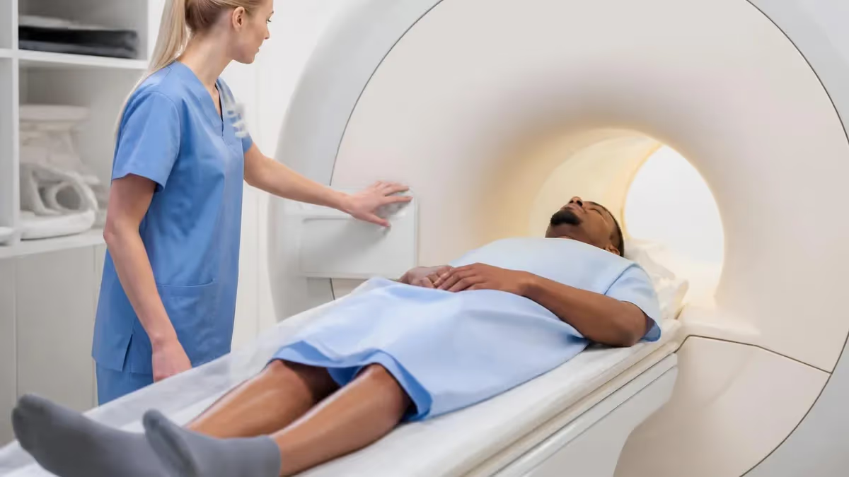
Understanding what does an MRI do is essential knowledge for patients preparing for a scan, students studying medical imaging, and anyone curious about how modern medicine peers inside the human body without a single incision. Magnetic Resonance Imaging — MRI for short — is a noninvasive diagnostic technique that uses powerful magnetic fields and radiofrequency pulses to generate detailed, high-contrast images of soft tissues, organs, joints, and the nervous system. Unlike X-rays or CT scans, MRI produces no ionizing radiation, making it a safer choice for many clinical scenarios, particularly when repeated imaging is required over time.
At its core, an MRI machine is a sophisticated physics instrument. The large cylindrical magnet at the center of the scanner creates a static magnetic field measured in Tesla units — typically 1.5T or 3T in clinical settings. When a patient enters this field, hydrogen atoms in the body's water molecules align with the magnetic field much the way compass needles align with Earth's magnetic poles.
A radiofrequency pulse is then transmitted through the patient, temporarily knocking those hydrogen protons out of alignment. As the protons relax back to their original orientation, they emit radiofrequency signals that the scanner's receiver coils detect and a computer translates into cross-sectional images.
The level of anatomical detail an MRI can reveal is remarkable. Clinicians can visualize the delicate white and gray matter structures of the brain, detect tiny tears in knee ligaments, identify inflammation in spinal discs, and spot early-stage tumors that other imaging modalities might miss entirely. The contrast between different tissue types — muscle, fat, fluid, cartilage, and bone marrow — is far superior in MRI than in conventional X-ray or ultrasound, which is why MRI has become the gold standard for neurological, musculoskeletal, and oncological imaging across the United States.
One of the most powerful aspects of MRI is its versatility. Technologists and radiologists can adjust scan sequences and parameters to highlight specific tissue characteristics. T1-weighted sequences excel at showing anatomy and fat-containing structures. T2-weighted sequences reveal fluid and pathology. Contrast agents containing gadolinium can be injected intravenously to further illuminate blood vessels, tumors, and areas of active inflammation. Functional MRI (fMRI) tracks blood flow in the brain during cognitive tasks, while diffusion-weighted imaging (DWI) detects acute strokes within minutes of onset by measuring the movement of water molecules at the cellular level.
For students and credentialing candidates preparing for the ARRT MRI registry exam, a thorough grasp of MRI physics and clinical applications is non-negotiable. The exam tests your understanding of everything from magnet safety classifications to pulse sequence optimization, contrast mechanism, and patient screening protocols. Resources like what does an mri do in practice on specific body regions help bridge theoretical knowledge and clinical reality, giving you concrete examples of how scan protocols are tailored to diagnose real pathology in patients every day.
Beyond exam preparation, understanding MRI fundamentals empowers technologists to communicate more confidently with patients, troubleshoot image artifacts, and collaborate effectively with radiologists when protocol modifications are needed. Patients who understand how MRI works tend to experience less scan anxiety, remain still during acquisition, and produce higher-quality diagnostic images — a direct benefit to clinical outcomes. This article covers the physics principles, clinical applications, safety considerations, and preparation steps that define what an MRI does from the moment a patient walks in to the moment images reach the interpreting physician.
Whether you are a radiology student building your knowledge base, a healthcare professional seeking a quick refresher, or a patient trying to make sense of your upcoming appointment, the sections below will give you a comprehensive, accurate, and practically useful understanding of MRI technology. From the electromagnetic science underlying image formation to the specific conditions MRI diagnoses most effectively, you will leave with a complete picture of why MRI has become one of the most powerful diagnostic tools in modern medicine.
MRI by the Numbers
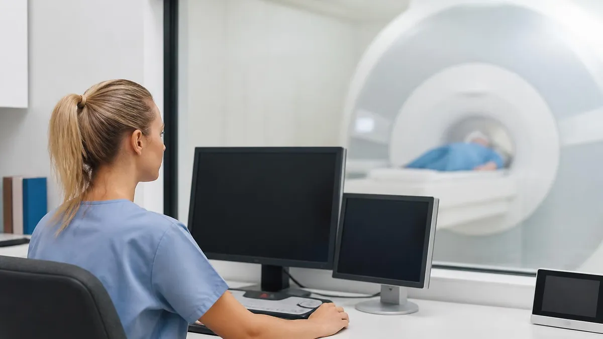
How an MRI Scan Works: Step by Step
Patient Screening and Preparation
Positioning and Coil Placement
Magnetic Field Alignment
Radiofrequency Pulse Transmission
Signal Detection and Relaxation
Image Reconstruction and Reporting
MRI's clinical reach spans virtually every organ system in the human body, but its greatest strengths lie in neurological, musculoskeletal, and oncological imaging. In the brain, MRI can detect ischemic stroke within minutes using diffusion-weighted sequences, identify demyelinating lesions characteristic of multiple sclerosis, locate intracranial tumors as small as a few millimeters, and diagnose hemorrhage, aneurysm, and vascular malformations with extraordinary precision. Functional MRI extends these capabilities by mapping brain activity, helping neurosurgeons plan tumor resections to avoid eloquent cortex responsible for speech or motor function.
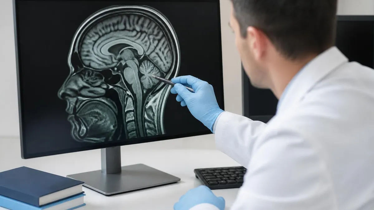
Spinal imaging is another domain where MRI delivers unmatched diagnostic value. The ability to visualize intervertebral discs, the spinal cord itself, nerve roots, and surrounding ligaments in multiple planes gives clinicians a comprehensive picture of the spine that plain X-rays simply cannot provide. Conditions like herniated discs, spinal stenosis, cord compression, syringomyelia, and spinal tumors are routinely diagnosed through MRI-based protocols. Sagittal T2-weighted sequences of the spine create a visual map that neurosurgeons use to plan decompression surgeries and interventional pain specialists rely on to guide targeted injections.
Musculoskeletal MRI has transformed orthopedic diagnosis and surgical planning. When a patient tears their anterior cruciate ligament, the injury can be graded precisely by an MRI that shows the degree of fiber disruption, associated bone bruising, and any co-existing meniscal pathology. Shoulder MRI differentiates full-thickness from partial-thickness rotator cuff tears, identifies labral pathology responsible for instability, and evaluates the rotator interval. For the hip, MRI arthrography using intra-articular gadolinium contrast is the gold standard for detecting acetabular labral tears, femoroacetabular impingement, and early avascular necrosis before plain radiographs show any abnormality.
In oncology, MRI plays a pivotal role at every stage of the cancer care continuum — from initial detection and staging to treatment planning and post-therapy surveillance. Breast MRI is recommended for high-risk patients and as a problem-solving tool when mammography and ultrasound are inconclusive. Prostate MRI using multiparametric protocols (mpMRI) has revolutionized the detection of clinically significant prostate cancer, guiding targeted biopsies that dramatically improve cancer detection rates while reducing unnecessary procedures. Liver MRI with hepatobiliary contrast agents can characterize lesions that remain indeterminate on CT, distinguishing benign hemangiomas from metastatic deposits or primary hepatocellular carcinoma.
Cardiac MRI represents one of the most technically demanding but diagnostically rewarding applications of the modality. By gating image acquisition to the cardiac cycle using electrocardiogram (ECG) triggers, cardiac MRI captures the beating heart in exquisite detail. It is considered the reference standard for measuring ventricular volumes, ejection fraction, and myocardial mass. Late gadolinium enhancement sequences reveal myocardial fibrosis and scar tissue, allowing precise characterization of cardiomyopathies, myocarditis, and infarct patterns that guide treatment decisions in heart failure and arrhythmia management.
Abdominal and pelvic MRI has grown significantly in clinical adoption, driven by advances in fast imaging sequences that reduce the impact of respiratory and peristaltic motion. Magnetic resonance cholangiopancreatography (MRCP) is a noninvasive technique for imaging the biliary tree and pancreatic duct, replacing diagnostic endoscopic retrograde cholangiopancreatography (ERCP) in many clinical situations. MRI of the pelvis is the primary staging modality for rectal cancer, cervical cancer, endometrial cancer, and ovarian pathology, providing detailed delineation of tumor extension into surrounding structures that determines surgical approach and radiation field planning.
Pediatric imaging represents a specialized but critically important application of MRI. Because children are far more sensitive to ionizing radiation than adults, the principle of ALARA — as low as reasonably achievable — pushes radiologists to prefer MRI over CT whenever possible. Pediatric neuroimaging using MRI identifies congenital brain malformations, perinatal hypoxic-ischemic injury, epileptic foci, and developmental anomalies with unmatched clarity. Sedation or general anesthesia is sometimes required for young children who cannot remain still during extended scan times, but advances in fast pediatric protocols have substantially reduced the need for anesthesia in many centers.
MRI Practice Test Questions
Prepare for the MRI - Magnetic Resonance Imaging exam with our free practice test modules. Each quiz covers key topics to help you pass on your first try.
MRI Knowledge
MRI Exam Questions covering Knowledge. Master MRI Test concepts for certification prep.
MRI Physics
Free MRI Practice Test featuring Physics. Improve your MRI Exam score with mock test prep.
MRI Anatomy and Pathology
MRI Test Prep for MRI Anatomy and Pathology. Practice MRI Quiz questions and boost your score.
MRI Anatomy and Positioning
MRI Questions and Answers on MRI Anatomy and Positioning. Free MRI practice for exam readiness.
MRI Contrast Agents
Free MRI Quiz on MRI Contrast Agents. MRI Exam prep questions with detailed explanations.
MRI Patient Care and Positioning
MRI Practice Questions for MRI Patient Care and Positioning. Build confidence for your MRI certification exam.
Types of MRI Sequences and What They Show
T1-weighted images are produced using short repetition time (TR) and short echo time (TE) parameters. Fat appears bright on T1, making these sequences excellent for depicting anatomy, fatty marrow, subacute hemorrhage, and post-contrast enhancement after gadolinium injection. The excellent anatomical detail of T1 sequences makes them the workhorse for contrast studies in the brain, spine, and abdomen, where enhancing lesions stand out sharply against the darker background tissue.
T2-weighted images use long TR and long TE, causing fluid to appear very bright. This sequence is highly sensitive for pathology because most disease processes — inflammation, edema, tumors, and infection — increase tissue water content. The bright fluid signal on T2 makes it invaluable for detecting spinal cord lesions in multiple sclerosis, joint effusions in orthopedic imaging, and cerebral infarcts in neurological emergencies. FLAIR (fluid-attenuated inversion recovery) is a T2 variant that suppresses free fluid, improving lesion detection in the brain's cortex and periventricular white matter.
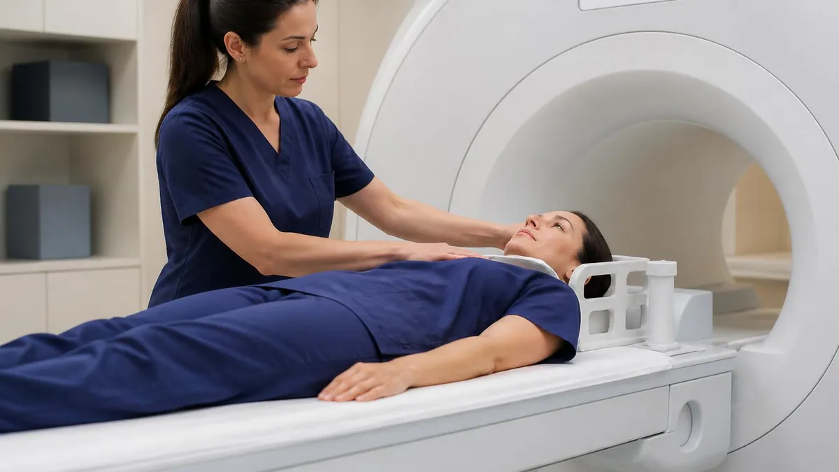
MRI Advantages and Limitations: What You Should Know
- +No ionizing radiation — safe for pregnant patients and repeat imaging over time
- +Superior soft tissue contrast compared to CT, X-ray, and ultrasound
- +Multiplanar imaging — axial, sagittal, coronal, and oblique views in a single exam
- +Highly versatile with dozens of sequences tailored to specific clinical questions
- +Functional capabilities including fMRI, DWI, spectroscopy, and perfusion imaging
- +Large field of view enabling whole-body and multi-region protocols
- −Long scan times (20–90 minutes) compared to CT scans (seconds to minutes)
- −Loud acoustic noise from gradient coil switching requires hearing protection
- −Claustrophobia affects approximately 5–10% of patients, sometimes requiring sedation
- −Strict contraindications for ferromagnetic implants including many pacemakers and cochlear implants
- −Gadolinium contrast carries risk of nephrogenic systemic fibrosis in severe kidney disease
- −High equipment and operational costs result in higher patient charges than CT or X-ray
Pros and Cons at a Glance
| Pros | Cons |
|---|---|
| No ionizing radiation — safe for pregnant patients and repeat imaging over time | Loud acoustic noise from gradient coil switching requires hearing protection |
| Superior soft tissue contrast compared to CT, X-ray, and ultrasound | Strict contraindications for ferromagnetic implants including many pacemakers and cochlear implants |
| Multiplanar imaging — axial, sagittal, coronal, and oblique views in a single exam | Gadolinium contrast carries risk of nephrogenic systemic fibrosis in severe kidney disease |
| Highly versatile with dozens of sequences tailored to specific clinical questions | High equipment and operational costs result in higher patient charges than CT or X-ray |
MRI Safety and Preparation Checklist for Patients
- ✓Disclose all metal implants, surgical hardware, and medical devices to your technologist before entering the MRI room.
- ✓Remove all jewelry, piercings, hair clips, and ferromagnetic accessories before entering Zone III or IV.
- ✓Inform staff if you have a pacemaker, neurostimulator, cochlear implant, or any implanted electronic device.
- ✓Tell your technologist and ordering physician if you are pregnant or suspect you may be pregnant.
- ✓Disclose any history of kidney disease or renal insufficiency if gadolinium contrast has been ordered.
- ✓Notify staff of any previous allergic reactions to MRI contrast agents or iodinated contrast.
- ✓Follow fasting instructions (typically 4–6 hours for abdominal/pelvic scans with contrast).
- ✓Wear comfortable, metal-free clothing or change into a hospital gown provided by the facility.
- ✓Practice relaxation or breathing techniques in advance if you experience claustrophobia or anxiety.
- ✓Ask your physician about sedation options if you are unable to remain still for extended scan durations.
MRI Is the Gold Standard for Soft Tissue Diagnosis
When a physician needs to evaluate soft tissue pathology — brain lesions, spinal cord compression, ligament tears, or organ tumors — MRI consistently outperforms every other imaging modality in sensitivity and specificity. Its ability to generate multiplanar images with exceptional contrast resolution, without using ionizing radiation, has made it the first-line imaging choice in neurology, orthopedics, oncology, and cardiology across major US medical centers.
Comparing MRI to other medical imaging modalities reveals both its exceptional strengths and the specific clinical situations where alternative technologies remain preferable. Plain radiography — the conventional X-ray — uses ionizing radiation to produce 2D projectional images that excel at depicting cortical bone, detecting fractures, and evaluating lung fields. X-rays are fast, inexpensive, and widely available, making them the first-line study for suspected fractures, pneumonia, and bowel obstruction. However, their inability to differentiate soft tissues limits their diagnostic utility for conditions affecting muscles, cartilage, tendons, and the nervous system — precisely the domain where MRI excels.
Computed tomography (CT) represents the primary competitor to MRI in many clinical scenarios. CT uses X-ray attenuation differences measured from multiple angles to generate cross-sectional images with excellent spatial resolution for calcified structures, dense organs, and vascular anatomy when combined with iodinated contrast. CT is faster than MRI — whole-body CT can be completed in under a minute — making it the preferred modality in trauma, stroke triage for hemorrhage exclusion, and acute abdominal pain evaluation. CT's radiation dose, however, is a significant consideration, particularly for pediatric patients and individuals requiring repeated imaging.
Ultrasound uses high-frequency sound waves to generate real-time images without radiation, making it safe for pregnant patients and ideal for dynamic imaging of moving structures like the heart, fetal anatomy, and vascular flow. Ultrasound is the first-line modality for evaluating the thyroid, gallbladder, ovaries, and uterus. Its limitations include operator dependence, limited penetration in obese patients, and inability to image through bone or air-containing structures. For deep retroperitoneal structures or intracranial pathology, MRI offers far superior visualization compared to ultrasound.
Nuclear medicine techniques including PET (positron emission tomography) and SPECT (single photon emission computed tomography) measure physiological and metabolic activity by imaging the biodistribution of radioactive tracers. PET-CT combining functional metabolic data with CT anatomy is the standard of care for staging most malignancies and evaluating treatment response in oncology. PET detects disease based on glucose metabolism, making it highly sensitive for metabolically active tumors and metastases. However, its spatial resolution for anatomical detail remains inferior to MRI, and the ionizing radiation dose from PET tracers is a limiting factor for certain patient populations.
In practice, multimodality imaging — combining MRI with CT, PET, or ultrasound — is common in complex cases where each modality contributes complementary information. For example, a patient with suspected liver malignancy might undergo ultrasound for initial detection, contrast-enhanced MRI for characterization, and PET-CT for staging. Understanding how MRI fits within this broader imaging ecosystem is critical for radiologists and technologists who must help referring clinicians select the right study for each clinical question, balancing diagnostic yield against cost, availability, radiation exposure, and patient tolerance.
Interventional MRI represents an emerging frontier where the diagnostic capabilities of MRI are combined with minimally invasive therapeutic procedures. MRI-guided biopsy allows sampling of lesions that are visible only on MRI and invisible on CT or ultrasound — particularly important in prostate and breast imaging. MRI-guided focused ultrasound (MRgFUS) is an entirely noninvasive treatment for uterine fibroids and essential tremor, using precisely focused acoustic energy to ablate tissue while MRI provides real-time thermal monitoring. These interventional applications further expand the clinical scope of what MRI can do beyond diagnosis into active treatment delivery.
The future of MRI includes ultra-high-field systems at 7T and beyond, which are transitioning from research into clinical practice for neuroimaging and musculoskeletal applications. Artificial intelligence and deep learning algorithms are being integrated into every phase of the MRI workflow — from automated patient screening and scan protocol selection to image reconstruction, artifact reduction, and AI-assisted lesion detection. These advances promise to make MRI faster, more accurate, and more accessible, extending its diagnostic reach into community hospitals and outpatient centers that currently rely on older-generation equipment.
MRI scanners generate extremely powerful magnetic fields that can turn ferromagnetic objects into dangerous projectiles. Patients and staff must never bring ferromagnetic items — oxygen tanks, IV poles, tools, wheelchairs — into Zone IV (the magnet room). Pacemakers, neurostimulators, and many older implanted devices are MRI-incompatible and may malfunction, heat, or migrate in the magnetic field. Always complete thorough patient screening before every scan, regardless of previous MRI history at your or other facilities.
For students pursuing ARRT MRI certification and registered technologists seeking to deepen their clinical expertise, a structured approach to MRI education combines physics theory, anatomy knowledge, clinical protocols, and hands-on scanning experience. The MRI certification examination administered by the American Registry of Radiologic Technologists (ARRT) covers content areas spanning patient care, safety, image production, and procedures — all of which demand both conceptual understanding and practical application skills. Building a solid foundation in MRI physics is the essential first step, because protocol optimization, artifact recognition, and image quality troubleshooting all flow from understanding how the scanner generates signal.
Physics mastery for the MRI registry requires fluency in electromagnetism fundamentals, Larmor frequency calculations, relaxation time constants (T1, T2, and T2*), k-space trajectories, and gradient function. Understanding how changing TR and TE affects image contrast, how gradient echo sequences differ from spin echo, and how parallel imaging acceleration factors reduce scan time are all topics that appear on the registry examination and arise daily in clinical practice. Candidates who struggle with physics often find that visual analogies — comparing proton relaxation to a spinning top losing energy — help translate abstract mathematics into intuitive understanding.
Anatomy and pathology knowledge is equally critical. MRI technologists must recognize normal anatomy on axial, sagittal, and coronal images of every body region, identify common pathological findings, and understand how clinical indications drive protocol selection. A referral for a patient with knee pain following a sports injury requires a different set of sequences than a referral for possible meniscal pathology in an older patient with osteoarthritis. Being able to communicate with radiologists about image quality issues, suggest protocol modifications for specific clinical questions, and recognize when a scan requires immediate physician attention are hallmarks of a competent, experienced MRI technologist.
Patient care skills in MRI encompass screening, communication, contrast administration, monitoring, and emergency response. Gadolinium-based contrast agents (GBCAs) require intravenous access, informed consent, and post-injection monitoring for rare hypersensitivity reactions. All gadolinium agents are classified by their retention profile — linear versus macrocyclic chelation chemistry affects how much gadolinium is retained in tissues after administration, a topic that has received increasing regulatory attention from the FDA since 2017. Technologists must be current on FDA guidance, institutional contrast policies, and the evidence regarding gadolinium deposition to counsel patients appropriately and practice safely.
MRI safety beyond contrast agents encompasses the Zone system established by the American College of Radiology (ACR) White Paper on MR Safety. Zone I is publicly accessible. Zone II is the screening area where patients transition from uncontrolled to controlled access. Zone III is the control room — restricted to screened MRI personnel.
Zone IV is the magnet room itself, where all ferromagnetic materials pose a projectile hazard and electronic devices may malfunction. All MRI facilities are required to implement and enforce these zone restrictions, and technologists must understand the rationale behind each restriction to uphold safety standards when unusual situations arise.
Continuing education and professional development keep MRI technologists at the forefront of a rapidly evolving field. New sequences, AI-assisted workflows, next-generation contrast agents, and expanding interventional applications emerge regularly. The ARRT requires continuing education credits for credential maintenance, and professional organizations including the Society for MR Radiographers and Technologists (SMRT) offer specialty education resources. Attending professional conferences, reviewing radiology literature, and participating in peer-learning activities within your department are all effective ways to sustain and expand your knowledge beyond what the credentialing examination alone requires.
For those just beginning their MRI education journey, practice tests aligned to ARRT examination content are among the most efficient preparation tools available. Practicing under timed conditions with registry-style questions builds the test-taking stamina and pattern recognition needed to perform well on exam day. Reviewing detailed explanations for both correct and incorrect answers reinforces conceptual understanding rather than rote memorization. Combined with clinical hours on the scanner and thorough study of MRI physics and anatomy, a well-designed practice test regimen gives candidates the confidence and knowledge they need to achieve registry certification on their first attempt.
Preparing effectively for any MRI-related examination — whether a credentialing exam, a continuing education module, or a clinical competency assessment — requires a deliberate, organized study strategy built around the core content domains. The most successful candidates do not simply read textbooks passively; they actively engage with material through practice questions, case reviews, self-quizzing, and peer discussion. Spacing your study sessions over weeks rather than cramming in the days before an exam leverages the neuroscience of spaced repetition, dramatically improving long-term retention of complex physics and anatomy content.
Start your study plan with an honest diagnostic assessment. Take a full-length practice exam under realistic conditions to identify your strongest and weakest content areas before investing significant time in comprehensive review. If your practice scores indicate strength in patient care but weakness in MRI physics, front-load your study schedule with physics topics — Larmor frequency, T1/T2 relaxation, gradient design, and artifact recognition — before moving into areas where you already perform well. This targeted approach maximizes the efficiency of your available study time and prevents the common mistake of over-reviewing comfortable material while neglecting challenging concepts.
Physics study for MRI benefits from visual and spatial learning strategies. Drawing diagrams of the magnetization vector during RF excitation and relaxation, sketching k-space trajectories, and mapping out the effects of TR and TE changes on tissue contrast all reinforce the three-dimensional, dynamic nature of MRI physics in a way that text reading alone cannot.
Many students find that explaining concepts aloud — the Feynman technique of teaching what you are learning — rapidly identifies gaps in understanding that silent reading obscures. Study groups, online forums, and tutoring sessions with experienced technologists or physicists can provide the external feedback that self-study lacks.
Anatomy review for the MRI registry should be conducted using actual MRI images rather than anatomical atlases alone. Normal brain MRI on T1 and T2 sequences, normal knee MRI showing all major ligaments and menisci, normal spine MRI with labeled disc levels and neural foramina — these are the visual templates that the exam will test you to recognize and name.
Using annotated MRI atlases, online image libraries, or educational DICOM viewers to review anatomy in all standard imaging planes builds the visual fluency that rapid image interpretation requires. Correlating imaging anatomy with clinical scenarios (which ligament is torn in anterior knee instability? which brain region is affected in expressive aphasia?) deepens understanding beyond simple labeling.
In the clinical setting, every scan you perform is an opportunity to reinforce your academic knowledge with real-world experience. When you position a patient for a brain MRI and select the imaging protocol, mentally connect the sequence choices — T1 post-contrast, T2 FLAIR, DWI — to the clinical indication. When you recognize an artifact on a gradient echo sequence, trace it back to its physical cause.
When a patient asks you what the MRI will show, practice explaining the physics and clinical purpose in plain language. This continuous habit of connecting theory to practice accelerates professional development and builds the kind of deep, integrated knowledge that sustains a long career in MRI.
Time management during the actual credentialing examination is a skill that requires deliberate practice. Candidates who have not practiced under timed conditions frequently find themselves rushing through the final quarter of the exam or leaving questions unanswered.
Building exam endurance through full-length timed practice tests, learning to quickly flag and return to uncertain questions, and developing a consistent pacing strategy — typically aiming to average no more than 60–75 seconds per question — prepares you for the mental demands of a multi-hour high-stakes examination. Anxiety management techniques including deep breathing, positive visualization, and pre-exam routines also contribute meaningfully to exam-day performance.
After achieving registry certification, the learning journey continues rather than concludes. MRI technology evolves continuously — new sequences enter clinical practice, new safety guidance is issued, and new clinical applications expand the scope of what MRI can diagnose and treat.
Engaging with ongoing professional development through SMRT membership, ARRT continuing education requirements, and institutional in-service training ensures that your knowledge remains current and your clinical practice remains evidence-based. The MRI technologists who build the most rewarding and impactful careers are those who approach their profession as a continuous learning endeavor, bringing the same curiosity and rigor to year twenty of practice that they brought to their first clinical rotation.
Sample MRI - Magnetic Resonance Imaging Practice Questions
Try these questions from our free MRI - Magnetic Resonance Imaging practice tests. The correct answer and an explanation follow each question.
This kind of magnetic shielding confines the main magnet's field by using other magnets and their corresponding magnetic fields.
- A. Active
- B. Passive
Answer: A. Active
Active magnetic shielding uses additional superconducting coils, placed around the main magnet, that generate opposing magnetic fields. These opposing fields effectively cancel out the main magnet's fringe field at a certain distance. This method significantly reduces the extent of the stray magnetic field outside the scanner, making it a more complex but often more effective and lighter shielding solution than passive methods.
What does the term 'repetition time' (TR) refer to in MRI pulse sequences?
- A. The time between the RF pulse and the echo
- B. The time between successive RF excitation pulses applied to the same slice
- C. The total scan duration
- D. The time for magnetization to return to equilibrium after T2 decay
Answer: B. The time between successive RF excitation pulses applied to the same slice
TR is the interval between the beginning of one RF excitation pulse and the beginning of the next for the same slice, controlling T1 contrast.
Substances in the presence of an applied magnetic field that show a modest rise in magnetic field. They exhibit both positive and low susceptibility.
- A. Superpapmagnetic
- B. Ferromagnetic
- C. Paramagnetic
- D. Diamagnetic
Answer: C. Paramagnetic
Paramagnetic materials exhibit a weak, positive susceptibility to an external magnetic field. Their unpaired electrons align with the field, causing a modest increase in the local magnetic field, but this effect is lost once the external field is removed. Gadolinium, a common MRI contrast agent, is a well-known paramagnetic substance.
In MRI, what does the term 'k-space' represent?
- A. The raw frequency-domain data before Fourier transformation into an image
- B. The final reconstructed image space
- C. The physical space inside the MRI bore
- D. A map of gradient field inhomogeneities
Answer: A. The raw frequency-domain data before Fourier transformation into an image
K-space is the mathematical frequency domain where raw MRI data is stored; a Fourier transform converts k-space data into the final spatial image.
Take the full MRI - Magnetic Resonance Imaging practice test
MRI Questions and Answers

MRI of Cervical Spine: Complete Guide to Imaging, Anatomy, Pathology, and Patient Preparation

Knee MRI Images: A Complete Guide to Reading, Understanding, and Interpreting Knee Scans
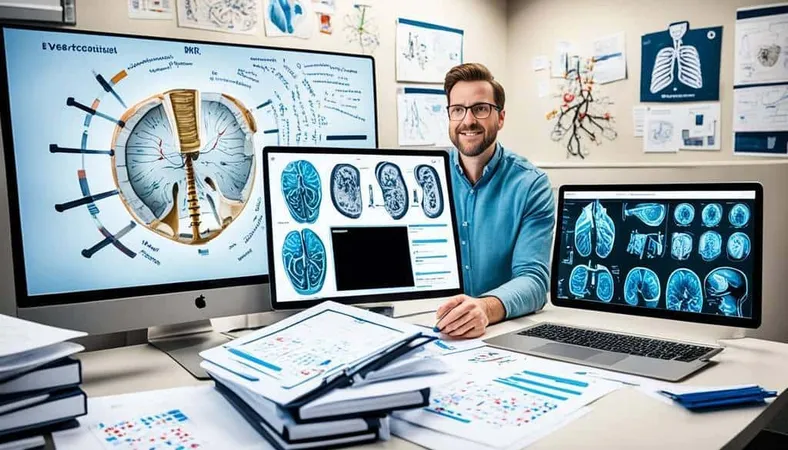
Ace Your Exam with MRI Practice Test 2026 Prep

MRI Tech School 2026 — Programs, Requirements, and What to Expect
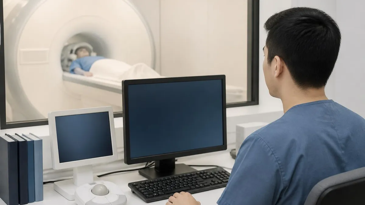
MRI Procedure: What to Expect Before, During and After Your Scan
About the Author

Medical Laboratory Scientist & Clinical Certification Expert
Johns Hopkins UniversityDr. Sandra Kim holds a PhD in Clinical Laboratory Science from Johns Hopkins University and is certified as a Medical Technologist (MT) and Medical Laboratory Scientist (MLS) through ASCP. With 16 years of clinical laboratory experience spanning hematology, microbiology, and molecular diagnostics, she prepares candidates for ASCP board exams, MLT, MLS, and specialist certification tests.
Join the Discussion
Connect with other students preparing for this exam. Share tips, ask questions, and get advice from people who have been there.
View discussion (8 replies)