
Do TIA Show on MRI? What Imaging Reveals About Transient Ischemic Attacks
Do TIA show on MRI? 🧠 Learn what imaging reveals, why DWI is critical, and how radiologists detect transient ischemic attacks before stroke strikes.
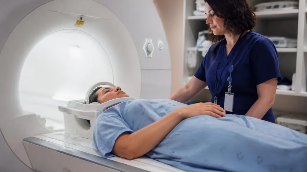
One of the most urgent questions emergency clinicians and patients face after a suspected mini-stroke is: do TIA show on MRI? The short answer is yes — but with critical nuance. A transient ischemic attack (TIA) produces temporary neurological symptoms that resolve within 24 hours, yet modern MRI technology, especially diffusion-weighted imaging, can detect the underlying tissue changes even after symptoms have disappeared. Understanding what shows up on MRI — and when — is essential for anyone studying neuroradiology or preparing for MRI registry examinations.
TIA has historically been called a "warning stroke" because it shares the same mechanism as ischemic stroke: a brief blockage of blood flow to part of the brain. Unlike a full stroke, the obstruction clears before permanent neuronal death occurs — but that does not mean MRI comes away empty-handed. Approximately 30 to 50 percent of clinically diagnosed TIA patients show evidence of acute ischemia on diffusion-weighted MRI, which is a far higher detection rate than older CT-based methods could achieve.
The timing of imaging relative to symptom onset matters enormously. If a patient arrives in the emergency department within the first six hours of symptom onset, the probability of a positive DWI finding is highest. After 24 hours, some lesions may begin to fade on certain sequences, while others become more apparent on FLAIR imaging. This dynamic window means that the choice of sequence, magnet strength, and imaging protocol directly determines whether the attack leaves a detectable footprint.
For MRI technologists and radiologists, recognizing the subtle signatures of TIA on multiple pulse sequences is a core competency. The lesions tend to be small — often less than 15 millimeters — and may appear in watershed territories, the cortex, or the deep white matter. They can be easily confused with chronic small vessel disease, which is extremely common in the older adult population most at risk for TIA. Distinguishing acute from chronic changes requires a systematic approach to signal characteristics and clinical correlation.
From an educational standpoint, TIA imaging is a high-yield topic on the ARRT MRI registry examination. Questions frequently probe which sequence best identifies acute ischemia, why apparent diffusion coefficient maps are necessary, and how imaging guides the risk stratification tools clinicians use — such as the ABCD2 score. Grasping these concepts not only improves patient care but also directly boosts exam performance.
This article explores the complete picture of tia on mri — from the underlying pathophysiology and optimal imaging protocols to the specific appearance on each MRI sequence and the clinical decisions that imaging informs. Whether you are a radiologic technologist, a neurology resident, or a patient trying to understand your imaging report, this guide delivers the depth and accuracy you need to navigate one of neurology's most time-sensitive scenarios.
Throughout this discussion, we will also address common pitfalls — including false-negative DWI scans, lesions that mimic TIA, and the role of 3-Tesla versus 1.5-Tesla magnets in detection sensitivity. By the end, you will have a thorough, exam-ready understanding of how MRI answers the question of whether a transient ischemic attack leaves its mark on the brain.
TIA and MRI: Key Numbers to Know
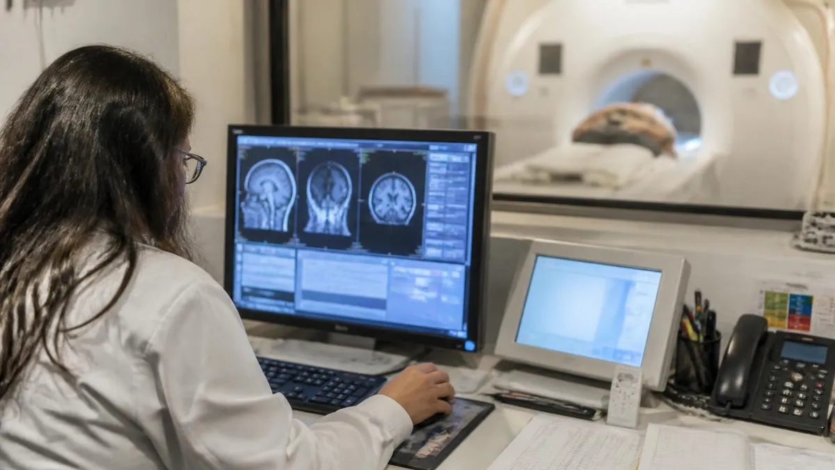
How TIA Progresses Through MRI Detection Windows
0–6 Hours: Peak DWI Positivity
6–24 Hours: FLAIR Begins to Activate
24–72 Hours: T2 and FLAIR Brighten
3–7 Days: Pseudonormalization Risk
Beyond 1 Week: Chronic Phase
Understanding which MRI sequences detect TIA — and why — is arguably the most important technical knowledge a neuroradiology technologist or registry candidate can possess. The workhorse of acute ischemia detection is diffusion-weighted imaging (DWI), which measures the random Brownian motion of water molecules in tissue. When cells swell due to energy failure from ischemia, intracellular water can no longer move freely, and this restricted diffusion produces a characteristic bright signal on DWI images paired with a dark region on the apparent diffusion coefficient (ADC) map.
The ADC map is not optional — it is indispensable. DWI signal can appear bright due to T2 shine-through, a phenomenon where naturally long T2 relaxation times in some tissues masquerade as restricted diffusion. The ADC map eliminates this ambiguity: true restricted diffusion causes the ADC value to drop, appearing dark, whereas T2 shine-through leaves the ADC normal or bright. Every TIA workup should include both the DWI trace image and the ADC map reviewed side by side, and MRI registry examinations frequently test this exact concept.
FLAIR (fluid-attenuated inversion recovery) plays a complementary role. This sequence suppresses the bright CSF signal seen on T2 images, making small cortical and periventricular lesions far more conspicuous. In TIA, FLAIR typically lags behind DWI by several hours — it may be negative in the first few hours when DWI is already positive. However, FLAIR becomes increasingly valuable after 24 hours and is the primary sequence for assessing chronic white matter burden, which provides important context for distinguishing new from old changes.
T2-weighted fast spin echo imaging offers high contrast between gray matter, white matter, and CSF, and remains useful for visualizing the overall extent of cerebrovascular disease. Susceptibility-weighted imaging (SWI) adds another layer by detecting microbleeds and venous structures. Identifying microbleeds in a patient presenting with TIA has direct clinical relevance: it may influence decisions about anticoagulation therapy, since patients with numerous microbleeds carry a higher risk of hemorrhagic transformation if given blood thinners.
MR angiography (MRA) — either time-of-flight or contrast-enhanced — is an essential adjunct that examines the cervical and intracranial vasculature for stenosis, occlusion, or arterial dissection. Many TIA events originate from atherosclerotic plaques in the carotid arteries or from cardioembolism, and identifying the culprit vessel is central to secondary stroke prevention. An MRI protocol that includes diffusion imaging, FLAIR, SWI, and MRA provides the most comprehensive evaluation of TIA in a single sitting.
Gadolinium-enhanced T1 imaging is not always included in TIA protocols but can reveal leptomeningeal enhancement, which suggests inflammatory or infectious mimics, or early blood-brain barrier disruption in acute ischemia. When the clinical picture is ambiguous — for example, when symptoms suggest stroke but the patient has fever and elevated inflammatory markers — contrast-enhanced sequences help broaden the differential diagnosis and guide appropriate management.
At 3 Tesla, the increased signal-to-noise ratio improves detection of small DWI lesions, particularly in the posterior fossa where susceptibility artifacts from bone interfaces degrade image quality at lower field strengths. For patients presenting with TIA symptoms referable to the brainstem or cerebellum, a 3-Tesla scanner provides a meaningful advantage over a 1.5-Tesla system. Technologists should be aware that higher field strength also increases susceptibility artifact, which can occasionally obscure small hemorrhages near air-tissue interfaces.
TIA on MRI: Sequence-by-Sequence Appearance
On diffusion-weighted imaging, an acute TIA-related infarct appears as a focal area of bright signal — hyperintense — due to restricted water diffusion inside swollen neurons and glia. The corresponding ADC map shows a dark (hypointense) region, confirming true restriction rather than T2 shine-through. Lesions are typically small, often less than 10 to 15 millimeters, and may be solitary or multifocal depending on the embolic source.
Detection sensitivity on DWI is highest within the first 24 hours, dropping modestly as pseudonormalization occurs between days three and seven. A critical registry exam point: a negative DWI does not absolutely exclude TIA, because some lesions — especially those in the brainstem due to susceptibility artifact — can be missed at 1.5 Tesla. Repeating DWI at 24 hours or upgrading to 3-Tesla improves sensitivity in clinically convincing cases.
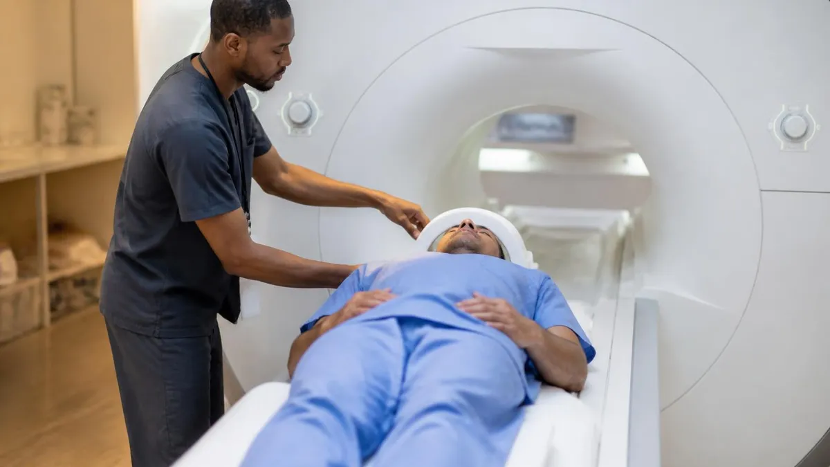
MRI vs. CT for TIA Evaluation: Which Is Better?
- +DWI detects acute ischemia in 30–50% of TIA cases where CT appears completely normal
- +FLAIR and T2 sequences visualize small cortical lesions invisible to CT due to bone artifact
- +MRA images cervical and intracranial vessels without ionizing radiation or iodinated contrast
- +SWI detects microbleeds that are clinically important for anticoagulation decisions
- +3-Tesla MRI provides superior resolution for posterior fossa and brainstem structures
- +Multiparametric protocol (DWI, FLAIR, SWI, MRA) answers most clinical questions in one session
- −MRI takes 30–60 minutes versus 5–10 minutes for CT, delaying initial assessment in unstable patients
- −Contraindicated in patients with certain metallic implants, pacemakers, or severe claustrophobia
- −Not available 24/7 at all community hospitals, limiting access in rural or lower-resource settings
- −DWI can miss posterior fossa TIA at 1.5 Tesla due to susceptibility artifact near the skull base
- −Pseudonormalization between days 3–7 can cause false-negative DWI in subacute presentations
- −Motion artifact from uncooperative or agitated patients degrades image quality significantly
MRI TIA Workup: Essential Imaging Checklist
- ✓Order MRI within 24 hours of symptom onset to maximize DWI sensitivity for acute ischemia
- ✓Always review both the DWI trace image and the ADC map together to confirm true restricted diffusion
- ✓Include FLAIR to detect subtle cortical lesions and assess chronic white matter burden
- ✓Add SWI to identify microbleeds that influence anticoagulant versus antiplatelet therapy decisions
- ✓Include MRA of the head and neck to evaluate carotid, vertebral, and intracranial vasculature
- ✓Note the DWI-FLAIR mismatch: positive DWI with negative FLAIR indicates hyper-acute ischemia
- ✓Evaluate the posterior fossa on 3-Tesla if available, since 1.5-Tesla misses brainstem lesions more often
- ✓Assess for ipsilateral carotid stenosis greater than 50% as a high-priority intervention target
- ✓Check for atrial fibrillation history — multifocal DWI lesions in multiple vascular territories suggest cardioembolic source
- ✓Correlate imaging findings with the ABCD2 score to stratify 2-day and 7-day stroke risk
DWI-FLAIR Mismatch Is a Critical Marker of Hyper-Acute Ischemia
When DWI shows a bright lesion but FLAIR appears negative in the same region, the time from symptom onset is almost certainly less than 4.5 hours. Clinicians use this mismatch to help identify patients who may still be candidates for thrombolytic therapy even when the exact onset time is unknown — such as wake-up strokes. MRI technologists who understand this principle can prioritize sequence selection and communicate findings more effectively to the clinical team.
False-negative MRI results in TIA are a recognized clinical challenge that every technologist and radiologist must understand. Studies show that standard 1.5-Tesla DWI misses acute ischemic lesions in approximately 17 to 20 percent of confirmed TIA cases. The posterior fossa — the region containing the brainstem, cerebellum, and cranial nerve nuclei — is the most common location for false negatives because susceptibility artifact from the surrounding bone creates signal dropout that obscures small infarcts. A patient presenting with sudden vertigo, diplopia, or facial numbness may have a real brainstem TIA that is simply invisible on a standard 1.5-Tesla scan.
Repeating the MRI within 24 hours can rescue some false-negative initial scans, as lesions may become more apparent as edema evolves. Upgrading to a 3-Tesla system is the most reliable solution: the higher signal-to-noise ratio and improved spatial resolution substantially reduce the artifact that plagues posterior fossa imaging at lower field strengths. Some academic medical centers now use dedicated thin-slice DWI protocols specifically designed for the brainstem, with slice thicknesses of 2 to 3 millimeters rather than the standard 5-millimeter slices used in routine brain imaging.
Mimics of TIA on MRI create the opposite problem: lesions that look like ischemia but are not. Complex migraine with aura can produce cortical spreading depression that appears as transient DWI positivity without an ischemic mechanism. Seizure activity — Todd's paralysis — can cause focal neurological deficits that prompt urgent MRI, and post-ictal changes occasionally produce subtle DWI signal that misleads less experienced readers. Metabolic disturbances such as hypoglycemia, hyponatremia, and Wernicke's encephalopathy produce characteristic MRI patterns that may be confused with ischemic injury.
Demyelinating lesions — particularly from multiple sclerosis — are perhaps the most common non-vascular mimic of TIA on MRI. An MS plaque that happens to be in an eloquent location can produce sudden transient symptoms, and on FLAIR it appears as a periventricular or juxtacortical hyperintense lesion that superficially resembles a TIA infarct. However, MS plaques typically do not restrict on DWI in the acute phase, which is a critical differentiating feature. The clinical history — patient age, prior episodes, and systemic risk factors — always informs the imaging interpretation.
Cerebral venous thrombosis (CVT) deserves special mention as a TIA mimic because it produces non-arterial distribution infarcts with hemorrhagic transformation that can confuse even experienced neuroradiologists. CVT should be considered in young patients, particularly women on oral contraceptives, who present with TIA-like symptoms but have infarcts that cross arterial territory boundaries. SWI and contrast-enhanced MR venography (MRV) are the key sequences for diagnosing CVT, and both should be added to the protocol when CVT is clinically suspected.
Reversible cerebral vasoconstriction syndrome (RCVS) causes recurrent thunderclap headaches with transient neurological symptoms and can produce small cortical infarcts visible on DWI. Unlike atherosclerotic TIA, RCVS typically affects younger patients without traditional cardiovascular risk factors and shows multifocal segmental arterial narrowing on MRA. Posterior reversible encephalopathy syndrome (PRES), associated with hypertensive emergency and immunosuppressive therapy, produces parieto-occipital T2/FLAIR hyperintensity that usually does not restrict on DWI — another key distinguishing feature from ischemic TIA.
Understanding these mimics is not merely academic — it has direct therapeutic consequences. A patient misdiagnosed with TIA and placed on antiplatelet therapy for an MS plaque loses the opportunity for appropriate disease-modifying therapy. Conversely, a TIA patient whose imaging is falsely reassuring as negative may not receive the urgent secondary prevention interventions that dramatically cut the near-term stroke risk. Rigorous sequence selection, systematic reading approach, and close clinical correlation remain the pillars of accurate TIA MRI interpretation.
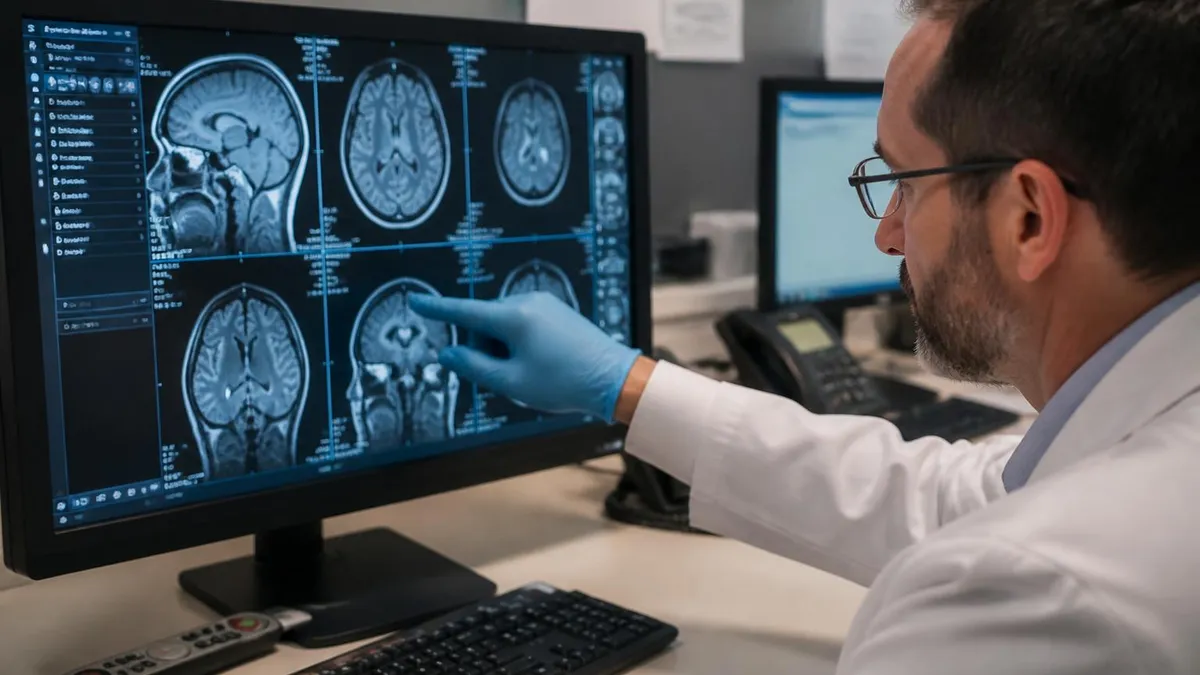
A transient ischemic attack is not a benign event. Population-based studies consistently show that the risk of a full ischemic stroke within 2 days of TIA is approximately 10 percent, and within 90 days it may reach 15 percent or higher in high-risk patients. Prompt MRI evaluation — ideally within 24 hours of symptom onset — combined with urgent secondary prevention measures (antiplatelet therapy, statin initiation, blood pressure control) reduces this risk by up to 80 percent. Delay in imaging directly translates to preventable strokes.
The clinical decisions that flow from TIA MRI findings are far-reaching and demonstrate why imaging accuracy is a life-or-death matter. When DWI confirms acute ischemia, clinicians immediately intensify secondary prevention therapy: dual antiplatelet therapy (aspirin plus clopidogrel) for 21 days, followed by long-term single antiplatelet therapy, is now the standard of care based on landmark trials including POINT and CHANCE. Statin therapy is initiated regardless of baseline cholesterol levels. Blood pressure is carefully managed — not lowered too aggressively in the acute phase, but brought under tight long-term control.
The vascular imaging findings from MRA drive surgical and interventional decisions. Ipsilateral carotid stenosis of 50 to 69 percent (symptomatic) warrants consideration of endarterectomy or stenting, with benefit greatest when surgery is performed within two weeks of the TIA. Stenosis of 70 percent or greater carries a stronger indication for revascularization. Intracranial stenosis identified on MRA may indicate candidacy for intensive medical management or, in selected cases, intracranial angioplasty. The imaging study does not merely confirm that a TIA occurred — it maps the road to preventing the next one.
Cardiac workup is initiated in parallel with imaging. Multifocal DWI lesions distributed across multiple vascular territories — for example, lesions in both the anterior and posterior circulation simultaneously — strongly suggest a cardioembolic source. This pattern triggers an urgent search for atrial fibrillation, including prolonged cardiac monitoring (at least 30 days), echocardiography to look for structural heart disease, and hematologic evaluation for hypercoagulable states. Anticoagulation with a direct oral anticoagulant (DOAC) replaces antiplatelet therapy when atrial fibrillation is confirmed, substantially reducing future stroke risk.
Risk stratification tools like the ABCD2 score integrate imaging findings with clinical variables. The score assigns points for age over 60, blood pressure over 140/90, clinical features (unilateral weakness scores highest), symptom duration, and diabetes. When the ABCD2 score is 4 or greater, or when DWI shows a positive lesion, guidelines recommend urgent hospital admission or a same-day TIA clinic evaluation rather than outpatient follow-up. Positive DWI independently increases stroke risk prediction beyond the clinical score alone, underscoring the value of early MRI access.
Anticoagulation decisions in TIA patients with microbleeds on SWI require individualized risk-benefit analysis. Cerebral amyloid angiopathy — characterized by lobar microbleeds — carries a higher risk of spontaneous intracerebral hemorrhage. When atrial fibrillation coexists with extensive lobar microbleeds, the prescribing clinician must weigh hemorrhagic risk against cardioembolic stroke prevention. The imaging findings from SWI directly enter this calculus, and the MRI technologist's role in acquiring high-quality susceptibility-weighted images is therefore clinically meaningful.
For MRI registry candidates, understanding how imaging guides these decisions reinforces why pulse sequence selection is not merely a technical exercise. The registry examination tests whether candidates understand which sequences to include, how to interpret findings, and why certain protocols are essential. Connecting technical knowledge to clinical application — knowing that a missed posterior fossa TIA lesion means a patient goes home at 10 percent stroke risk without proper therapy — gives weight and purpose to exam preparation that pure memorization cannot provide.
Long-term follow-up imaging after TIA also has a defined role. Patients with confirmed TIA and significant white matter disease may undergo repeat MRI at 3 to 6 months to assess disease progression and response to risk factor modification. In cases where the initial etiology was unclear, follow-up vessel wall MRI — an advanced technique that images the arterial wall rather than just the lumen — can detect atherosclerotic plaque, dissection, or vasculitis that were not apparent on the initial MRA study. The imaging conversation in TIA does not end at the emergency department discharge.
Many readers pair this guide with the mri pelvis cpt code to check their readiness before exam day.
For MRI technologists and registry candidates, a practical, systematic approach to TIA scanning is the foundation of consistent high-quality results. Start by confirming the clinical indication with the ordering provider: knowing whether the patient presents within hours of symptom onset versus days later should influence the protocol selected and the sequences prioritized. Acute presentations demand the fastest possible DWI acquisition; subacute or unclear-onset cases benefit most from a comprehensive protocol that includes FLAIR, SWI, and MRA in addition to diffusion imaging.
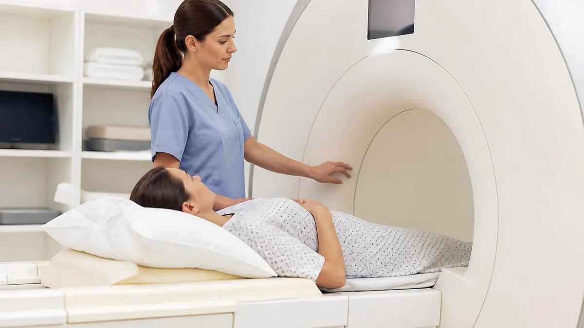
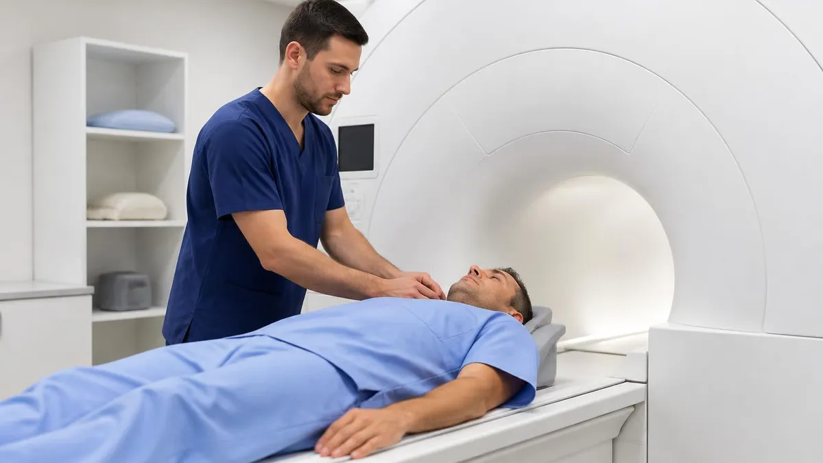
Patient preparation reduces the most avoidable source of image degradation: motion. Elderly patients presenting with TIA may be anxious, in pain, or cognitively impaired. A brief, calm explanation of the procedure — what sounds to expect, the importance of holding still, and the approximately 30-minute duration — pays dividends in image quality. Offer foam earplugs or music through MRI-compatible headphones. Position the patient in a comfortable, supported position with a head coil that minimizes head movement, and confirm that the neck is properly supported to enable high-quality MRA acquisition without vessel saturation artifact.
When scanning, apply the protocol systematically: DWI first, because it is the highest-priority sequence for acute TIA and takes only 2 to 3 minutes on modern scanners. Then FLAIR, T2, and SWI, followed by MRA of the intracranial circulation and then the cervical vessels. If gadolinium is ordered — for example, to evaluate vessel wall or leptomeningeal enhancement — perform non-contrast sequences first, then inject the contrast agent and acquire post-contrast T1 sequences. This order ensures that contrast does not confound the DWI or FLAIR signal interpretation.
Quality control during the scan is as important as protocol selection. After acquiring DWI, briefly review the images on the scanner console before moving to the next sequence. Look for motion artifact (blurring, ghosting), susceptibility artifact in the posterior fossa, and adequate signal in the cerebellum and brainstem. If the posterior fossa appears degraded, consider repeating DWI with a smaller field of view, thinner slices, or an adjusted echo time to reduce artifact. Catching a poor posterior fossa acquisition during the scan allows immediate correction; discovering it after the patient leaves does not.
Document the scan parameters used, including field strength, slice thickness, echo time, and any protocol modifications made for the individual patient. This documentation supports meaningful comparison if the patient returns for follow-up imaging. For registry examinations, understanding why specific scan parameters affect image quality — why a shorter echo time reduces susceptibility artifact, why thin slices improve detection of small lesions, why parallel imaging accelerates acquisition — transforms rote protocol knowledge into flexible, problem-solving expertise.
Stay current with evolving evidence on TIA imaging. Newer techniques including ultra-high-b-value DWI (b=3000), readout-segmented DWI (RESOLVE), and synthetic MRI are being validated for improved lesion detection in TIA. Vessel wall MRI — using black-blood sequences to visualize arterial wall pathology — is increasingly used at comprehensive stroke centers to characterize the culprit lesion in cryptogenic TIA. Understanding these advances positions the technologist as a knowledgeable partner in the clinical team, not merely an operator of equipment.
Finally, communication matters. When a technologist identifies a striking DWI lesion in a patient presenting with transient symptoms, proactive notification of the supervising radiologist ensures expedited interpretation and rapid relay to the clinical team. Protocols for urgent radiologist notification should be established in every department that performs acute neuroimaging. Time from imaging to treatment decision is a quality metric in stroke care, and the technologist's efficiency and communication directly contribute to meeting that benchmark — making every registry concept studied a real-world tool for patient safety.
Sample MRI - Magnetic Resonance Imaging Practice Questions
Try these questions from our free MRI - Magnetic Resonance Imaging practice tests. The correct answer and an explanation follow each question.
On MRI of the liver, a simple hepatic cyst is expected to show which signal characteristics?
- A. T1 high, T2 low
- B. T1 low, T2 high, no enhancement
- C. T1 high, T2 high, with peripheral enhancement
- D. T1 low, T2 low
Answer: B. T1 low, T2 high, no enhancement
Simple hepatic cysts have low T1 signal, very high T2 signal due to pure fluid content, and show no contrast enhancement.
What does a high b-value in diffusion-weighted imaging (DWI) indicate?
- A. Shorter echo time and less diffusion weighting
- B. Stronger diffusion-sensitizing gradients and greater sensitivity to restricted diffusion
- C. Lower magnetic field strength required
- D. Faster acquisition with less motion sensitivity
Answer: B. Stronger diffusion-sensitizing gradients and greater sensitivity to restricted diffusion
The b-value reflects the strength and duration of diffusion-sensitizing gradients; higher b-values increase diffusion contrast but also reduce SNR.
What is the primary advantage of parallel imaging techniques (e.g., GRAPPA, SENSE) in MRI?
- A. They increase SNR at the same scan time
- B. They reduce scan time or allow higher spatial resolution by using multiple coil elements to under-sample k-space
- C. They eliminate the need for gradient coils
- D. They improve T1 contrast
Answer: B. They reduce scan time or allow higher spatial resolution by using multiple coil elements to under-sample k-space
Parallel imaging exploits spatial sensitivity differences among phased-array coil elements to reconstruct fully sampled images from under-sampled k-space, reducing scan time by the acceleration factor (R).
What is the main advantage of parallel imaging techniques (e.g., GRAPPA, SENSE) in MRI?
- A. Reduced acquisition time by undersampling k-space using multiple coil elements
- B. Improved SNR by adding signals from multiple coils
- C. Elimination of susceptibility artifacts
- D. Increased spatial resolution without penalty
Answer: A. Reduced acquisition time by undersampling k-space using multiple coil elements
Parallel imaging uses the spatial sensitivity profiles of phased-array coils to reconstruct missing k-space lines, allowing faster acquisitions at the cost of some SNR.
Take the full MRI - Magnetic Resonance Imaging practice test
MRI Questions and Answers
DWI MRI: Diffusion-Weighted Imaging Explained (2026 Guide)
Side Effects of Brain MRI: Understanding Risks, Safety, and What Every Patient Should Know
MRI Lumbar Spine Without Contrast: Complete Guide to the Scan, What It Shows, and How to Prepare
MRI History: From Nuclear Resonance to Modern Scanners

MRI of Cervical Spine: Complete Guide to Imaging, Anatomy, Pathology, and Patient Preparation
About the Author

Medical Laboratory Scientist & Clinical Certification Expert
Johns Hopkins UniversityDr. Sandra Kim holds a PhD in Clinical Laboratory Science from Johns Hopkins University and is certified as a Medical Technologist (MT) and Medical Laboratory Scientist (MLS) through ASCP. With 16 years of clinical laboratory experience spanning hematology, microbiology, and molecular diagnostics, she prepares candidates for ASCP board exams, MLT, MLS, and specialist certification tests.
Join the Discussion
Connect with other students preparing for this exam. Share tips, ask questions, and get advice from people who have been there.
View discussion (8 replies)