Sedative for MRI: Complete Guide to Sedation Options, Safety, and What to Expect
Need a sedative for MRI? 🧠 Learn about sedation options, medications, safety tips, and how to prepare for a stress-free MRI scan.

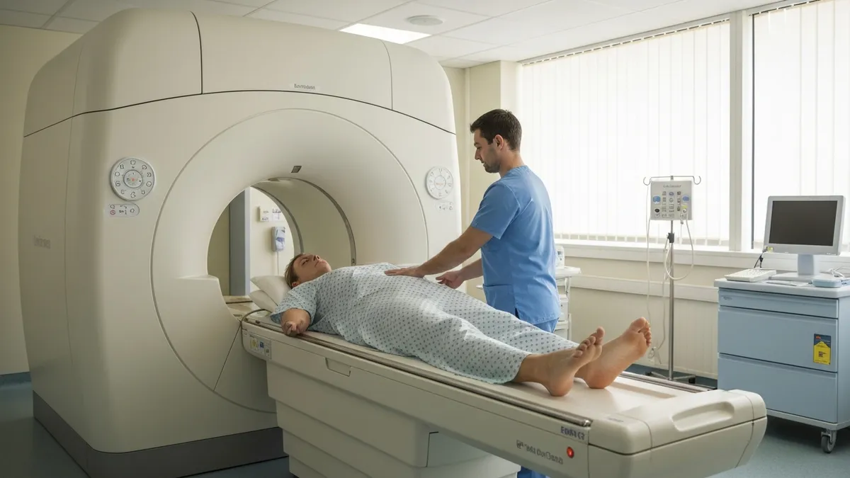
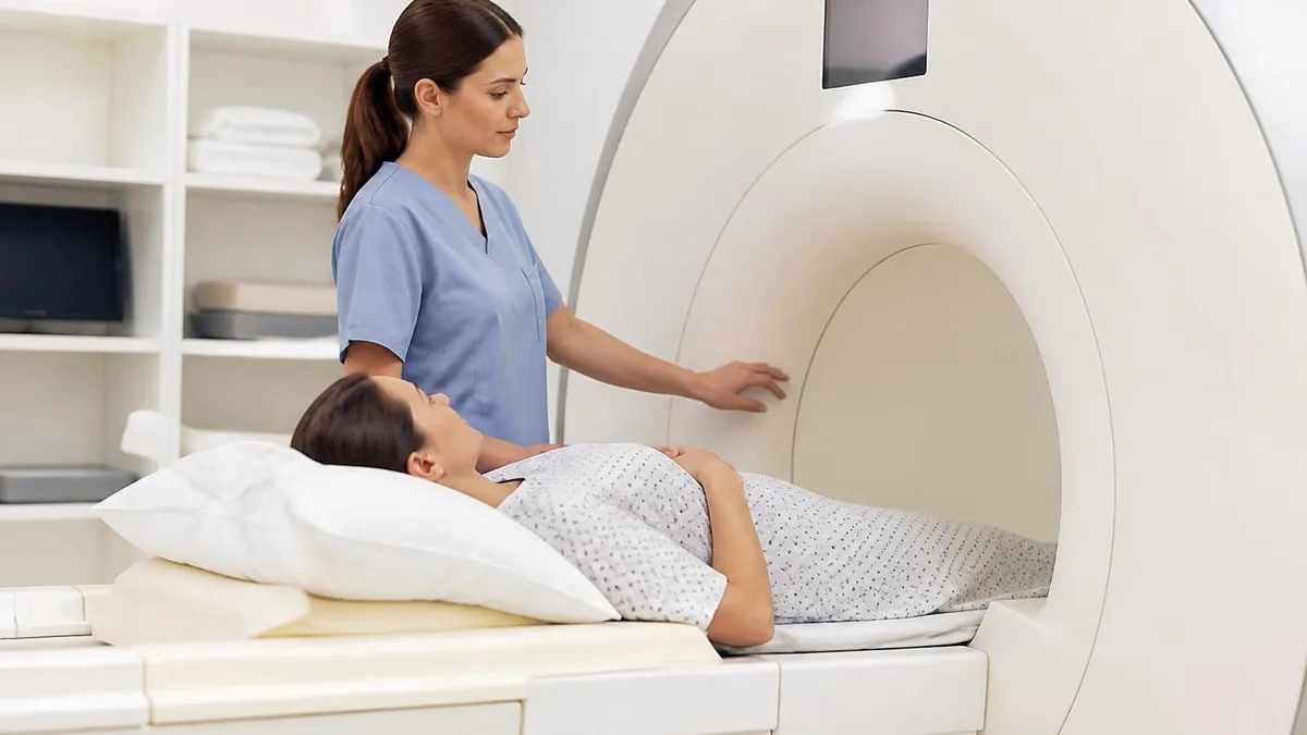
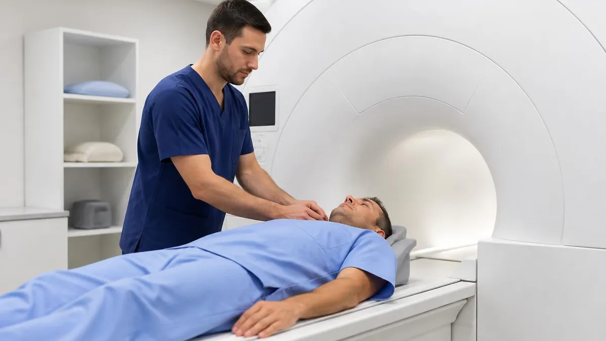
A sedative for MRI is one of the most commonly requested accommodations in diagnostic imaging, and for good reason. Magnetic resonance imaging scans can last anywhere from 20 minutes to over an hour, requiring patients to remain completely still inside a narrow, loud tube. For individuals with claustrophobia, anxiety disorders, chronic pain, or sensory processing challenges, this experience can feel overwhelming — even impossible.
Understanding your sedation options before your scheduled scan can make the difference between completing a critical diagnostic exam and having to reschedule. Whether you are a patient, a caregiver, or a healthcare student, this guide covers everything you need to know.
MRI sedation is not a one-size-fits-all solution. Physicians and radiologists tailor sedation protocols to each patient's age, weight, medical history, anxiety severity, and the complexity of the imaging study being performed. A child undergoing a brain MRI may require general anesthesia administered by a pediatric anesthesiologist, while an adult with mild claustrophobia might only need a low-dose oral benzodiazepine taken about an hour before the scan.
The level of sedation prescribed ranges from minimal anxiolysis — where you remain fully conscious but relaxed — all the way to deep sedation or general anesthesia, where you are completely unaware of your surroundings throughout the procedure.
The most frequently used medications for MRI sedation in adults include midazolam (Versed), diazepam (Valium), lorazepam (Ativan), and hydroxyzine (Vistaril). Each of these drugs works differently in the body and carries its own set of benefits and potential side effects. Midazolam is particularly popular because of its rapid onset, short duration of action, and amnesic properties — meaning many patients have little to no memory of the scan afterward. Hydroxyzine is a non-benzodiazepine antihistamine that provides mild sedation and is sometimes preferred for patients who need to avoid controlled substances due to personal or professional reasons.
Safety is the top priority whenever sedation is involved in a medical procedure. Before prescribing any sedative for MRI, your physician will review your full medication list to screen for dangerous drug interactions, assess your respiratory health, and determine whether you have any history of adverse reactions to sedatives or anesthetics.
Patients with obstructive sleep apnea, severe obesity, or certain cardiac conditions may require closer monitoring during the scan. You will typically be asked to arrive with a fasting period of four to six hours if moderate or deep sedation is planned, to reduce the risk of aspiration during the procedure.
Pediatric MRI sedation deserves special mention because children — especially those under seven years old — often cannot cooperate with the stillness required for high-quality imaging. Pediatric protocols frequently use agents like propofol, dexmedetomidine, or ketamine, administered intravenously by a board-certified anesthesiologist or nurse anesthetist. In some children's hospitals, child life specialists use non-pharmacological distraction techniques, including customized MRI environments with projected visuals and music, that can reduce or eliminate the need for sedation altogether. Researching what your facility offers beforehand can save time and reduce anxiety for both child and parent.
Recovery from MRI sedation is another important consideration that patients often overlook when scheduling their scan. Even with a mild oral benzodiazepine, you will not be permitted to drive yourself home after the procedure. Plan to have a responsible adult accompany you, and expect to spend at least 30 to 60 minutes in a monitored recovery area if you received intravenous sedation.
Cognitive impairment — including slowed reaction times and difficulty concentrating — can persist for several hours after the sedative wears off, so avoid operating heavy machinery or signing important legal documents on the day of your scan. Staying well-hydrated and eating a light meal after clearance from the clinical staff can help speed your recovery.
If you are preparing for a career in MRI technology or radiology, understanding sedation protocols is an essential part of your clinical knowledge base. MRI technologists work alongside anesthesiologists, nurses, and radiologists to ensure sedated patients remain safe throughout the scan. You need to be familiar with MRI-conditional monitoring equipment, emergency protocols, and the specific contraindications that apply when sedated patients are inside high-field magnets. Resources like sedative for mri preparation guides can help you build the foundational knowledge you need for certification exams and clinical practice.
MRI Sedation by the Numbers
Types of Sedation Used in MRI Scans
Minimal Sedation (Anxiolysis)
Moderate Sedation (Conscious Sedation)
Deep Sedation
General Anesthesia
Non-Pharmacological Techniques
The process of obtaining a sedative for an MRI scan begins with a conversation between you and your ordering physician or radiologist. During this conversation, your provider will ask detailed questions about your anxiety history, previous sedation experiences, current medications, allergies, and any underlying health conditions that might affect how your body processes sedative drugs. This intake process is critically important because sedatives interact with dozens of commonly prescribed medications — including opioid pain relievers, antidepressants, antiepileptics, and antifungal agents — and using them together without appropriate screening can produce dangerous respiratory depression or prolonged sedation.
For adults requesting mild oral sedation, the prescription workflow is relatively straightforward. Your physician writes a prescription for a short-acting benzodiazepine such as diazepam 5 mg or lorazepam 1 mg, and instructs you to take it approximately 30 to 60 minutes before your scheduled scan time.
You will need to arrange transportation before you even fill the prescription, since driving after taking these medications is not safe regardless of how alert you feel. Some imaging centers provide the medication on-site immediately before the scan, which eliminates the risk of patients taking it too early, too late, or at an incorrect dose at home.
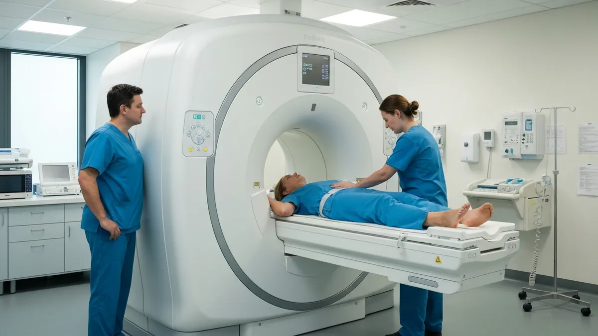
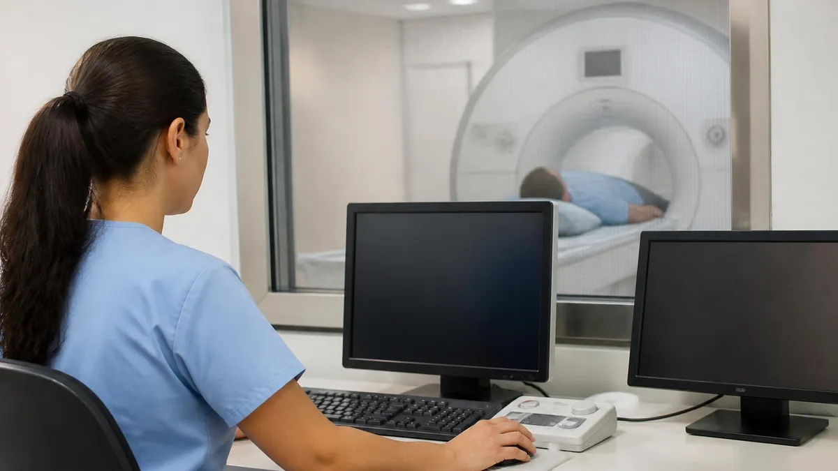
Intravenous sedation protocols are considerably more complex and require specialized staffing and equipment. When moderate or deep sedation is planned, you will typically arrive at the imaging center 60 to 90 minutes before your scan time to complete pre-sedation assessments. A nurse or anesthesiologist will start an IV line, review your vital signs, confirm your fasting status, and obtain informed consent for the sedation procedure. During the scan, your oxygen saturation, heart rate, blood pressure, and respiratory rate will be monitored continuously using MRI-compatible equipment that does not interfere with image quality.
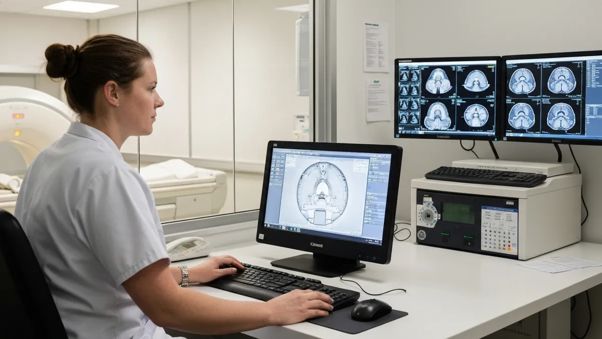
The radiologist and anesthesia team must also consider how sedation affects image quality itself. While sedation eliminates motion artifact by keeping patients still, certain drugs can influence blood flow, respiratory patterns, or cardiac function in ways that subtly alter MRI signal characteristics — particularly in functional MRI, cardiac MRI, and certain perfusion studies. This is why communication between the ordering clinician, the radiologist, and the sedation provider is so important: everyone involved needs to understand what is being imaged and how the chosen sedative might interact with those particular tissue signals or physiological measurements.
Dosing for pediatric patients follows weight-based protocols that differ substantially from adult dosing. For example, oral midazolam syrup dosed at 0.3 to 0.5 mg per kilogram of body weight (with a maximum of 20 mg) is a widely used pediatric pre-procedure anxiolytic. However, pediatric patients metabolize drugs at different rates than adults, and small children have narrower margins between therapeutic doses and doses that cause respiratory compromise. For this reason, any sedation of a child for MRI should occur in a facility equipped with pediatric emergency resuscitation equipment and staffed by providers with pediatric sedation training and current PALS certification.
Reversal agents are an important safety backstop in any sedation protocol. Flumazenil rapidly reverses the effects of benzodiazepines by competitively blocking their receptors in the brain. It can restore consciousness within one to two minutes, though its duration of action is shorter than most benzodiazepines, meaning re-sedation can occur if the patient is not monitored for a sufficient period after reversal. Naloxone (Narcan) serves a similar reversal function for opioid-based sedation agents. Every MRI facility that provides sedation services should have both of these agents immediately available, along with the staff training to administer them correctly in an emergency.
Documentation and follow-up care are the final pieces of a complete sedation protocol. After your scan, the clinical team will record the agents administered, doses given, your vital signs throughout the procedure, any adverse events that occurred, and your level of consciousness at discharge.
This record becomes part of your permanent medical file and is essential if you undergo another sedated procedure in the future. If you experienced any unexpected reactions — excessive drowsiness, paradoxical agitation, nausea, or allergic responses — report these to your care team before you leave so they can be documented and considered when planning future imaging studies.
Sedation Options: Benefits, Risks, and Alternatives Compared
Oral benzodiazepines such as lorazepam (Ativan) and diazepam (Valium) are the most commonly prescribed medications for mild MRI anxiety in adult patients. These drugs are inexpensive, widely available, and generally well-tolerated by healthy adults. A typical dose takes effect within 30 to 60 minutes and provides anxiolysis for two to four hours, which is sufficient for most standard MRI examinations. The primary advantages include ease of administration, predictable onset, and no need for IV placement or continuous monitoring in low-risk patients.
The limitations of oral sedatives include variable absorption rates — food, gastric motility disorders, and certain medications can all affect how quickly and completely the drug enters the bloodstream. Some patients experience paradoxical reactions, particularly older adults, who may become agitated or confused rather than calm after benzodiazepine administration. Hydroxyzine is a useful non-controlled alternative for patients who cannot use benzodiazepines, offering mild antihistamine-based sedation with a lower risk of dependence or abuse potential, though its sedative effect is generally weaker.
Pros and Cons of Using a Sedative for MRI
- +Allows patients with severe claustrophobia to complete essential diagnostic scans
- +Reduces motion artifact and improves overall image quality for more accurate diagnoses
- +Short-acting agents wear off quickly, allowing most patients to recover within a few hours
- +IV sedation can be adjusted in real time based on the patient's response during the scan
- +Amnesic properties of midazolam reduce post-procedural psychological trauma and distress
- +Pediatric sedation protocols allow young children to receive critical imaging without distress
- −Patient cannot drive for the remainder of the day, requiring a dedicated transportation escort
- −Benzodiazepines carry risks of paradoxical agitation, especially in elderly and pediatric patients
- −Fasting requirements for moderate and deep sedation add inconvenience and scheduling complexity
- −Deep sedation and general anesthesia require additional staffing and specialized MRI-safe equipment
- −Drug interactions with common medications including SSRIs, opioids, and antiepileptics can occur
- −Insurance may not cover the added cost of anesthesia services, creating unexpected out-of-pocket expenses
Preparing for a Sedated MRI: Step-by-Step Checklist
- ✓Talk to your ordering physician about your anxiety level and request a sedation consultation at least one week before your scan.
- ✓Provide a complete list of all prescription medications, over-the-counter drugs, supplements, and herbal remedies to your care team.
- ✓Confirm your fasting instructions — typically nothing by mouth for 4 to 6 hours before moderate or deep sedation.
- ✓Arrange a responsible adult driver to escort you to and from the imaging facility on the day of your scan.
- ✓Wear comfortable, loose-fitting clothing without metal zippers, snaps, or underwire that might need to be removed.
- ✓Remove all jewelry, piercings, and hair accessories before arriving; leave valuables at home.
- ✓Arrive at least 60 to 90 minutes early if IV sedation is planned to allow time for pre-sedation assessment and IV placement.
- ✓Inform the clinical team immediately if you develop any new symptoms — cold, fever, or illness — before your appointment.
- ✓Ask whether your facility uses MRI-compatible monitoring equipment and confirm that reversal agents are on-site.
- ✓Plan to rest at home for the remainder of the day and avoid important decisions or commitments until fully recovered.
Always Disclose Sleep Apnea Before Sedated MRI
Patients with untreated obstructive sleep apnea face elevated respiratory risk during MRI sedation because sedatives further suppress the airway reflexes that are already compromised during sleep. Always inform your physician and the imaging team about any sleep apnea diagnosis — even if you consider it mild or well-controlled with a CPAP device — so your sedation plan can be adjusted accordingly with appropriate airway monitoring in place.
Pediatric MRI sedation represents one of the most technically demanding areas of pediatric anesthesia and radiology practice. Young children — particularly those between the ages of one and seven — are developmentally incapable of understanding why they must remain motionless inside a loud, confined machine for extended periods.
Even a cooperative six-year-old who is trying their best will inevitably move during a 45-minute brain MRI if they are not sedated. Motion degradation in pediatric MRI is not merely an inconvenience; in neurological emergencies such as suspected stroke, tumor, or vascular malformation, a motion-degraded scan can delay life-saving diagnosis and treatment by hours.
The most widely used agents for pediatric MRI sedation include oral midazolam syrup, intranasal dexmedetomidine, intravenous propofol, and intramuscular ketamine. Each has its place depending on the child's age, weight, medical history, anticipated scan duration, and the imaging protocol being used. For short scans in otherwise healthy toddlers, oral midazolam syrup at 0.3 to 0.5 mg per kilogram provides reliable sedation within 15 to 30 minutes. For longer or more complex studies, intravenous propofol infusion allows precise depth-of-sedation control and rapid recovery — most children are awake and eating within 30 to 60 minutes of propofol being discontinued.
Intranasal dexmedetomidine has emerged as a preferred option in many pediatric centers because it avoids the need for IV placement before sedation — a significant source of distress for needle-phobic children. Administered via a mucosal atomizer device into the nasal cavity, dexmedetomidine typically produces adequate sedation within 45 to 60 minutes with a duration of 60 to 90 minutes. This timeline requires careful scheduling coordination: the child needs to receive the nasal spray far enough in advance that sedation is at its peak when MRI scanning begins. If the scan runs longer than expected, supplemental dosing may be needed.
Special populations beyond young children also require tailored sedation approaches for MRI. Adults with severe autism spectrum disorder (ASD) may be unable to tolerate the sensory experience of an MRI scan regardless of verbal coaching or incentives, and may require deep sedation or general anesthesia even for routine imaging.
Similarly, patients with dementia or acquired brain injuries may be unable to follow instructions or remain still, necessitating sedation that would not be required in a cognitively intact adult of the same age. Individuals with severe intellectual disabilities often have complex medication profiles that require careful review before any sedative is added.
Patients with needle phobias represent another special consideration that is frequently underestimated by clinical teams. Someone with severe needle phobia may refuse IV placement so strongly that IV sedation becomes practically impossible without first addressing the phobia itself. In these cases, oral or intranasal agents provide the best path to procedural completion. Topical anesthetic creams (such as EMLA or LMX) applied to the IV site 45 to 60 minutes before placement can significantly reduce the pain of insertion, and some patients do well with vibration-based pain distraction devices used simultaneously with needle procedures.
Patients who are pregnant require especially careful consideration before any MRI sedation is planned. While MRI itself is generally considered safe throughout pregnancy when medically indicated (avoiding gadolinium-based contrast in the first trimester where possible), sedative medications cross the placental barrier and can affect fetal development or suppress neonatal respiratory drive if delivered close to term. If sedation is truly essential for a pregnant patient, the risk-benefit discussion should involve the patient's obstetrician, the radiologist, and a maternal-fetal medicine specialist to ensure that imaging benefits clearly outweigh the sedation-related fetal risks.
Patients with renal or hepatic impairment metabolize sedative drugs more slowly than healthy adults, meaning standard doses can produce prolonged sedation, unexpected drug accumulation, or toxic metabolite buildup. Dose reductions, extended monitoring periods, and careful agent selection are all necessary in this population. For example, midazolam is hepatically metabolized, and its effects can last significantly longer in patients with liver disease. Lorazepam and oxazepam, which undergo simpler conjugation reactions in the liver, are often preferred in patients with hepatic dysfunction because their metabolism is less dependent on cytochrome P450 enzyme activity, making their pharmacokinetics more predictable.
Never take a prescribed oral sedative before your MRI unless you have already confirmed that a responsible adult driver is physically present and ready to accompany you. Driving under the influence of a benzodiazepine — even at low doses — is illegal, dangerous, and can result in serious accidents. Patients who arrive at the imaging facility without a confirmed driver may be turned away for their own safety, delaying their imaging and potentially impacting their medical care.
Recovery after MRI sedation is a phase of care that deserves as much attention as the sedation itself. The moment the scan ends, the clinical focus shifts from keeping you still inside the magnet to safely returning you to baseline neurological function.
For patients who received only a mild oral benzodiazepine, recovery is usually simple: you will rest in a recliner for 30 to 45 minutes while a nurse monitors your vital signs and assesses your orientation and coordination before clearing you for discharge. Most patients who received oral lorazepam or diazepam are ready to leave the facility within an hour of their scan completing, provided they have a driver waiting.
Patients who received intravenous or deep sedation require a more structured post-anesthesia care unit (PACU) level of monitoring. You will be transferred to a monitored recovery bay where pulse oximetry, blood pressure, heart rate, and respiratory rate are recorded at regular intervals until you meet discharge criteria. These criteria typically include sustained oxygen saturation above 95% on room air, stable vital signs, ability to follow simple commands, ability to ambulate with minimal assistance, tolerance of oral fluids without nausea or vomiting, and adequate pain control. Most patients meet these criteria within 60 to 90 minutes of IV sedation being discontinued.
Nausea and vomiting after sedation (PONV) is one of the most common and uncomfortable recovery complaints. It occurs in roughly 20 to 30% of patients receiving propofol-based sedation and is even more common with opioid-containing protocols. Prophylactic antiemetic medications such as ondansetron (Zofran) or dexamethasone are routinely administered before or during the procedure in higher-risk patients to reduce PONV incidence. If you have a history of severe post-procedural nausea, inform your care team in advance so they can optimize your antiemetic regimen before your scan rather than treating nausea reactively afterward.
Cognitive recovery — the return of full mental clarity, memory consolidation, and executive function — takes longer than physical recovery from sedation. Research on benzodiazepine effects on cognition consistently shows that fine motor coordination and complex decision-making can be impaired for up to 24 hours after a single moderate dose of midazolam or lorazepam, even if the patient feels subjectively alert and clearheaded.
This is why the post-sedation instructions given at discharge are so important: avoid alcohol, avoid important legal or financial decisions, avoid childcare responsibilities that require split-second reactions, and do not return to work until the following day if your job involves safety-sensitive tasks.
Hydration plays an underappreciated role in sedation recovery. Many sedated patients arrive at the imaging facility already mildly dehydrated due to the required fasting period, and sedative medications can further impair the thirst response. Drinking adequate fluids — water, diluted juice, or clear broth — in the hours after your scan helps your kidneys clear the metabolized drug byproducts and supports the normalization of blood pressure if it dropped slightly during the procedure. IV fluid administration during the scan itself helps counteract fasting-related dehydration in patients who received intravenous sedation.
Follow-up communication after sedated MRI is an area where patient advocacy makes a real difference. Before you leave the imaging center, make sure you understand how and when you will receive your scan results, whether you need to follow up with your referring physician within a specific timeframe, and what symptoms might indicate a delayed adverse reaction that warrants immediate medical attention.
Signs that should prompt a call to your doctor or a visit to the emergency department include prolonged confusion lasting more than four to six hours after sedation, difficulty breathing, chest pain, severe and unremitting nausea, or an allergic reaction such as hives, facial swelling, or throat tightness.
For healthcare professionals and MRI students, understanding the full arc of sedated MRI — from pre-procedure screening through intra-procedure monitoring to post-procedure recovery and discharge — is a core clinical competency. The MRI technologist is often the first team member to notice that a sedated patient is moving unexpectedly, showing signs of airway obstruction, or displaying an abnormal vital sign trend on the monitoring screen.
Your ability to recognize these signals and communicate them immediately to the anesthesia provider or radiologist can directly impact patient safety and outcome. Practicing case-based scenarios and reviewing institutional sedation protocols should be a regular part of your professional development as an MRI technologist or radiology resident.
Practical preparation strategies can dramatically improve the sedated MRI experience for both patients and clinical teams. One of the most consistently effective interventions is a pre-procedure phone call or video consultation between the patient and the MRI technologist or sedation nurse.
During this call, the care team can walk through exactly what the patient will experience — from the sounds the machine makes (clicking, banging, and buzzing at varying frequencies and intensities) to the temperature of the room, the feeling of the headcoil being placed, and how long each sequence will last. Patients who receive detailed pre-procedure preparation report significantly lower anxiety scores on the day of their scan compared to patients who receive only written materials.
Selecting the right scanner for an anxious patient is another underutilized tool in reducing sedation need. Wide-bore (70 cm diameter) 1.5T and 3T scanners offer substantially more space than older standard-bore (60 cm) machines, and short-bore designs reduce the length of the tunnel the patient must remain inside.
Open-bore low-field scanners (typically 0.3T to 1.0T) eliminate the tunnel entirely by using a C-shaped magnet configuration, but they sacrifice image quality compared to high-field closed-bore systems. For patients who need high-resolution imaging (such as brain tumor surveillance or small joint pathology), the wide-bore closed scanner typically offers the best balance between patient comfort and diagnostic quality.
Music and audio interventions are low-cost, high-impact tools that imaging centers should offer to every patient regardless of anxiety level. MRI-compatible pneumatic headphones or piezoelectric earphones allow patients to listen to music, podcasts, or guided meditation during their scan without introducing any ferromagnetic metal into the magnetic field. Studies consistently show that music therapy reduces cortisol levels, lowers heart rate, and decreases self-reported fear during MRI procedures. Giving patients a choice of music genre or audio content — rather than simply assigning generic classical music — further improves the effect by supporting the patient's sense of control over their environment.
Communication techniques used by the MRI technologist during the scan also matter enormously. Speaking to the patient in calm, unhurried tones through the intercom system, providing countdowns before each sequence begins, and narrating what sounds the patient is about to hear can prevent the startle and disorientation that often trigger panic attacks in claustrophobic individuals.
Periodic check-ins — even brief ones like asking the patient to squeeze the emergency squeeze ball twice if they are doing well — maintain connection and reduce the sense of isolation that many patients describe as the worst part of the MRI experience, even more distressing than the noise or the confined space.
For patients who need sedation, optimizing the timing of medication administration is a frequently overlooked practical detail. Oral sedatives taken too early (more than 90 minutes before the scan begins) may peak before the patient even enters the scanner, leaving them groggy during transportation but insufficiently sedated during the actual imaging.
Conversely, medications taken too late may not have reached full effect when the scan begins, leading to motion artifact from residual anxiety and agitation. Working with your imaging facility to understand the expected wait time after your scheduled appointment start time allows you and your physician to calculate the optimal medication timing with precision.
Post-sedation care at home should include having a friend or family member stay with you for at least four to six hours after discharge, particularly if you received moderate or deep sedation. Keeping the home environment calm, avoiding screens and mentally demanding tasks for the first few hours, and eating light foods (crackers, toast, broth) before attempting heavier meals can all support a smoother recovery.
Some patients experience mild short-term memory gaps — particularly around events that occurred close to the time of midazolam administration — and this is a normal expected effect of the drug's amnesic properties, not a sign of neurological damage. These memory gaps typically resolve completely within 24 hours.
Finally, the financial aspect of MRI sedation deserves candid discussion. The cost of anesthesia services — which can range from several hundred to over a thousand dollars depending on the duration, agent used, and facility — is not always covered by health insurance, even when the MRI itself is a covered procedure. Some insurance plans require prior authorization for sedation services, and obtaining this authorization can take several business days.
Starting the authorization process as early as possible, and working with the imaging facility's billing department to understand your estimated out-of-pocket exposure before your appointment, prevents unpleasant financial surprises that can add stress to an already anxiety-provoking healthcare experience.
MRI Questions and Answers
DWI MRI: Diffusion-Weighted Imaging Explained (2026 Guide)
Side Effects of Brain MRI: Understanding Risks, Safety, and What Every Patient Should Know

MRI Lumbar Spine Without Contrast: Complete Guide to the Scan, What It Shows, and How to Prepare
MRI History: From Nuclear Resonance to Modern Scanners

MRI of Cervical Spine: Complete Guide to Imaging, Anatomy, Pathology, and Patient Preparation
About the Author
Medical Laboratory Scientist & Clinical Certification Expert
Johns Hopkins UniversityDr. Sandra Kim holds a PhD in Clinical Laboratory Science from Johns Hopkins University and is certified as a Medical Technologist (MT) and Medical Laboratory Scientist (MLS) through ASCP. With 16 years of clinical laboratory experience spanning hematology, microbiology, and molecular diagnostics, she prepares candidates for ASCP board exams, MLT, MLS, and specialist certification tests.
Join the Discussion
Connect with other students preparing for this exam. Share tips, ask questions, and get advice from people who have been there.
View discussion (4 replies)