Pituitary MRI: Complete Guide to Scan Protocol, Findings, and Patient Prep
🏆 Pituitary MRI explained: scan protocol, dynamic contrast technique, microadenoma vs macroadenoma findings, prep steps, and what radiologists look for.

A pituitary MRI is a focused, high-resolution scan of the sella turcica and the small bean-shaped gland that sits inside it. Radiologists do not treat it like a routine brain MRI. The pituitary is tiny, sometimes only 6 to 8 millimeters tall in adults, and the lesions that change a patient's life can be smaller still. That difference in scale is why the protocol, the slice thickness, and the timing of contrast all matter more here than in almost any other neuro study.
If you are a tech learning the sequence, a student preparing for boards, or a patient who just got the order in your portal, this guide walks through everything: why the scan is ordered, how it is performed, what the radiologist is hunting for, and how to read the report without panicking. The MRI category hub on Practice Test Geeks has more cross-anatomy reading you can pair with this one.
Pituitary imaging sits at the crossroads of endocrinology and neuroradiology. An order for the scan almost always follows a hormone abnormality: prolactin too high, growth hormone runaway, ACTH out of range, or pituitary failure causing fatigue and low libido. Sometimes it is a visual field defect or a stubborn headache. Whatever the trigger, the radiologist is being asked a precise question, not a vague one, and the protocol is built to answer it.
Why a Pituitary MRI Gets Ordered
Endocrinologists order a pituitary MRI for a short, focused list of reasons. Hyperprolactinemia is the most common single trigger. When prolactin sits above 100 ng/mL with no obvious medication explanation, a prolactinoma is suspected, and imaging confirms it. Acromegaly workups, Cushing syndrome workups, and the puzzle of hypopituitarism all eventually point to the same scanner.
Visual symptoms are another lane. Bitemporal hemianopsia (loss of outer vision on both sides) is the classic textbook sign of an expanding pituitary mass pushing on the optic chiasm. Neuro-ophthalmologists order pituitary MRI quickly when that pattern shows up on a visual field test. Headache alone is a weaker indication, but it gets pushed up the priority list when combined with hormone changes or vision loss.
Then there is incidental discovery. Patients come in for sinus imaging or a head trauma scan, and a small bright nodule appears in the sella. These pituitary incidentalomas are common: autopsy studies suggest up to one in ten adults carry a small clinically silent adenoma. Most never need treatment. The pituitary MRI is then ordered to characterize the lesion, measure it, and decide whether to follow or intervene.
Pituitary MRI by the Numbers
How a Pituitary MRI Differs From a Routine Brain MRI
A standard brain MRI uses 4 to 5 mm slices and covers everything from the foramen magnum to the vertex. That works for stroke, tumor screening, demyelination, and most general questions. It is terrible for pituitary work. A 5 mm slice can completely miss a 3 mm microadenoma. The radiologist needs thin slices, high in-plane resolution, and targeted coverage focused on the sella turcica.
The dedicated pituitary protocol typically includes T1 sagittal pre-contrast, T1 coronal pre-contrast, T2 coronal, dynamic post-contrast T1 coronal, and delayed post-contrast T1 sagittal and coronal sequences. Slice thickness usually drops to 2 or 3 mm, sometimes 1 mm on 3T scanners with the right hardware. The field of view shrinks to focus on the sella, optic chiasm, cavernous sinuses, and the pituitary stalk.
Dynamic contrast is the part most non-radiologists do not appreciate. The normal pituitary enhances quickly and brightly after gadolinium injection. Most adenomas enhance more slowly. By scanning every 15 to 30 seconds during the first 90 seconds after contrast, the radiologist captures the moment when the normal gland lights up but the adenoma still looks dark. That brief window is when small lesions reveal themselves. After 5 to 10 minutes, the adenoma catches up and the contrast trick disappears.
Patient Preparation: What Actually Matters
Patient prep for pituitary MRI is straightforward but not trivial. There are no food restrictions for a non-contrast scan. For a contrast study, most centers ask patients to skip food for 2 to 4 hours to reduce nausea risk from gadolinium. Hydration is encouraged before and after to support contrast clearance through the kidneys.
Metal screening is the same as any MRI: pacemakers, certain aneurysm clips, cochlear implants, and old metal foreign bodies in the eye are absolute or relative contraindications. The screening form should be filled in honestly. Tattoos, dental work, and surgical clips placed in the last 20 years are usually safe at 1.5T and 3T, but the radiology team needs to know about them.
Kidney function matters because gadolinium clears renally. eGFR below 30 raises the risk of nephrogenic systemic fibrosis with older linear agents. Modern macrocyclic contrast agents are much safer, but radiology departments still check eGFR before injecting patients with chronic kidney disease. Pregnancy is another flag; MRI without contrast is generally considered safe in pregnancy, but contrast is avoided unless absolutely necessary.
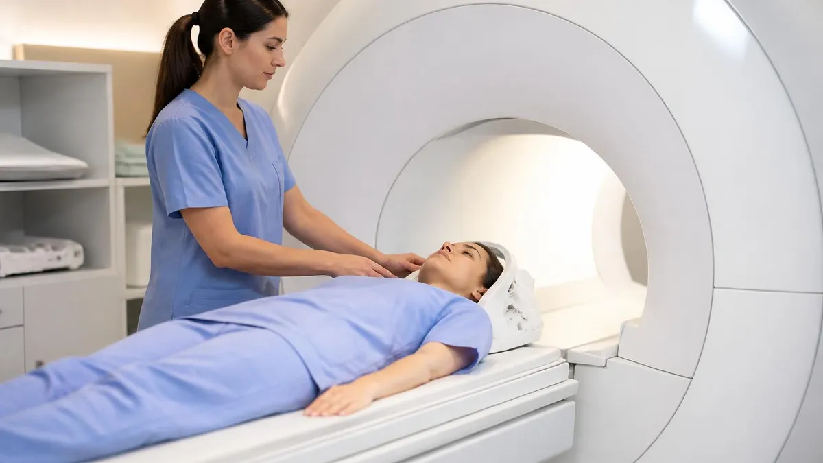
Patients who are claustrophobic should mention it during scheduling. Pituitary MRI takes 25 to 40 minutes inside a closed bore. A mild oral anxiolytic (often lorazepam) prescribed by the ordering doctor or a wide-bore scanner makes the experience tolerable for most people. If you are studying imaging or radiologic technology, the brain MRI workflow guide covers more general patient prep that overlaps with pituitary cases.
Why the Dynamic Sequence Matters
Microadenomas often look identical to normal pituitary tissue on regular post-contrast images. The dynamic sequence catches the lesion in the brief window where the normal gland enhances but the tumor lags behind. Without this sequence, small prolactinomas and ACTH-secreting tumors can be missed entirely.
What Radiologists Actually Look For
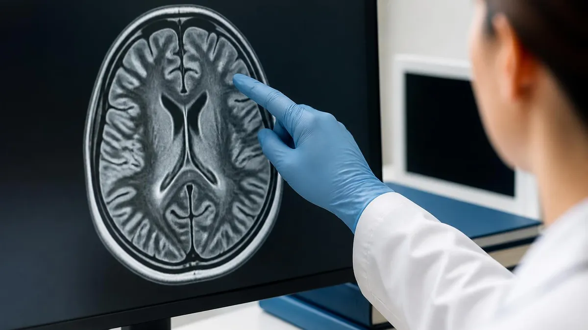
The radiologist works through the scan in a predictable order. First, gland size and shape. The normal adult pituitary is around 6 to 8 mm in height in men and slightly taller in women, especially during pregnancy when it can swell to 10 or 12 mm physiologically. A gland that bulges upward against the optic chiasm or sags into an empty sella is flagged immediately.
Second, signal intensity. The posterior pituitary normally shows a bright T1 spot from stored vasopressin. Loss of that bright spot is a clue toward diabetes insipidus or stalk transection. The anterior pituitary is usually homogeneous on T1 and T2. Any focal area of different signal earns a closer look.
Third, the pituitary stalk. It should be midline and roughly 2 to 3 mm thick. A thickened stalk raises suspicion for inflammation, granulomatous disease, lymphocytic hypophysitis, or metastasis. A stalk that deviates sideways often points toward a small adenoma on the opposite side, pushed there by the mass effect.
Fourth, the cavernous sinuses and surrounding structures. Macroadenomas can invade the cavernous sinus, encase the internal carotid artery, or extend up into the suprasellar cistern. The Knosp grading scale classifies cavernous sinus invasion from grade 0 (no invasion) to grade 4 (carotid fully encased). Surgical planning depends heavily on Knosp grade.
Common Pituitary Lesion Types
Less than 10 mm in diameter. Often hypoenhancing on dynamic sequences. Most commonly prolactinomas in women of reproductive age.
10 mm or larger. Often heterogeneous with cystic or hemorrhagic areas. Can compress optic chiasm and invade cavernous sinus.
Non-enhancing cystic lesion between anterior and posterior lobes. Usually incidental, rarely causes hormonal disturbance.
Suprasellar tumor with calcifications and cystic components. Common in children and adults over 50. Bimodal age distribution.
Microadenoma vs Macroadenoma: The 10 mm Rule
The single most useful number in pituitary radiology is 10 millimeters. Lesions smaller than 10 mm are called microadenomas. Lesions equal to or larger than 10 mm are macroadenomas. That cutoff matters because microadenomas almost never cause mass effect on the chiasm or hormonal failure from compression. Their symptoms come from hormone hypersecretion, not from physical pressure.
Macroadenomas behave differently. By the time a pituitary tumor reaches a centimeter, it starts pushing against neighbors. The optic chiasm sits directly above the sella, so visual field loss is one of the first signs. Larger tumors can invade the cavernous sinus laterally, trapping cranial nerves III, IV, V1, V2, and VI. Patients can develop double vision, facial numbness, or droopy eyelids.
Macroadenomas can also crush the normal pituitary against the bone, causing panhypopituitarism. Patients become tired, infertile, cold, and sometimes critically adrenally insufficient. The radiologist measuring a macroadenoma will note suprasellar extension in millimeters, mention chiasm contact or compression, and grade cavernous sinus invasion. Neurosurgeons read those details to plan transsphenoidal resection.
Reading the Report Without Panic
Patients who get a pituitary MRI report often see phrases that sound alarming and are actually routine. "Heterogeneous enhancement pattern" can mean a normal physiological variant. "Slight asymmetric enhancement" is sometimes called pars intermedia cyst, which is harmless. "Empty sella" sounds dramatic but is frequently incidental and asymptomatic.
Truly important phrases include: "hypoenhancing focus measuring X mm," which suggests a possible adenoma; "suprasellar extension with mass effect on the optic chiasm," which means the tumor is pressing on vision; "deviation of the pituitary stalk," which hints at a small adenoma even when one is not clearly seen; and "loss of normal posterior pituitary bright spot," which raises concern for posterior pituitary dysfunction.
Patients should always discuss the report with the ordering physician, not Google. Endocrinologists know how to weigh imaging against hormone levels. A 3 mm bright spot in someone with normal prolactin probably needs no treatment. The same finding in someone with prolactin of 300 ng/mL is the diagnosis. Context drives management.
Key Pituitary MRI Numbers
Pituitary MRI Sequence Breakdown
Sudden severe headache, vision loss, and altered consciousness in a patient with a known or suspected pituitary tumor is a neurosurgical emergency. MRI must be done urgently with attention to T2 and SWI sequences to detect hemorrhage. Pituitary apoplexy can cause acute adrenal crisis and death within hours if untreated.
Common Pitfalls and False Positives
Pituitary imaging has more false positives than most neuroradiologists like to admit. Pars intermedia cysts, small Rathke cleft remnants, and physiological gland hypertrophy during pregnancy or puberty can all mimic adenomas on a quick read. A pituitary that looks plump in a 16-year-old girl is usually normal, not pathological. The radiologist must correlate with age, sex, and clinical context.
Medications also distort the picture. Patients on dopamine agonists for prolactinoma can see their tumors shrink dramatically, sometimes vanishing completely on follow-up MRI. Estrogen therapy and pregnancy can enlarge the gland. Steroid suppression can shrink normal pituitary tissue. The history form filled out before the scan is not bureaucracy; it changes how the images are read.
Motion is another enemy. Pituitary protocols depend on millimeter precision. A patient who moves between sequences ruins the dynamic contrast timing and forces the tech to repeat acquisitions. Patients with tremor, anxiety, or pain sometimes need sedation to get a usable study. Modern motion-correction software helps, but it is not magic.
After the Scan: Next Steps
For most patients, the scan is just one piece of a longer workup. Endocrinologists pair imaging with hormone panels: prolactin, IGF-1, ACTH, cortisol, TSH, free T4, LH, FSH, testosterone or estradiol depending on sex. A pituitary lesion seen on MRI without any hormonal abnormality is usually watched, not treated. A lesion seen with classic prolactinoma chemistry is treated with cabergoline or bromocriptine first, surgery only if medical therapy fails.
Surgical candidates are referred to neurosurgeons specializing in transsphenoidal approaches. Modern endoscopic transsphenoidal surgery can remove tumors through the nostril with minimal external scarring. Outcomes depend heavily on tumor size, Knosp grade, and surgeon volume. High-volume centers report cure rates above 80% for microprolactinomas and 60% for non-invasive macroadenomas.
Radiation therapy enters the conversation for residual or recurrent tumors after surgery. Stereotactic radiosurgery (Gamma Knife or CyberKnife) delivers focused doses with relative pituitary sparing. Fractionated external beam is reserved for larger residual disease. Both carry small but real risks of secondary hypopituitarism years later, so patients need lifelong endocrine follow-up.

Pituitary MRI Patient Checklist
- ✓Bring prior imaging on CD or USB for comparison
- ✓List all medications, especially dopamine agonists or steroids
- ✓Note allergies to gadolinium contrast or kidney disease history
- ✓Fast 2 to 4 hours if contrast is planned
- ✓Remove all metal, including jewelry, hairpins, and dental flippers
- ✓Mention pregnancy or possible pregnancy
- ✓Tell the tech about claustrophobia for sedation options
- ✓Hydrate well before and after the contrast scan
Cost, Access, and Insurance
A pituitary MRI in the United States typically costs $1,200 to $3,500 without insurance, depending on whether contrast is used and whether the scan is performed at a hospital or freestanding imaging center. Hospital-based scans usually run 2 to 3 times higher than outpatient imaging centers for the exact same protocol. Insurance generally covers the scan when ordered for a documented hormone abnormality or visual field defect.
Pre-authorization is required by most commercial insurers and Medicare Advantage plans. Endocrinologists are familiar with the paperwork. Patients who pay out of pocket can shop around. Some imaging centers post cash-pay prices online; transparent pricing is becoming common after federal price disclosure rules took effect. Self-pay rates of $500 to $900 are achievable in many markets.
3T scanners produce better images than 1.5T machines for pituitary work, especially for microadenoma hunting. Not every facility has a 3T magnet. Patients with negative 1.5T scans and high clinical suspicion sometimes need referral to a tertiary center for a repeat 3T study. The extra cost is usually justified when the diagnosis would change management.
What Studying for Imaging Boards Looks Like
Radiology residents and MRI tech students spend a disproportionate amount of board prep time on the pituitary because the questions test knowledge density. A single coronal T1 image can hide three or four testable findings: gland size, posterior bright spot, stalk position, and adenoma signal. Practice tests force you to scan systematically rather than guessing.
Tech-focused programs cover protocol parameters: TR, TE, slice thickness, FOV, and the timing of dynamic contrast. Resident programs add the differential diagnosis, the Knosp scale, and clinical correlation. Both groups benefit from working through high-yield cases repeatedly. The MRI category page lists current practice tests and study materials covering pituitary, brain, spine, and musculoskeletal protocols.
Pituitary MRI Strengths and Limitations
- +Highest soft-tissue contrast of any imaging modality
- +No ionizing radiation, safe for repeat scans
- +Dynamic contrast catches microadenomas other scans miss
- +Clear visualization of optic chiasm and cavernous sinus
- +Excellent for surgical and radiation planning
- −Expensive compared to CT
- −Long scan time, often 30 to 40 minutes
- −Contrast risks in chronic kidney disease
- −Motion sensitivity ruins thin-slice protocols
- −Cannot be performed on patients with most older pacemakers
Frequently Misunderstood Findings
An empty sella is one of the most reported and most misunderstood findings on pituitary MRI. The term describes a sella turcica that appears filled mostly with cerebrospinal fluid, with the pituitary flattened against the floor. Primary empty sella is usually incidental and asymptomatic. Secondary empty sella follows surgery, radiation, or tumor infarction and can be associated with hormone deficiencies. Both look similar on a static image; the clinical history separates them.
Pituitary hyperplasia is another tricky finding. The gland can swell to 12 mm or more during pregnancy, lactation, and severe primary hypothyroidism. Untreated primary hypothyroidism in particular causes a striking pituitary enlargement from thyrotroph hyperplasia that mimics a macroadenoma. Treating the thyroid shrinks the gland and avoids unnecessary surgery. Missing this diagnosis is one of the more painful errors in pituitary imaging.
Stalk thickening above 4 mm raises a defined differential: lymphocytic hypophysitis, neurosarcoidosis, Langerhans cell histiocytosis, germinoma, and metastasis. Each has its own clinical clues and management path. The radiology report flags the finding; the endocrinologist works the differential.
The Bottom Line
Pituitary MRI is one of the most rewarding studies in neuroradiology when done well and one of the most dangerous when done sloppily. The difference between a 3 mm prolactinoma and a normal gland often comes down to whether the dynamic contrast sequence was timed correctly and whether the slices were thin enough.
For patients, the takeaway is to insist on a dedicated pituitary protocol when symptoms point to the gland, not a generic brain MRI. For students and techs, the takeaway is that protocol details matter more here than almost anywhere else in imaging. Master the basics, practice on real cases, and review the anatomy until you can sketch it from memory.
Pituitary MRI Report Red Flags
- ✓Hypoenhancing focus measuring X mm on dynamic sequence
- ✓Suprasellar extension with mass effect on optic chiasm
- ✓Cavernous sinus invasion Knosp grade 3 or 4
- ✓Pituitary stalk deviation more than 2 mm from midline
- ✓Loss of normal posterior pituitary bright spot
- ✓Heterogeneous T2 signal with hemorrhagic components
- ✓Thickened stalk above 4 mm with infiltrative pattern
- ✓Bony erosion of sellar floor on bone-window sequences
MRI Safety Contraindications to Disclose
- ✓Older non-MRI-conditional cardiac pacemaker or ICD
- ✓Cochlear implant or other implanted hearing device
- ✓Aneurysm clip placed before 1995 or unknown type
- ✓Metallic foreign body in eye (especially welders)
- ✓Implanted neurostimulator or drug-infusion pump
- ✓Severe kidney disease (eGFR below 30) if contrast planned
- ✓Known prior reaction to gadolinium-based contrast agents
- ✓First-trimester pregnancy if contrast is being considered
- ✓Permanent eye makeup or tattoo containing metallic pigment
- ✓Recent surgical clip placement (within last 6 weeks)
MRI Questions and Answers
About the Author
Medical Laboratory Scientist & Clinical Certification Expert
Johns Hopkins UniversityDr. Sandra Kim holds a PhD in Clinical Laboratory Science from Johns Hopkins University and is certified as a Medical Technologist (MT) and Medical Laboratory Scientist (MLS) through ASCP. With 16 years of clinical laboratory experience spanning hematology, microbiology, and molecular diagnostics, she prepares candidates for ASCP board exams, MLT, MLS, and specialist certification tests.
Join the Discussion
Connect with other students preparing for this exam. Share tips, ask questions, and get advice from people who have been there.
View discussion (6 replies)