MRI for Muscle Knots: How Magnetic Resonance Imaging Diagnoses Myofascial Trigger Points in 2026
MRI muscle knot imaging explained: 🎯 how MRI detects myofascial trigger points, elastography techniques, costs, accuracy, and when to order a scan.
An MRI muscle knot evaluation has become one of the most requested musculoskeletal imaging studies in 2026, as patients and clinicians push beyond physical palpation to confirm what causes that stubborn, painful lump beneath the skin. Magnetic resonance imaging offers unparalleled soft-tissue contrast, allowing radiologists to visualize the dense, contracted bands of muscle fibers known clinically as myofascial trigger points. For chronic pain sufferers who have exhausted massage, dry needling, and physical therapy, the MRI scan provides objective evidence of pathology rather than relying on subjective complaints alone, transforming how clinicians approach treatment planning.
A muscle knot, technically called a myofascial trigger point, is a hyperirritable spot in skeletal muscle associated with a palpable taut band of muscle fibers. While these knots have traditionally been diagnosed through manual palpation by experienced clinicians, MRI now offers a non-invasive way to objectively visualize and measure them. Specialized sequences like magnetic resonance elastography (MRE) and T2 mapping can detect the subtle changes in tissue stiffness and water content that characterize active trigger points, making MRI a powerful adjunct in chronic pain workups for patients with fibromyalgia, post-whiplash syndrome, or persistent regional myofascial pain.
The technology behind muscle knot imaging has evolved rapidly since 2020, with 3 Tesla scanners now standard at most academic medical centers and 7T research systems revealing microstructural details previously invisible. Researchers at institutions like Mayo Clinic, Johns Hopkins, and the National Institutes of Health have published landmark studies demonstrating that trigger points appear on MRI as focal areas of increased T2 signal with measurably greater stiffness on elastography. These findings have helped validate myofascial pain syndrome as a genuine, measurable pathology rather than a diagnosis of exclusion based purely on patient self-report.
Despite these advances, MRI is not the first-line tool for every patient with sore shoulders or a tight neck. Most muscle knots resolve with conservative treatment within a few weeks, and the cost of an MRI scan, ranging from $400 to $3,500 depending on location and insurance, makes it impractical for routine cases. However, when symptoms persist longer than six weeks, when neurological signs appear, or when the diagnosis is unclear, MRI becomes invaluable. It helps rule out more serious conditions like tumors, abscesses, hematomas, or muscle tears that can mimic the presentation of a stubborn knot.
This comprehensive guide walks you through everything you need to know about using MRI to evaluate muscle knots in 2026, from the underlying physics of how the scan detects tissue abnormalities to practical advice on what to expect during your appointment. We will examine the specific MRI sequences radiologists use, how to interpret findings on your scan report, when insurance is likely to approve the study, and what treatment options follow once a diagnosis is confirmed. Whether you are a patient researching options or a clinician refining your imaging algorithm, this article delivers actionable, evidence-based information.
Understanding the relationship between MRI technology and myofascial pain requires familiarity with both imaging principles and musculoskeletal anatomy. Throughout this guide, we will reference current research, professional guidelines from the American College of Radiology, and real-world clinical scenarios drawn from leading pain centers. By the end, you should have a clear sense of when MRI is appropriate for muscle knot evaluation, what the limitations are, and how to discuss the option intelligently with your physician or radiologist during your next appointment.
Before diving deeper, it helps to know the broader context of how MRI fits into modern diagnostic medicine. If you are new to the technology entirely, our companion guide on what is an MRI test covers the foundational principles in plain language. For now, we will focus specifically on how this powerful imaging modality applies to one of the most common yet poorly understood causes of chronic pain in the United States today: the persistent, painful muscle knot that just will not go away no matter what you try.
MRI Muscle Knot Imaging by the Numbers
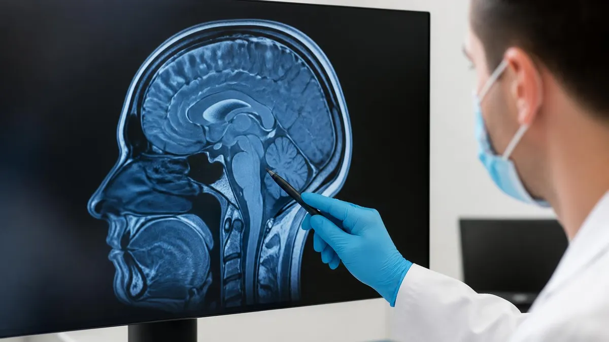
How MRI Visualizes a Muscle Knot
Active trigger points show increased T2 signal because of localized edema and inflammation. Radiologists look for focal hyperintensities within otherwise normal-appearing muscle fibers, often measuring just 5-10 millimeters in diameter.
MRE measures tissue stiffness by analyzing how mechanical shear waves propagate through muscle. Knots appear as discrete areas of elevated stiffness, sometimes three to five times stiffer than surrounding healthy muscle tissue.
DTI maps the direction and magnitude of water diffusion within muscle fibers. Trigger points disrupt the normally orderly diffusion pattern, creating measurable changes in fractional anisotropy that correlate with clinical symptoms.
MR spectroscopy can detect metabolic changes in active trigger points, including elevated lactate, acetylcholine, and inflammatory mediators. This biochemical fingerprint helps distinguish active from latent knots.
STIR and fat-suppressed T2 sequences enhance the conspicuity of edematous tissue by removing fat signal. This makes subtle muscle abnormalities, including small trigger points, much easier to detect against a darkened background.
To understand why MRI is so effective at detecting muscle knots, you need to know what is actually happening inside the tissue at a microscopic level. A myofascial trigger point is not a tangled bundle of fibers as the colloquial name suggests. Rather, it represents a localized region where individual sarcomeres, the contractile units of muscle, have become persistently shortened. This sustained contraction increases local metabolic demand while simultaneously compressing the tiny blood vessels that supply oxygen and nutrients, creating a self-perpetuating cycle of dysfunction.
This pathological state produces several distinctive features that MRI can detect. First, the hypercontracted muscle fibers create a measurable increase in tissue stiffness, which magnetic resonance elastography can quantify with remarkable precision. Second, the metabolic stress leads to mild but detectable edema, increasing local water content in ways that show up as bright signal on T2-weighted sequences. Third, inflammatory mediators including substance P, bradykinin, and serotonin accumulate at the trigger point, sometimes producing chemical shifts visible on spectroscopy.
Researchers have used MRI to validate distinctions between active and latent trigger points. Active knots produce spontaneous pain and characteristic referral patterns when palpated, while latent ones are tender only when pressed. On imaging, active trigger points consistently show greater T2 signal abnormalities, higher stiffness on elastography, and more pronounced metabolic changes than latent ones. This objective differentiation, impossible with palpation alone, helps clinicians prioritize which knots to treat aggressively and which to monitor conservatively over time.
The neurological dimension of myofascial pain adds another layer of complexity that imaging is beginning to address. Functional MRI studies have shown that patients with chronic myofascial pain syndromes display altered activity in pain-processing brain regions including the insula, anterior cingulate cortex, and somatosensory cortex. This central sensitization explains why some muscle knots persist long after the original injury has healed and why peripheral treatments alone sometimes fail to provide lasting relief for chronic sufferers seeking durable solutions today.
Different muscle groups present unique imaging challenges that radiologists must navigate carefully. The trapezius, upper back, and gluteal muscles are common sites for trigger points and are relatively easy to image because of their size and accessibility. Smaller muscles like the rhomboids, piriformis, or scalenes require higher-resolution protocols and sometimes dedicated surface coils to visualize properly. Deep paraspinal muscles can be particularly challenging because of their proximity to vertebrae, which create artifacts that can obscure subtle soft-tissue abnormalities on certain sequences.
The duration and intensity of symptoms influence what MRI is likely to show. Acute muscle knots that develop after recent overuse or injury often display prominent edema and may be confused with grade I muscle strains. Chronic trigger points present for months or years may show more subtle findings, with stiffness changes outweighing signal abnormalities. In some long-standing cases, atrophy of surrounding muscle tissue or fatty infiltration becomes the dominant finding, indicating that the pathological process has progressed beyond simple muscle contracture into structural degeneration.
Understanding the MRI report requires familiarity with radiology terminology, which can feel intimidating to patients reading their results for the first time. Terms like signal intensity, edema, hyperintense, hypointense, and contractile band each carry specific meanings. Our guide to the MRI medical abbreviation standards used by radiologists nationwide can help you decipher the technical language and ask informed questions during your follow-up appointment with your ordering physician or musculoskeletal specialist.
MRI Practice Test Questions
Prepare for the MRI - Magnetic Resonance Imaging exam with our free practice test modules. Each quiz covers key topics to help you pass on your first try.
MRI Knowledge
MRI Exam Questions covering Knowledge. Master MRI Test concepts for certification prep.
MRI Physics
Free MRI Practice Test featuring Physics. Improve your MRI Exam score with mock test prep.
MRI Anatomy and Pathology
MRI Test Prep for MRI Anatomy and Pathology. Practice MRI Quiz questions and boost your score.
MRI Anatomy and Positioning
MRI Questions and Answers on MRI Anatomy and Positioning. Free MRI practice for exam readiness.
MRI Contrast Agents
Free MRI Quiz on MRI Contrast Agents. MRI Exam prep questions with detailed explanations.
MRI Patient Care and Positioning
MRI Practice Questions for MRI Patient Care and Positioning. Build confidence for your MRI certification exam.
MRI Sequences Used for Muscle Knot Detection
T2-weighted imaging serves as the workhorse sequence for evaluating muscle pathology because water-rich tissues like edematous muscle appear bright while normal muscle stays dark. This high contrast makes even small areas of inflammation visible to the trained eye. Most musculoskeletal protocols include T2 in at least two orthogonal planes, typically axial and coronal for trunk muscles or axial and sagittal for limb muscles.
Short Tau Inversion Recovery, or STIR, is a fat-suppressed variant that further enhances sensitivity to edema by nulling the bright fat signal that can obscure subtle muscle changes. STIR is particularly valuable in regions with abundant intermuscular fat, such as the buttocks, thighs, or paraspinal muscles in older adults. The combination of T2 and STIR provides excellent baseline detection of active trigger points and inflammatory muscle conditions.
Should You Get an MRI for Your Muscle Knot?
- +Provides objective documentation of trigger points that palpation alone cannot quantify
- +Rules out serious mimics like tumors, abscesses, hematomas, and structural muscle tears
- +Allows precise mapping of multiple knots for targeted treatment planning sessions
- +Helps establish medical necessity for insurance coverage of advanced therapies
- +Tracks treatment response over time with quantitative elastography measurements
- +Identifies associated pathology like nerve impingement or joint involvement
- +Validates patient symptoms with objective evidence reducing psychological distress
- −High cost ranging from several hundred to several thousand dollars per scan
- −Often requires prior authorization that can delay imaging by weeks
- −Most insurance plans will not cover MRI for uncomplicated muscle knot complaints
- −Elastography is not widely available outside major academic medical centers
- −Findings may not change treatment if conservative therapy has not been tried
- −Claustrophobia and acoustic noise make the experience unpleasant for some patients
- −Incidental findings can lead to unnecessary follow-up tests and patient anxiety
MRI Muscle Knot Pre-Scan Preparation Checklist
When to Image and When to Wait
Most muscle knots resolve with conservative treatment within four to six weeks. Current 2026 American College of Radiology guidelines recommend MRI only after failure of initial conservative therapy, persistence beyond six weeks, or presence of red flag symptoms like progressive weakness, sensory loss, unexplained weight loss, or fever. Premature imaging often leads to incidental findings that cause anxiety without changing management.
The financial reality of getting an MRI for muscle knot evaluation in 2026 varies dramatically depending on where you live, what facility you choose, and how your insurance is structured. Cash prices at large hospital systems can exceed $3,500 for a single body region, while independent imaging centers in competitive metropolitan markets sometimes offer the same study for under $500. Medicare reimbursement rates hover around $300 to $450 for most musculoskeletal MRI codes, providing a benchmark that some self-pay patients can negotiate toward when paying out of pocket.
Insurance coverage for muscle knot MRI depends heavily on clinical documentation. Most commercial plans require prior authorization that includes evidence of failed conservative treatment lasting at least four to six weeks, neurological symptoms, or concerning physical exam findings. Coding the study correctly matters enormously. A scan ordered for non-specific muscle pain is often denied, while the same study ordered to rule out a specific pathology like a muscle tear, nerve impingement, or inflammatory myositis is more likely to be approved by the insurance reviewer.
High-deductible health plans have changed the calculus for many patients. With deductibles averaging over $2,500 for individuals and $5,000 for families, an MRI scan often falls entirely on the patient before insurance contributes anything. This has driven increased interest in cash-pay imaging centers, medical tourism to lower-cost states, and even cross-border imaging in Mexico or Canada where high-quality scans can cost a fraction of US prices. Some employers now contract directly with imaging networks to offer steerage discounts.
The location of the scan significantly impacts both cost and quality. Hospital-based outpatient imaging departments typically charge two to four times more than independent facilities for identical studies, though they may offer faster turnaround for urgent cases. Academic medical centers provide access to advanced sequences like elastography and spectroscopy that community facilities lack, but at premium prices. Mobile MRI units traveling to rural areas have expanded access but may not offer the most sophisticated protocols needed for trigger point evaluation specifically.
Magnet strength affects diagnostic quality for subtle findings like trigger points. The 1.5 Tesla scanners common at smaller facilities produce excellent images for most clinical questions but may miss the small T2 signal changes characteristic of myofascial pathology. The 3 Tesla scanners standard at larger centers provide roughly double the signal-to-noise ratio, dramatically improving detection of small abnormalities. Research-grade 7 Tesla systems offer even finer detail but remain limited to a handful of institutions and are not typically available for routine clinical scans.
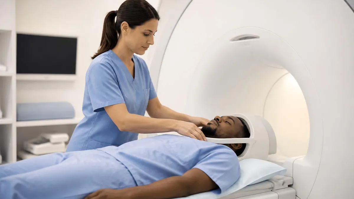
Open MRI scanners, while more comfortable for claustrophobic patients, generally use lower field strengths and may not detect subtle trigger point findings. If your physician specifically wants high-resolution imaging of muscle pathology, request a closed-bore 3T scanner if possible. For patients who absolutely cannot tolerate enclosed scanners, oral sedation with medications like lorazepam or alprazolam, IV sedation, or open upright mri MRI alternatives may make diagnostic-quality imaging feasible. Discuss these options with your ordering provider well in advance of your scheduled appointment date.
Understanding the history and evolution of MRI technology helps patients appreciate why certain scanners produce better images than others. Our article on the history of MRI traces the remarkable journey from the original 1977 prototype that took five hours to produce a single image to today's machines that complete comprehensive musculoskeletal studies in under thirty minutes with detail unimaginable to the technology's pioneers just a few decades ago.
Seek immediate evaluation rather than waiting for routine MRI scheduling if you experience progressive muscle weakness, loss of sensation, bowel or bladder dysfunction, unexplained weight loss, fever, night sweats, or pain that wakes you from sleep. These symptoms can indicate serious pathology like tumors, infections, or nerve compression requiring urgent diagnostic workup beyond simple muscle knot evaluation.
Once your MRI confirms the presence of a muscle knot or rules out alternative diagnoses, treatment planning becomes more focused and evidence-based. The imaging findings guide selection of interventions, identifying which knots are most active, which are deep enough to require imaging guidance for injection, and which are associated with neighboring pathology that needs separate attention. This precision approach often produces faster and more durable results than empirical treatment based on palpation findings alone, particularly in patients with chronic widespread pain syndromes.
Dry needling and trigger point injections benefit substantially from MRI-confirmed targets. Practitioners can plan their needle trajectory to reach deep knots safely, avoiding nerves, blood vessels, and lung tissue in dangerous anatomical regions like the upper thoracic paraspinals. Ultrasound guidance during the actual procedure complements pre-procedure MRI mapping, with the imaging study providing the strategic plan while real-time ultrasound provides tactical visualization during needle placement. This combined approach has dramatically reduced complications in advanced interventional pain practice settings.
Physical therapy programs are increasingly tailored using MRI findings. Therapists can prioritize manual treatment of imaging-confirmed active knots, design specific stretching protocols for affected muscle groups, and use imaging to demonstrate progress to skeptical patients. Some progressive pain centers now perform follow-up scans after treatment to document objective improvement in stiffness measurements, providing powerful evidence of treatment efficacy that motivates continued patient engagement with home exercise programs and lifestyle modifications recommended by their care team.
Medications play a supporting role in comprehensive trigger point management. Muscle relaxants like cyclobenzaprine or tizanidine can reduce hypercontraction at the level of individual fibers. Anti-inflammatory drugs address the local edema visible on T2 imaging. For chronic cases with central sensitization, medications like duloxetine, gabapentin, or low-dose tricyclic antidepressants target the neurological amplification revealed by functional imaging studies of pain-processing brain regions in chronic myofascial pain patients across multiple research cohorts.
Newer interventional approaches are emerging from research enabled by advanced imaging. Botulinum toxin injections into MRI-confirmed deep trigger points have shown promise for refractory cases, with the imaging ensuring accurate placement of this expensive medication. Focused ultrasound, currently in clinical trials, uses MRI guidance to deliver therapeutic acoustic energy directly to trigger points without any needle penetration. Regenerative medicine approaches including platelet-rich plasma and prolotherapy are also benefiting from precise pre-procedure imaging characterization of target tissues.
Lifestyle and ergonomic interventions remain foundational regardless of how sophisticated the imaging or interventional treatment becomes. MRI findings often reveal patterns of muscle dysfunction related to posture, workplace ergonomics, sleep position, or recreational activities. A scan showing bilateral upper trapezius knots in a desk worker, for example, points clearly toward workstation evaluation and postural retraining. Without addressing these underlying drivers, even successful focal treatment often fails to provide lasting relief as new knots form to replace the treated ones.
Patient education emerges as perhaps the most underrated benefit of MRI for muscle knot evaluation. Seeing concrete visual evidence of their pathology helps many patients understand and accept their condition, reducing the frustration and self-doubt that often accompanies chronic pain diagnoses. Reviewing the images with a knowledgeable provider transforms abstract complaints into a tangible problem with identifiable solutions, dramatically improving engagement with long-term treatment plans and self-management strategies that ultimately determine which patients achieve lasting relief from chronic myofascial pain.
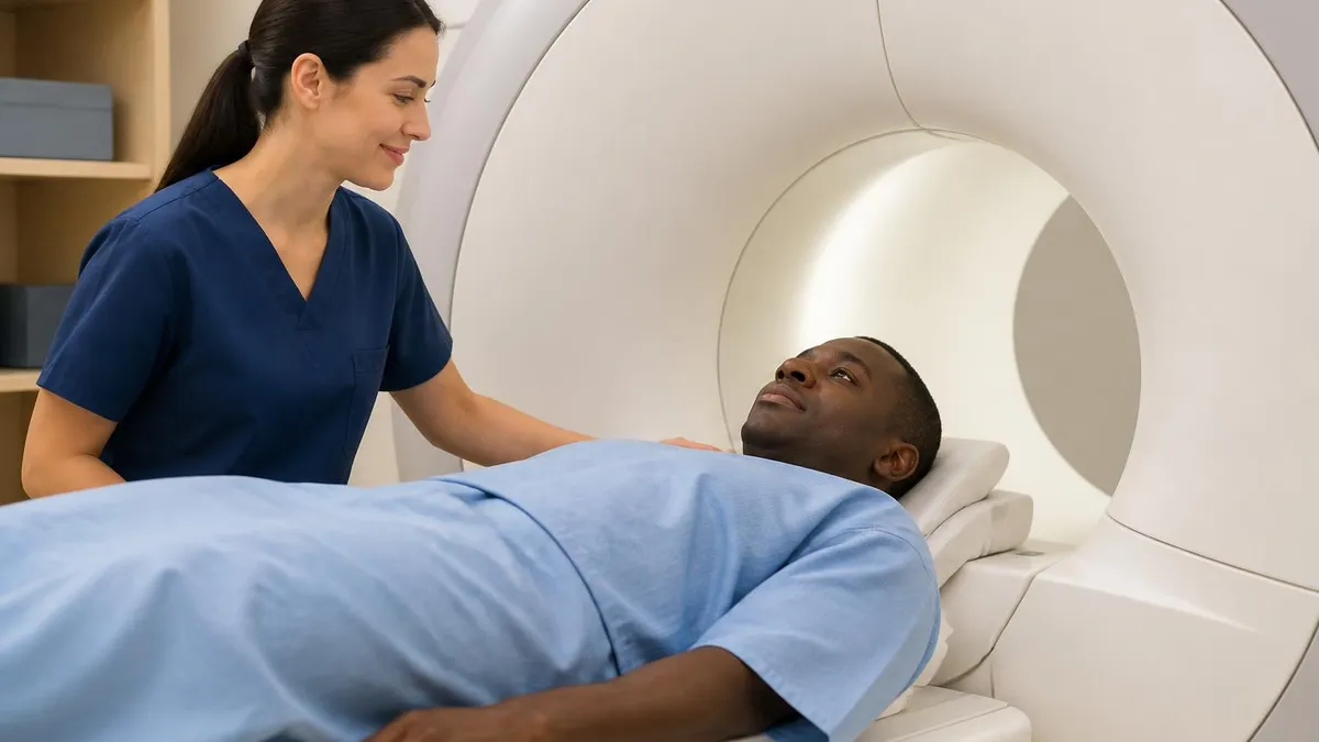
Preparing intelligently for your MRI muscle knot appointment can make the difference between a diagnostic-quality study and a wasted insurance authorization. Start by gathering all relevant medical records, including notes from previous physical therapy, chiropractic care, massage therapy, prior injections, and any imaging done in the past two years. Bring this documentation to your appointment with the ordering physician so they can write a comprehensive history that justifies the study to insurance reviewers and helps the radiologist understand the clinical context.
Communication with the imaging facility before your appointment date helps avoid surprises. Confirm the specific protocol that will be performed, asking whether elastography or other advanced sequences are available if your physician has requested them. Verify whether contrast will be used, as this affects pre-scan preparation including fasting and kidney function testing. Ask about the scanner type and field strength being used, since some facilities have multiple machines and you may have the option to request the higher-resolution unit if available at no extra cost.
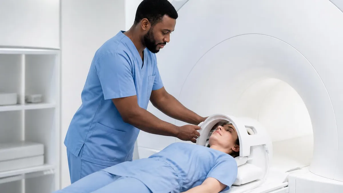
On the day of your scan, manage your comfort proactively. Arrive well-rested and well-hydrated unless specifically instructed otherwise. Eat a light meal a few hours before the appointment to avoid hunger discomfort during the typically thirty to sixty minute scan. Wear comfortable, metal-free clothing or plan to change into a hospital gown. Bring a friend or family member to drive you home if sedation will be used. Mention any pain you experience in specific positions so the technologist can adjust your positioning if possible during the study.
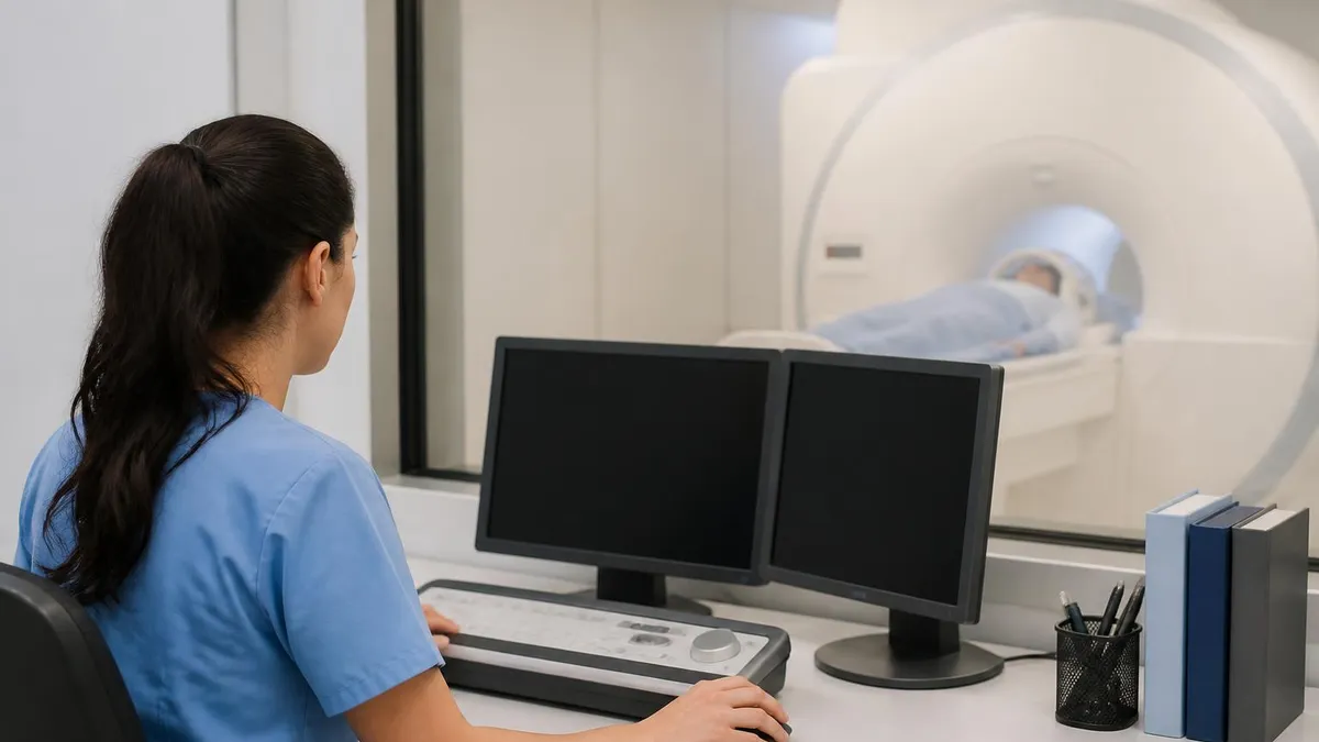
During the scan itself, communicate openly with the technologist through the intercom system. They cannot see subtle facial expressions but can respond to verbal feedback about discomfort, claustrophobia, or unusual sensations. The famous loud knocking sounds of an MRI scanner can be disturbing if you are unprepared. Hearing protection is mandatory and effective, but the noise still penetrates somewhat. Our guide to the mri machine noise operation explains why these sounds occur and offers strategies for managing them comfortably during your scan.
After the scan, do not expect immediate results in most cases. The radiologist needs time to review images carefully, compare them to any prior studies, and dictate a comprehensive report. This typically takes 24 to 72 hours for routine cases and longer if specialized sequences like elastography require dedicated post-processing. Some facilities provide preliminary findings to the ordering physician within hours for urgent cases, but the formal report and your detailed discussion with the ordering provider usually wait several days.
Interpreting your results requires partnership with a knowledgeable clinician. Avoid the temptation to Google individual phrases from the report, which often produces frightening worst-case scenarios that rarely apply to your actual situation. Schedule a follow-up appointment with the ordering provider specifically to review findings and discuss treatment implications. Bring written questions and consider asking for copies of the images on a CD or via online patient portal for your own records and potential future second opinions or specialty consultations.
Finally, remember that imaging is just one tool in comprehensive muscle pain management. A normal MRI does not invalidate your symptoms, and an abnormal MRI does not necessarily explain all of your pain. The most successful patients combine objective imaging information with subjective symptom tracking, work closely with multidisciplinary care teams, maintain realistic expectations about treatment timelines, and engage actively with self-management strategies including exercise, stress management, sleep optimization, and ergonomic improvements that address the lifestyle factors driving their chronic muscle pain condition.
MRI Questions and Answers
What Is an MRI Test? How Magnetic Resonance Imaging Scans Diagnose Disease in 2026

MRI Medical Abbreviation: What MRI Stands For and Why It Matters

The History of MRI: From Discovery to Modern Medicine

Knee MRI Images: A Complete Guide to Reading, Understanding, and Interpreting Knee Scans

Noise of MRI Machine: Why MRI Scanners Are So Loud and What to Expect
About the Author
Medical Laboratory Scientist & Clinical Certification Expert
Johns Hopkins UniversityDr. Sandra Kim holds a PhD in Clinical Laboratory Science from Johns Hopkins University and is certified as a Medical Technologist (MT) and Medical Laboratory Scientist (MLS) through ASCP. With 16 years of clinical laboratory experience spanning hematology, microbiology, and molecular diagnostics, she prepares candidates for ASCP board exams, MLT, MLS, and specialist certification tests.
Join the Discussion
Connect with other students preparing for this exam. Share tips, ask questions, and get advice from people who have been there.
View discussion (6 replies)