
MRI vs MRA: Differences, Uses, and How They Work 2026 September
💡 MRI vs MRA explained: scan purpose, contrast use, prep, timing, and what each test reveals about your body.
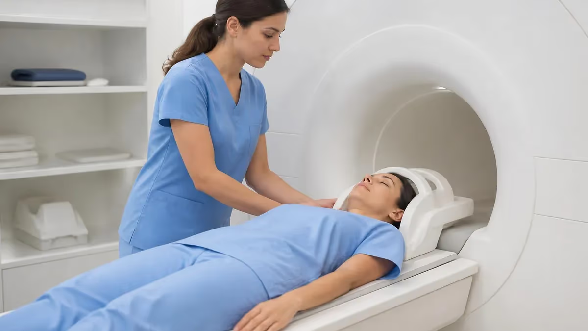
If your doctor has ordered an imaging test and you keep seeing the letters MRI and MRA, you are not alone in feeling a bit lost. The names look almost identical, both machines look the same from the outside, and the paperwork rarely explains the difference clearly.
The short version is this: an MRI takes detailed pictures of soft tissue, like the brain, muscles, organs, joints, and spinal cord. An MRA, which is short for magnetic resonance angiography, uses the same machine and the same magnet but focuses on blood vessels, the arteries and veins that carry blood through your body.
Think of it this way. An MRI is the wide-angle photographer that captures the whole landscape. An MRA is the macro lens that zooms into the rivers and streams flowing through that landscape.
Both are valuable. Sometimes you need only one. Sometimes your physician will order both during the same visit because the questions they answer overlap but do not match perfectly. This guide breaks down the real differences, the small but important details about contrast dye, how long each scan takes, what to expect on the table, and how to prepare.
By the end, you should be able to walk into your appointment knowing which test you are getting, why, and what the radiologist is hunting for. Knowledge calms nerves, and a calm patient produces sharper images. Less motion blur means a more confident report, fewer follow-up scans, and faster answers.
MRI vs MRA at a glance
Numbers help, but they only tell part of the story. The scan time depends on the body part, the strength of the magnet, the protocol your radiologist designed, and whether you can hold still without fidgeting. A brain MRI on a modern 3 Tesla machine can wrap in under thirty minutes when the patient cooperates.
A spinal MRI with multiple sequences can run closer to an hour. MRA studies tend to be faster because the focus is narrower, but a head and neck MRA with contrast can still take forty minutes from start to finish.
Neither test exposes you to ionizing radiation. That alone makes both MRI and MRA preferable for many patients, especially children, pregnant women in their second and third trimesters when clinically necessary, and anyone who has already had multiple CT scans during the year. The trade-off is time and noise. MRI machines are loud, and you will need to lie still, sometimes inside a tube, for the full duration of the study.
Plan for the whole appointment to last roughly twice the actual scan time, because intake paperwork, IV placement for contrast, and changing into a gown all add minutes that the technologist cannot skip.
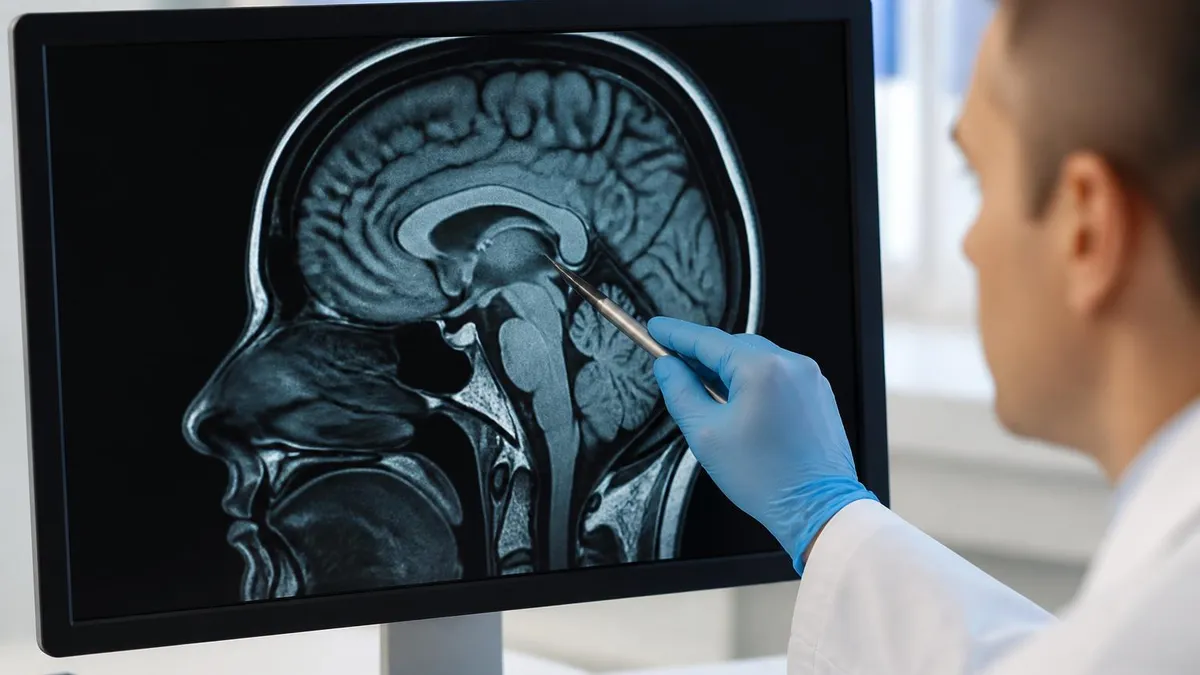
The key difference: MRI images the tissue you can squeeze, like brain matter or a torn meniscus, while MRA images the pipes that feed that tissue, the arteries and veins, so doctors can spot narrowing, blockages, aneurysms, or malformations.
That single sentence is worth memorizing because it answers nine out of ten patient questions before they are even asked. Your neurologist suspects a stroke and wants to see whether brain tissue has been damaged? MRI. Your neurologist suspects a stroke and wants to know whether a carotid artery is blocked and feeding into the problem? MRA. Both? Both.
The combination is so common in neurology, cardiology, and vascular surgery that most imaging centers schedule them back to back when the referral asks for one or the other. The same logic applies to other body regions.
A renal MRI looks at the kidney itself, the cortex, the medulla, any cysts or tumors. A renal MRA looks at the renal arteries feeding the kidney, hunting for stenosis that might be driving stubborn high blood pressure. A musculoskeletal MRI shows the ligament tear in your knee. An MRA of the same knee, which is far less common, would map the popliteal artery if a vascular injury is suspected.
Once you understand that MRI is for tissue and MRA is for vessels, the rest of the decisions, contrast or not, oral medication or not, breath-hold or no breath-hold, all start to make sense rather than feeling random.
What each test sees
Soft tissue contrast that no other imaging modality matches. Brain gray and white matter, spinal cord, muscle, cartilage, ligaments, tendons, liver, kidneys, pelvic organs, and bone marrow all show up in extraordinary detail. Multiple sequences can be combined to detect inflammation, infection, tumors, and old bleeding.
Three-dimensional maps of arteries and veins without inserting a catheter. Carotid and vertebral arteries, the circle of Willis, the renal arteries, the aorta, and the peripheral arteries in the legs are all routinely studied. Doctors can grade narrowing as a percentage and measure aneurysm sizes.
Calcifications, small bone fractures, and acute bleeding within the first few hours are sometimes easier to spot on a CT scan. Lung tissue is also notoriously tricky on MRI because of motion artifact and the lack of signal from air-filled spaces.
Very small vessels, like distal coronary arteries or tiny brain perforators, may still need catheter angiography for the best resolution. Heavy calcification in older patients can also mimic narrowing on MRA, especially with time-of-flight sequences.
Every imaging tool has strengths and blind spots, and good radiologists know when to recommend a different study rather than force the wrong test to answer a question it was not designed for. If you ever wonder why your doctor switched from MRA to a catheter angiogram, or from MRI to CT, the answer almost always lives in this table.
The body part, the speed of the bleeding, the size of the vessel, and the presence of metal hardware all change the math. Patients with pacemakers, certain cochlear implants, older aneurysm clips, or shrapnel near the eyes may not be able to enter the MRI room at all.
The technologist will run through a long screening form before you step inside, and that form exists for one reason: the magnet is always on, even when no one is in the room. Forgetting to remove a hairpin is annoying. Forgetting to disclose an implant can be dangerous.
MRI vs MRA: side by side
MRI evaluates organs, soft tissue, and structures. MRA evaluates the lumen and walls of blood vessels, looking specifically for narrowing, aneurysms, dissections, or arteriovenous malformations. The clinical question your physician is asking determines which test gets ordered, and a referral that says both means both are clinically warranted.
Notice how the same magnet does very different jobs depending on which sequences the technologist runs. That flexibility is why MRI machines are such workhorses in modern hospitals. The same fifteen-minute setup can be used to image a brain tumor in the morning and a carotid artery in the afternoon.
The software does the heavy lifting once the patient is on the table. For patients, the practical takeaway is simple. Ask your physician which sequences are planned.
If gadolinium is on the list, mention any kidney problems beforehand because the drug is cleared by the kidneys and rare reactions can happen in advanced renal disease. If breath-holds are part of the protocol, practice them at home for ten or fifteen seconds at a time so the actual scan feels familiar rather than panicked.
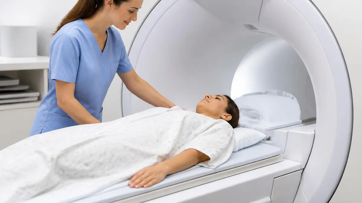
Pacemakers, defibrillators, neurostimulators, insulin pumps, cochlear implants, certain prosthetic heart valves, aneurysm clips, and retained metal fragments can all interact with the magnet. Even tattoos with iron-based ink can warm up. Disclose everything, even old surgeries you barely remember.
The screening form looks long because it has to be. The magnet inside an MRI scanner is roughly thirty thousand times stronger than the earth's natural magnetic field. Loose metal objects can become projectiles. Implanted metal can heat up, twist, or stop working.
Most modern devices, including many pacemakers manufactured in the last decade, are labeled as MR conditional and can be safely scanned with the right precautions. Older devices may rule out MRI entirely, in which case your physician will switch to CT angiography or ultrasound.
Pregnancy is another conversation worth having. MRI without gadolinium is generally considered safe during pregnancy when clinically necessary, but gadolinium crosses the placenta and is usually avoided unless the benefit clearly outweighs the risk. Always tell the booking team if there is any chance you are pregnant. They are not judging, they are protecting both of you.
Day-of-scan checklist
- ✓Leave jewelry, watches, hairpins, and piercings at home if possible
- ✓Wear clothing without metal zippers, snaps, or underwire bras
- ✓Bring a list of every implant, surgery, and metal you have inside you
- ✓Ask whether contrast is being used and disclose kidney problems
- ✓Mention claustrophobia in advance so a mild sedative can be arranged
- ✓Eat normally unless your doctor told you to fast for an abdominal MRA
- ✓Arrive twenty minutes early to complete the screening paperwork
- ✓Bring earplugs or use the headphones the clinic provides for the noise
Preparation is half the battle. Patients who arrive on time, dressed in scrub-style clothing without metal, and ready with their medication list breeze through the screening process. Patients who show up in jeans with rivets, wearing a smartwatch, and unsure whether their old knee replacement is titanium often end up rescheduled.
The clinic is not trying to inconvenience anyone, the staff is simply protecting equipment that costs more than a luxury home and a patient population that deserves the safest possible scan.
If claustrophobia is a real concern, talk to your physician at least a few days before the appointment. Open-bore MRI scanners exist for larger patients and for those who struggle inside a tube. A mild oral sedative taken thirty minutes before the scan also works wonders, although you will need a ride home afterward.
Some clinics offer prism glasses that let you see outside the tube, music through MR-safe headphones, or even movie projection on a small overhead screen. Ask.
Choosing between MRI and MRA
- +MRI offers unmatched soft tissue detail for organs, brain, joints, and spine
- +MRA maps arteries and veins in three dimensions without a catheter
- +Neither test uses ionizing radiation, making them safer for repeat imaging
- +Both can often be performed without contrast when kidneys are weak
- +Modern scanners have shortened total exam times significantly
- −Both tests are loud, long, and can feel claustrophobic inside the bore
- −Patients with certain implants may be ineligible for either study
- −Gadolinium contrast carries small risks, especially in advanced kidney disease
- −Out-of-pocket cost is higher than ultrasound or CT in most regions
- −Scheduling can take days or weeks at busy imaging centers
No imaging test is perfect, and the right choice depends on the clinical question, the patient's body, and the available equipment. A young athlete with knee pain and no risk factors does not need an MRA. An older patient with new-onset stroke symptoms almost certainly needs both an MRI of the brain to see damaged tissue and an MRA of the neck and head vessels to find the source.
Trust your physician's reasoning, but also feel free to ask, in plain language, why this test and not that one.
Cost is a real factor for many patients, and it varies wildly by country, insurance plan, and even by which hospital department orders the test. In the United States, an MRI without contrast can range from a few hundred dollars at a standalone imaging center to several thousand at a major hospital.
MRA pricing is similar. If you are paying out of pocket, call ahead and ask for a cash price. Many centers offer significant discounts for self-pay patients who pay at the time of service.
MRI Questions and Answers
One question that comes up constantly is whether the choice between MRI and MRA changes when the scan is for surveillance rather than for an initial diagnosis. Surveillance simply means follow-up imaging to track something already known.
A patient with a treated brain aneurysm, for example, may need an MRA every year or two to confirm the coil or clip is still doing its job. A patient with a known meningioma may need an MRI on the same schedule to watch for tumor growth. Same magnet, same machine, very different reasons for being there.
The radiologist reading a surveillance study compares the new images side by side with the previous ones, looking for any change, however subtle. That is one reason imaging centers archive your prior scans in a DICOM database. If you ever switch hospitals, ask the records department to transfer your previous studies on a disc or via electronic image exchange. Walking into a new appointment with prior images saves the radiologist hours of guesswork.
Insurance plays a role in surveillance scheduling too. Some carriers will not approve annual MRA without documented medical necessity, and prior authorization can take a week or more. Plan ahead, especially if your usual specialist is booked far in advance.
Bring your prior scans. Walking into a follow-up appointment with previous MRI or MRA images on disc or shared electronically saves the radiologist hours of guesswork and produces a sharper comparison report.
Beyond contrast and scheduling, the actual experience inside the scanner varies more than most patients expect. Some machines are wide-bore, meaning the tunnel is closer to twenty-eight inches across rather than the older twenty-four inch designs. The larger opening reduces the closed-in feeling dramatically and makes a real difference for patients with broad shoulders, claustrophobia, or higher body weight.
Other facilities offer open MRI scanners, which have no tunnel at all and instead use a C-shaped magnet. Open scanners are slower and produce images with slightly lower resolution, but for some patients the trade-off is worth it.
Audio comfort matters more than most people realize. Modern coils run quieter than the bone-rattling clatter of older systems, and most centers now offer noise-cancelling headphones that play your music of choice. Some clinics even let you bring a USB stick of your own playlist.
The mirror system above your eyes can also be angled so you see the technologist or the room beyond the bore, which short-circuits the feeling of being sealed inside a tube. Ask before you climb on the table, because once the scan starts, adjustments require pausing the entire sequence.
Inside the scanner: comfort tips
- ✓Ask whether the facility uses a wide-bore or open MRI scanner
- ✓Request noise-cancelling headphones and bring favorite music
- ✓Use the prism mirror to see outside the tube and reduce anxiety
- ✓Confirm you can squeeze the call ball at any moment
- ✓Practice slow nasal breathing for thirty seconds before the table moves
- ✓Keep your eyes closed during loud sequences to minimize sensory overload
- ✓Tell the technologist if you need a pause between sequences
Children deserve their own paragraph because the MRI experience is different for them in nearly every way. Infants and toddlers cannot reliably hold still for the thirty to sixty minutes a quality study requires, so pediatric centers often use feed-and-swaddle techniques for the youngest patients or light sedation for older children.
The room is often decorated with calming murals, the technologist explains the noises in playful language, and parents are usually invited to stay nearby for reassurance. Some centers even use mock scanners, basically cardboard tunnels with sound effects, so children can practice the experience before the real thing.
Adolescents and teenagers tolerate scans much like adults but appreciate having a sense of control. Letting a teen choose the music, the order of sequences within reason, and whether a parent stays in the room can transform a stressful exam into a manageable one.
For families weighing MRI versus other imaging options for a child, the lack of radiation is a powerful argument in favor of MRI, especially when multiple follow-up scans are likely to be needed over the coming years. The cumulative dose from repeated CT scans is something every pediatric radiologist takes seriously.
Imaging across the life cycle
Feed-and-swaddle techniques work for many infants. Light sedation may be needed for longer protocols. Pediatric coils improve image quality at small body sizes and reduce total scan time.
Mock scanners and child-life specialists help kids rehearse the experience. Letting them pick the music inside the bore turns a frightening visit into a manageable one.
Adolescents tolerate scans much like adults. Privacy and a sense of control matter most. Allowing a parent in the room is optional, not mandatory, and the teen should decide.
Joint stiffness, claustrophobia, and prior implants all become more common with age. Open or wide-bore scanners and careful positioning make studies more comfortable for older patients.
Walking into an imaging suite with a basic mental map of MRI versus MRA makes a real difference. You will recognize the words the technologist uses. You will know why the form asks about kidney function. You will understand why the same machine that scanned your shoulder last year is now scanning your carotid arteries today.
That fluency turns a stressful appointment into a routine medical task, and routine tasks rarely produce panic.
Both technologies continue to advance every year. Faster sequences, machine-learning reconstruction, and contrast-free angiography are all narrowing the gap between what radiology can show and what surgeons need to see.
Five years from now, scans that take forty minutes today will likely take fifteen, and image quality will keep climbing. For now, the basics still apply. MRI looks at tissue. MRA looks at vessels. The same magnet does both, the same precautions apply to both, and the same calm preparation makes both go smoothly.
If you have an MRI or MRA scheduled in the next few weeks, take a few minutes the night before to review the checklist above, lay out clothing without metal, and write down any questions you want to ask the technologist. Show up rested, hydrated, and ready to hold still.
That small bit of preparation is the difference between a clean diagnostic study and a repeat scan a week later because motion blurred the images. Your radiologist, your physician, and your future self will all thank you for the effort.
About the Author

Medical Laboratory Scientist & Clinical Certification Expert
Johns Hopkins UniversityDr. Sandra Kim holds a PhD in Clinical Laboratory Science from Johns Hopkins University and is certified as a Medical Technologist (MT) and Medical Laboratory Scientist (MLS) through ASCP. With 16 years of clinical laboratory experience spanning hematology, microbiology, and molecular diagnostics, she prepares candidates for ASCP board exams, MLT, MLS, and specialist certification tests.
Join the Discussion
Connect with other students preparing for this exam. Share tips, ask questions, and get advice from people who have been there.
View discussion (8 replies)