MRI vs CT Scan for Brain: Which Imaging Test Is Better? 2026 July
Which is better MRI or CT scan for brain imaging? Compare accuracy, 🧠 cost, speed, safety, and clinical uses to choose the right brain scan.
When patients and clinicians ask which is better MRI or CT scan for brain imaging, the honest answer depends entirely on what condition is being investigated, how quickly results are needed, and what the patient can safely tolerate inside the scanner. Both modalities produce cross-sectional images of the brain, but they use completely different physics, deliver different kinds of contrast, and excel in different clinical scenarios. Understanding those differences helps avoid unnecessary radiation, wasted time, and missed diagnoses.
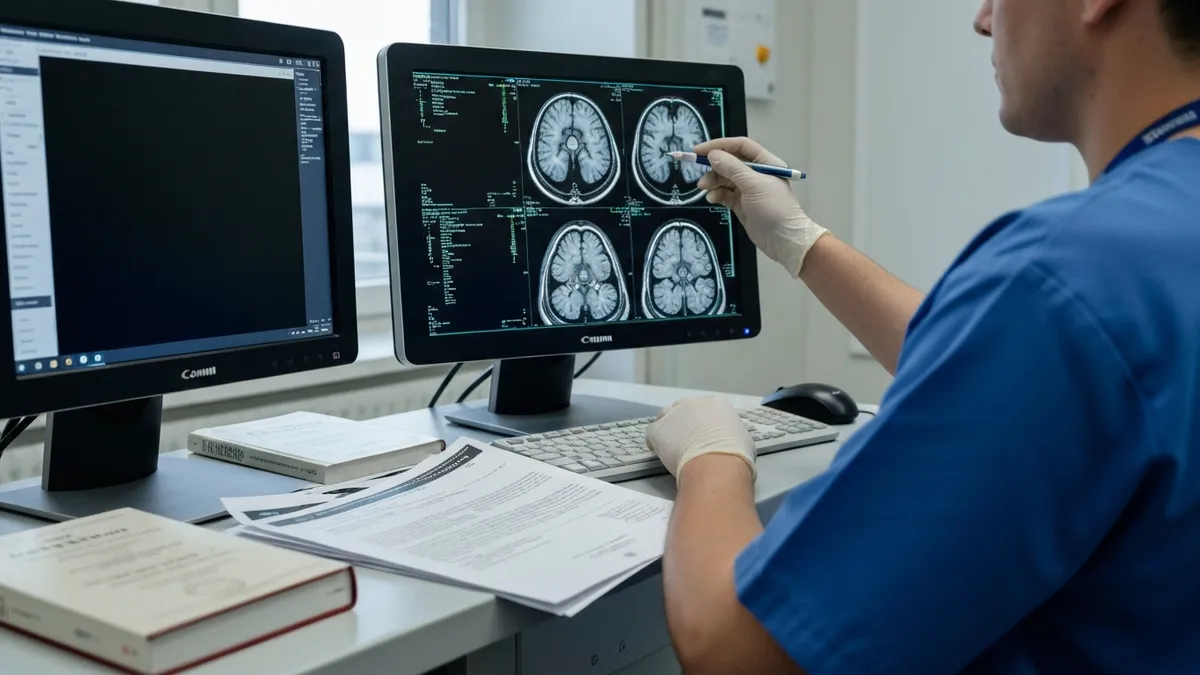
Computed tomography (CT) uses rotating X-ray beams and detectors to build a three-dimensional map of tissue density. It is fast, widely available, and exceptionally good at showing acute bleeding, fresh skull fractures, and large mass effects. Magnetic resonance imaging (MRI) uses a powerful static magnetic field combined with radiofrequency pulses to excite hydrogen protons inside the body. The resulting signals are reconstructed into images that show exquisite soft-tissue detail without any ionizing radiation.
For most emergency department visits where a stroke, head trauma, or sudden severe headache is suspected, a non-contrast head CT is the first-line study. It can be completed in under two minutes, costs roughly a third of what an MRI does, and rules out the hemorrhages that change treatment within the first hour. After that initial screen, however, MRI often takes over because it sees ischemic stroke, small tumors, demyelination, and infection days before a CT can.
For a deeper dive on how the magnetic resonance technique itself developed and why it became the soft-tissue gold standard, see The History of MRI: From Discovery to Modern Medicine. That background helps explain why MRI dominates outpatient neurology while CT remains king in the trauma bay. Neither test is universally superior — they are complementary tools.
Radiologists routinely read both studies on the same patient, and protocols at most academic centers have evolved to use CT for the first 24 hours and MRI for follow-up, characterization, and surgical planning. Knowing the strengths and limitations of each modality empowers patients to ask better questions when their doctor orders a brain scan.
This guide walks through the physics, clinical indications, contraindications, cost, speed, and patient experience for each scan. By the end, you will know exactly when an MRI beats a CT, when a CT beats an MRI, and when both are needed to reach a confident diagnosis. We will also cover contrast agents, safety considerations, and how to prepare for either exam.
Whether you are a patient awaiting a scan, a student studying neuroimaging, or a technologist preparing for the registry, the comparisons below reflect current 2026 clinical practice in the United States. Pricing reflects national Medicare averages, and protocols match the American College of Radiology Appropriateness Criteria most recently updated for cerebrovascular and neoplastic indications.
Brain Imaging by the Numbers

How Each Scan Works Step by Step
CT: X-ray Source Rotates
CT: Density Map Reconstructed
MRI: Magnetic Alignment
MRI: Multi-Sequence Imaging
Image Reconstruction
Diagnostic accuracy is the single most important variable when deciding which is better MRI or CT scan for brain pathology. The two modalities have fundamentally different sensitivity profiles, and choosing the wrong one for the clinical question wastes time, money, and sometimes lives. Acute intracranial hemorrhage is the classic CT win — fresh blood appears hyperdense (bright) on non-contrast CT within minutes and can be detected with sensitivity above 95% in the first six hours.
Acute ischemic stroke flips the script entirely. A CT performed within three hours of symptom onset is normal in roughly 60% of confirmed ischemic strokes because the dead tissue has not yet swelled enough to change density. Diffusion-weighted MRI, by contrast, detects restricted water diffusion within minutes and reaches sensitivity above 95% for cortical and deep gray matter infarcts. This is why comprehensive stroke centers perform both scans during a code stroke workup.
For tumor detection and characterization, MRI dominates. A contrast-enhanced MRI brain study reveals lesions as small as 2–3 millimeters and distinguishes solid from cystic components, identifies peritumoral edema, and shows the relationship of the mass to adjacent eloquent cortex. CT picks up most large tumors but routinely misses small metastases, low-grade gliomas, and posterior fossa lesions hidden by beam-hardening artifact from the dense temporal bones.
White matter disease is another MRI-only territory. Multiple sclerosis plaques, leukoencephalopathy, and small-vessel ischemic changes are nearly invisible on CT but light up clearly on FLAIR and T2-weighted MRI sequences. Neurodegenerative conditions like Alzheimer's disease are also better assessed with MRI volumetric protocols that quantify hippocampal atrophy. If the contrast question matters, our guide on MRI With and Without Contrast: How It Works, What to Expect explains when gadolinium is added.
Bone and calcification questions reverse the advantage. Skull fractures, calcified meningiomas, calcified granulomas, and base-of-skull anatomy are all displayed more clearly on CT. Bone windows on a head CT can identify hairline fractures and pneumocephalus that an MRI would either miss or render ambiguously. Trauma protocols almost always start with CT for this reason.
Vascular imaging splits the difference. CT angiography (CTA) with iodinated contrast provides crisp arterial and venous maps in under a minute of scan time and is the workhorse for acute stroke triage and aneurysm screening. MR angiography (MRA) can be performed without contrast using time-of-flight technique, making it the safer choice for patients with poor renal function or contrast allergies, though spatial resolution is slightly lower.
Subacute and chronic hemorrhage is detected better by MRI than CT. After about a week, blood breakdown products like hemosiderin and ferritin remain visible on MRI susceptibility-weighted imaging (SWI) for years, while CT density fades back toward normal brain attenuation. SWI also detects microbleeds associated with amyloid angiopathy and traumatic axonal injury that CT cannot see at all.
MRI Practice Test Questions
Prepare for the MRI - Magnetic Resonance Imaging exam with our free practice test modules. Each quiz covers key topics to help you pass on your first try.
MRI Knowledge
MRI Exam Questions covering Knowledge. Master MRI Test concepts for certification prep.
MRI Physics
Free MRI Practice Test featuring Physics. Improve your MRI Exam score with mock test prep.
MRI Anatomy and Pathology
MRI Test Prep for MRI Anatomy and Pathology. Practice MRI Quiz questions and boost your score.
MRI Anatomy and Positioning
MRI Questions and Answers on MRI Anatomy and Positioning. Free MRI practice for exam readiness.
MRI Contrast Agents
Free MRI Quiz on MRI Contrast Agents. MRI Exam prep questions with detailed explanations.
MRI Patient Care and Positioning
MRI Practice Questions for MRI Patient Care and Positioning. Build confidence for your MRI certification exam.
Clinical Uses: MRI vs CT for Brain by Condition
For suspected stroke, the standard US workflow starts with a non-contrast head CT to rule out hemorrhage, which is the single fact that changes immediate treatment. If the CT is negative for blood, the patient remains a candidate for thrombolytics or thrombectomy depending on the time window and clinical picture.
MRI with diffusion-weighted imaging then confirms ischemic infarction, defines the core and penumbra, and locates the vascular territory involved. Many comprehensive stroke centers now use rapid MRI protocols that complete in under ten minutes, allowing them to triage extended-window thrombectomy candidates up to 24 hours from last known well.
MRI vs CT for Brain: Side-by-Side Trade-offs
- +MRI provides far superior soft-tissue contrast for brain parenchyma
- +MRI uses no ionizing radiation — safe for repeated and pediatric imaging
- +MRI detects acute ischemic stroke within minutes via diffusion-weighted imaging
- +MRI characterizes tumors, demyelination, and infection with high specificity
- +MRI offers non-contrast vascular imaging (time-of-flight MRA) for renal-impaired patients
- +MRI sees microhemorrhages, white matter disease, and small cortical lesions CT misses
- +MRI provides functional and metabolic data with fMRI, DTI, and spectroscopy
- −MRI takes 30–60 minutes versus 30–60 seconds for CT
- −MRI costs 2–4 times more than a comparable head CT
- −MRI is contraindicated with many pacemakers, ferromagnetic implants, and metal foreign bodies
- −MRI claustrophobia limits roughly 5–10% of patients without sedation
- −MRI is less sensitive than CT for acute hemorrhage in the first few hours
- −MRI is less available than CT, especially in rural and emergency settings
- −MRI is more sensitive to patient motion, often requiring sedation in children
Patient Preparation Checklist for Brain MRI or CT
- ✓Tell your provider about pregnancy, kidney disease, or contrast allergies before scheduling
- ✓Remove all metal jewelry, watches, hearing aids, and hairpins before entering the MRI suite
- ✓Disclose pacemakers, cochlear implants, aneurysm clips, or any implanted device
- ✓Fast for four hours before contrast scans if instructed by your facility
- ✓Bring a current list of medications and prior imaging on a CD or USB
- ✓Wear loose, metal-free clothing or change into the hospital gown provided
- ✓Arrange a driver if you will receive sedation for claustrophobia
- ✓Drink water before the exam to help kidneys clear iodinated or gadolinium contrast
- ✓Notify staff immediately if you feel anxious, dizzy, or warm during the scan
- ✓Plan for 60–90 minutes total facility time even though scanning is shorter
CT first for blood and bone, MRI for everything else
In emergency settings where acute hemorrhage, fracture, or rapidly evolving mass effect must be ruled out within minutes, CT is the right first scan. Once those conditions are excluded — or when soft-tissue questions like stroke, tumor, demyelination, or infection dominate — MRI provides dramatically more diagnostic information without any ionizing radiation.
Cost, speed, and access are practical factors that often determine which scan a patient actually receives, regardless of which would be diagnostically ideal. In the United States in 2026, a non-contrast head CT typically bills between $300 and $1,500 depending on whether it is performed in a hospital outpatient department, freestanding imaging center, or emergency room. The same study at an independent outpatient facility can cost less than half of the hospital price for self-pay patients.
A non-contrast brain MRI typically bills between $1,200 and $4,000, with contrast adding another $300–$800 for the gadolinium agent and additional time. Insurance negotiated rates are much lower, but high-deductible plan patients can still face substantial out-of-pocket costs. Many patients are surprised to learn that the same MRI ordered by the same physician can vary by a factor of five depending on where it is performed.
Speed matters enormously in acute settings. A modern 64- or 128-slice CT scanner can image the entire brain in less than ten seconds of actual scan time, with the patient on the table for under two minutes. This is why CT is universally the first scan for any patient who is unstable, agitated, or in a time-critical situation like suspected stroke or trauma. Reports are typically available within 30 minutes in well-staffed emergency departments.
MRI scan times depend on the protocol. A focused acute stroke MRI can be completed in 6–10 minutes at experienced centers, while a comprehensive tumor protocol with contrast may take 45–60 minutes. Routine outpatient brain MRI usually requires 30–45 minutes on the table plus 15 minutes of setup. The patient must remain still throughout — even small movements can blur images and force repeated sequences.
Access varies dramatically by geography. Nearly every US hospital with an emergency department has a CT scanner available 24/7, and many rural hospitals share an MRI scanner with neighboring facilities or use mobile MRI trucks that visit on scheduled days. Wait times for non-emergent outpatient MRI can range from same-day in urban markets to several weeks in underserved areas. Independent imaging centers often have shorter waits and lower prices.
For patients shopping price and convenience, comparing outpatient facilities is worthwhile. See our overview of MRI Imaging Centers: Complete Guide to Independent Outpatient MRI Facilities for what to look for when choosing a non-hospital location. Many independent centers are accredited by the American College of Radiology and produce images of identical quality at substantially lower cost.
Insurance pre-authorization is another practical consideration. Most commercial insurers require prior authorization for brain MRI but not for emergency CT, which can delay non-urgent MRI by several days. Medicare generally does not require prior authorization for either study when ordered by a physician, but it does enforce medical necessity through audit. Working with your ordering physician to document indications upfront prevents denials.
Both CT and MRI have special considerations in pregnancy and childhood. CT exposes the fetus to ionizing radiation and should generally be avoided unless the diagnostic benefit clearly outweighs the risk. MRI is preferred in pregnancy but gadolinium contrast is usually avoided in the first trimester. In pediatric patients, MRI's lack of radiation is a major advantage, though sedation may be required to ensure the child remains still.
Safety profiles differ fundamentally between the two modalities, and understanding the contraindications is essential to deciding which is better MRI or CT scan for brain imaging in your specific situation. CT delivers ionizing radiation in doses of roughly 1.5–2 millisieverts for a standard head study — equivalent to about eight months of natural background radiation. While the absolute risk from a single scan is low, cumulative dose matters, particularly for children and patients who require repeated imaging.
MRI has no ionizing radiation, but its magnetic field creates unique safety concerns. The static field on a 1.5T or 3T scanner is roughly 30,000–60,000 times stronger than Earth's magnetic field and never turns off. Ferromagnetic objects can become deadly projectiles, and implanted devices may heat, move, or malfunction. Every patient must be screened with a detailed metal questionnaire before entering the scanner room.
Common MRI contraindications include older pacemakers and implantable cardioverter-defibrillators (though many newer devices are now MRI-conditional), cochlear implants, certain aneurysm clips made of ferromagnetic alloys, retained metal foreign bodies in the eyes, and some neurostimulators. Tattoos and permanent makeup with ferrous pigments can cause skin irritation but rarely prevent scanning. Patients with shrapnel, bullet fragments, or industrial metal exposure need orbital X-rays before MRI.
Contrast agents carry distinct risk profiles. Iodinated contrast used in CT can cause anaphylactoid reactions in roughly 0.6% of patients and contrast-induced nephropathy in patients with pre-existing kidney disease. Gadolinium-based MRI contrast historically caused nephrogenic systemic fibrosis in severely renal-impaired patients, though newer macrocyclic agents have dramatically reduced this risk. Both contrast types are generally safe when used appropriately.
Claustrophobia affects an estimated 5–10% of MRI patients severely enough to require intervention. Open MRI scanners, wide-bore systems, and oral anxiolytics can help, but in some cases CT remains the only practical option. Modern wide-bore 3T scanners with 70 cm openings have substantially reduced claustrophobic refusal rates compared to older 60 cm systems.
Acoustic noise is another MRI-specific issue. Gradient coils generate sound levels of 90–110 decibels during scanning — comparable to a jackhammer — requiring earplugs or headphones. Some patients find this distressing despite hearing protection. CT scanners are essentially silent by comparison, producing only the soft whir of the rotating gantry. For sensory-sensitive patients, this difference can be decisive.
Pediatric considerations deserve special attention. Children under age six often require sedation for MRI because the scan duration exceeds their tolerance for stillness. CT can usually be completed without sedation, which is one reason emergency departments lean on it for young trauma patients. However, the lifetime radiation exposure concern is greatest in children, so non-urgent imaging should default to MRI whenever a child can cooperate or be safely sedated.
Putting it all together for everyday decisions: when your physician orders a brain scan, ask which question they are trying to answer. The clinical question — not the patient's preference or the facility's convenience — should drive the modality choice. A new severe headache with suspicion for hemorrhage gets a CT. A six-month history of progressive memory loss gets an MRI. A code stroke alert gets both, in sequence, as fast as possible.
If you are scheduling an outpatient scan and have a choice between facilities, ask about scanner field strength for MRI. A 3T magnet provides higher signal-to-noise and faster scanning than a 1.5T magnet, though both produce diagnostic-quality images. For CT, ask about slice count — modern 64-slice or higher scanners produce thinner, more detailed images with less radiation than older 16-slice machines. Accreditation by the American College of Radiology is a reliable quality indicator.
Prepare for either scan by following the facility's instructions carefully. For MRI, that means removing all metal and completing the safety screening honestly. For CT with contrast, that means staying hydrated and disclosing kidney problems or prior contrast reactions. Bringing prior imaging on disc allows the radiologist to compare studies and identify subtle changes that might otherwise be missed.
During the scan itself, communication matters. Both modalities provide a panic button or squeeze ball. Use it if you feel unwell, panicked, or need to stop. Technologists can pause the scan, talk you through the next sequence, or in many cases hold your hand from the operator console via intercom. Most facilities allow a family member to sit in the scanner room during MRI provided they have also been screened for metal.
After the scan, ask when results will be available and how you will receive them. Emergency results are typically read within an hour. Outpatient results may take 24–72 hours depending on the facility's workflow. Patient portals now provide direct access to both the radiologist's report and the images themselves through online viewers, which can be valuable when seeking a second opinion or transferring care.
If you receive a report with unfamiliar terminology, ask your ordering physician to walk you through the findings in plain language. Phrases like 'no acute intracranial abnormality' are reassuring, while terms like 'enhancing lesion,' 'mri brain findings,' or 'mass effect' usually warrant explanation. Don't hesitate to ask whether additional imaging — for example an MRI follow-up to a CT, or vice versa — is recommended for further clarification.
For technologists and students preparing for the ARRT MRI registry or other certification exams, the comparisons above represent the kind of clinical reasoning that appears throughout board questions. Knowing why a stroke alert gets a non-contrast CT followed by DWI MRI, or why a calcified meningioma is easier to identify on CT than MRI, demonstrates the integrated knowledge that distinguishes a competent technologist from a great one.
MRI Questions and Answers

The History of MRI: From Discovery to Modern Medicine

MRI Medical Abbreviation: What MRI Stands For and Why It Matters

MRI With and Without Contrast: How It Works, What to Expect
Shoulder MRI: What It Shows, Procedure, and Reading the Report
MRI Imaging Centers: Complete Guide to Independent Outpatient MRI Facilities
About the Author
Medical Laboratory Scientist & Clinical Certification Expert
Johns Hopkins UniversityDr. Sandra Kim holds a PhD in Clinical Laboratory Science from Johns Hopkins University and is certified as a Medical Technologist (MT) and Medical Laboratory Scientist (MLS) through ASCP. With 16 years of clinical laboratory experience spanning hematology, microbiology, and molecular diagnostics, she prepares candidates for ASCP board exams, MLT, MLS, and specialist certification tests.
Join the Discussion
Connect with other students preparing for this exam. Share tips, ask questions, and get advice from people who have been there.
View discussion (6 replies)