
MRI Software: Image Processing, Analysis, and Workflow Tools 2026
MRI software overview: scanner control software, image processing, advanced analysis tools, AI-assisted reading, and PACS integration.
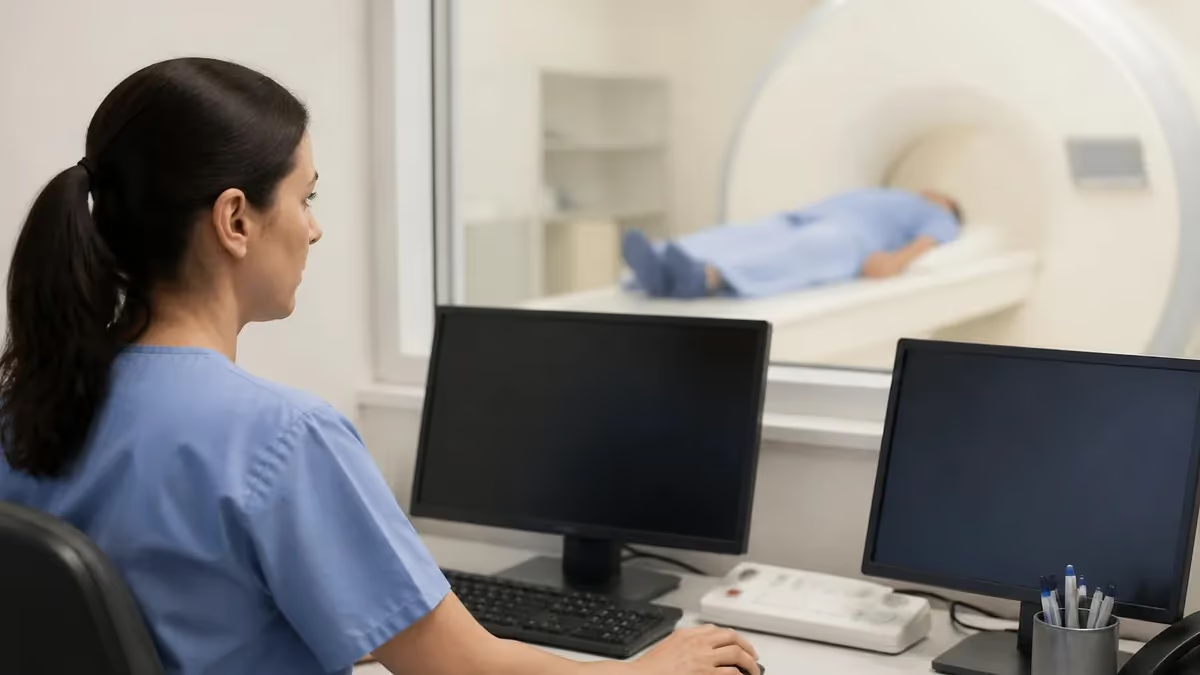
MRI software encompasses the complex ecosystem of computer programs that control MRI scanners, process raw data into diagnostic images, support radiologist interpretation, manage workflow across imaging departments, and enable advanced analysis beyond standard imaging. Modern MRI systems are as much software products as hardware products — the magnetic systems and gradient coils generate raw data, but software transforms that data into the high-quality images radiologists actually interpret. The sophistication of MRI software has advanced dramatically over recent decades, enabling clinical applications that earlier hardware-focused MRI systems couldn't achieve regardless of physical capabilities. Practice with the free MRI - Magnetic Resonance Imaging practice test on PracticeTestGeeks (200+ questions with answer explanations).
Understanding the MRI software landscape matters for radiologists, technologists, IT professionals supporting medical imaging, healthcare administrators managing radiology services, and software developers building products in this market. The software stack includes scanner control software (operating the scanner during examinations), image reconstruction software (transforming raw data into images), image processing and analysis software (specialized tools for specific clinical applications), PACS integration software (managing storage and distribution), reading workstation software (radiologist interpretation tools), AI-assisted analysis software (emerging area with rapid development), and various other specialized applications. Each category has distinct development paths, leading vendors, and clinical applications.
MRI: upcoming dates and official changes
- Aug 17, 2026: ARRT launched the entry-level Imaging Assistant, Magnetic Resonance (I.A.) credential with an initial testing window from October 15 to November 30, 2026.[3]
- The postprimary pathway ARRT MRI examination consists of 220 total questions, including 200 scored questions and 20 unscored pilot questions.[1]
- Candidates taking the postprimary pathway ARRT MRI examination receive 210 minutes of test time within a 230-minute total appointment.[1]
- To pass the ARRT MRI examination, candidates must achieve a scaled score of 75 on a score scale ranging from 1 to 99.[2]
- The application fee for the ARRT Magnetic Resonance Imaging postprimary pathway examination is $225, or $450 when applying with an NMTCB or ARDMS supporting credential.[3]
All verified MRI facts and sources
Numbers in brackets link to the official sources listed under Sources & last verified. Dates that have passed are removed automatically.
Free MRI - Magnetic Resonance Imaging Study Tools
Interactive MRI - Magnetic Resonance Imaging resources on PracticeTestGeeks, free to start:
- Free MRI Knowledge Questions and Answers — 11 questions with instant scoring and answer explanations
- MRI - Magnetic Resonance Imaging flashcards — review key terms and concepts card by card
- MRI - Magnetic Resonance Imaging study guide — topic-by-topic overview of what the exam covers
- MRI - Magnetic Resonance Imaging cheat sheet — one-page printable summary for last-minute review
MRI Software Quick Facts
Major scanner vendors: Siemens (syngo), GE (SIGNA), Philips (Ingenia), Canon, Hitachi each have proprietary software ecosystems. Image storage: PACS (Picture Archiving and Communication Systems) — major vendors include GE, Philips, Sectra, Agfa, Fuji. Reading workstations: Mostly vendor-agnostic with leading products from Visage, Philips, GE, Sectra. AI integration: Growing rapidly with FDA-approved tools for specific applications. Standards: DICOM (Digital Imaging and Communications in Medicine) governs interoperability.
Scanner control software is the proprietary software running on each MRI scanner, controlling pulse sequences, gradient operations, RF transmission, signal acquisition, and image reconstruction during examinations. Each major MRI manufacturer (Siemens, GE Healthcare, Philips, Canon, Hitachi) develops their own scanner software with proprietary user interfaces, sequence libraries, and workflow tools. Technologists trained on one vendor's software typically need additional training to operate other vendors' equipment, though core MRI principles transfer across systems. Recent versions emphasize automated workflow, intelligent protocols that adjust based on patient characteristics, and integration with hospital information systems.

Major Categories of MRI Software
Proprietary software running scanners. Major vendors: Siemens syngo, GE SIGNA, Philips Ingenia software. Controls pulse sequences and acquisition.
Software transforming raw k-space data into images. Standard reconstructions plus advanced options like compressed sensing acceleration.
Specialized analysis tools for specific clinical applications: cardiac analysis, oncology measurement, neurological volumetry, vascular analysis.
Picture Archiving and Communication Systems for image storage, distribution, and access. Mission-critical IT infrastructure for radiology.
Software radiologists use to interpret studies. Optimized displays, hanging protocols, measurement tools, comparison features.
Emerging area with FDA-approved tools for stroke detection, lesion identification, automated measurement. Growing rapidly.
Image reconstruction software transforms the raw frequency-domain data (k-space) acquired by MRI scanners into the spatial-domain images radiologists interpret. Standard reconstruction uses Fourier transformation and various correction algorithms. Advanced reconstructions include parallel imaging methods (SENSE, GRAPPA, ASSET) that accelerate acquisition by exploiting coil array information, compressed sensing methods that exploit signal sparsity for faster acquisition, deep learning-based reconstruction that uses neural networks for image generation from undersampled data, and various motion correction algorithms for challenging acquisitions. Recent developments have produced dramatic acceleration of imaging while maintaining or improving image quality through software advances rather than hardware changes.
Post-processing software addresses specific clinical applications beyond standard image generation. Cardiac MRI analysis software measures ventricular volumes, ejection fraction, myocardial tissue characteristics, and flow patterns. Oncology software supports tumor measurement, assessment over time, and treatment response evaluation. Neurology software performs brain volumetry for dementia assessment, lesion analysis for multiple sclerosis, and various functional and diffusion analyses. Vascular software processes MR angiography images for vessel assessment. Each specialty has its own software ecosystem with various commercial and academic offerings serving different needs.
PACS (Picture Archiving and Communication Systems) provide the IT backbone for radiology operations, handling image storage, distribution, retrieval, and security across medical imaging. Major PACS vendors include GE Healthcare (Centricity), Philips (IntelliSpace), Sectra, Agfa, Fuji, and several others. PACS software integrates with hospital information systems (HIS), radiology information systems (RIS), and electronic health records (EHRs) to provide comprehensive imaging workflow support. PACS represents substantial IT investment for healthcare organizations — typically multimillion-dollar systems with significant infrastructure requirements and ongoing maintenance costs.
Scanner control software typically includes: Patient information management. Pulse sequence libraries with manufacturer-defined and custom sequences. Real-time monitoring during scans. Image quality assessment tools. Protocol management for different examination types. Workflow integration with hospital systems. User interfaces for technologists. Image reconstruction integrated with acquisition. Quality assurance tools for system performance. Each major manufacturer has their own software ecosystem (Siemens syngo, GE SIGNA, Philips Ingenia) requiring vendor-specific training and certification.
The DICOM (Digital Imaging and Communications in Medicine) standard governs interoperability across medical imaging software. DICOM specifies image format, communication protocols, and integration profiles that allow software from different vendors to work together. While DICOM solved many interoperability problems, real-world implementation still has challenges due to vendor-specific extensions, incomplete implementations, and version differences. Healthcare IT professionals working with imaging software regularly encounter DICOM-related issues despite the standard's existence. Understanding DICOM fundamentals supports troubleshooting and effective software selection decisions.
Cloud-based MRI software has emerged as alternative to traditional on-premises deployments. Cloud PACS, cloud reading workstations, and cloud-based AI analysis tools all gain market share. Advantages include reduced local IT infrastructure requirements, easier deployment of new applications, automatic updates and maintenance, and remote access for radiologists working from home or other locations. Concerns include security and privacy compliance, data sovereignty issues, internet bandwidth dependence, and potential vendor lock-in. Healthcare organizations balance these factors based on their specific situations, regulatory requirements, and operational needs.
The economics of MRI software vary substantially across categories. Scanner software is bundled with scanner purchases (multimillion-dollar transactions). PACS systems typically cost $1-10 million for typical hospital deployments with annual maintenance running 15-20% of initial cost. Reading workstation software ranges from free (some open-source options) to substantial commercial licenses ($10,000-$100,000+ per workstation depending on features). AI software is rapidly expanding market with various pricing models including per-study, per-month, and per-license. Total imaging software investment for major healthcare organizations runs into tens of millions of dollars annually.
Don't focus only on initial cost — total cost of ownership over 5-10 years typically dwarfs initial license cost. Don't ignore integration requirements — software that doesn't integrate with existing systems creates operational problems. Don't underestimate training requirements — switching software systems requires substantial radiologist and technologist training. Don't choose based on AI hype — verify AI capabilities address your actual clinical needs versus marketing promises.
AI integration in MRI software has accelerated rapidly with FDA-approval of specific algorithms for various clinical applications. Stroke detection AI rapidly identifies acute ischemic injury, supporting time-sensitive treatment decisions. Tumor measurement AI provides reproducible measurements supporting treatment response assessment. Brain volumetry AI supports dementia assessment with quantitative measurements. Various other applications continue receiving FDA approval. Clinical integration of AI tools requires careful workflow design — AI works best as augmentation of radiologist interpretation rather than replacement, with clear protocols for handling AI findings and limitations.
The quality and reliability of AI tools vary substantially across products. Major vendors with significant resources (large medical device companies, specialized radiology AI companies) typically produce higher-quality AI than newer entrants. Validation studies, FDA approval status, real-world performance data, and integration quality all affect actual clinical value. Healthcare organizations evaluating AI products benefit from rigorous review of available evidence rather than purely vendor-provided claims. Some markets see proliferation of AI products with varying quality — selection requires careful evaluation of options.
Open-source MRI software exists alongside commercial offerings, particularly in research and academic settings. Tools like FSL (FMRIB Software Library), FreeSurfer, AFNI, ITK-SNAP, 3D Slicer, OsiriX (free version), and many others provide capabilities comparable to commercial software for specific applications. Academic medical centers often combine open-source and commercial software based on specific needs. Open-source advantages include cost savings, transparency for research, and customization for specific projects. Disadvantages include limited support, regulatory compliance complexity, and integration challenges with clinical systems.
Evaluating MRI Software Solutions
- ✓Verify integration with existing scanner and PACS systems
- ✓Confirm regulatory compliance (FDA approval if applicable)
- ✓Assess total cost of ownership over 5-10 years
- ✓Evaluate training requirements for radiologists and technologists
- ✓Review actual performance evidence beyond vendor claims
- ✓Consider workflow impact on operational efficiency
- ✓Verify support quality and vendor stability
- ✓Assess data migration requirements and ongoing data portability
For radiologists, software preferences often develop over years of practice with specific systems. Familiarity with specific software interfaces, hanging protocols, and workflow patterns produces efficient interpretation. Switching software systems often reduces productivity initially as radiologists adapt to new interfaces. Healthcare organizations changing PACS or reading software experience temporary productivity declines that can extend for months. The disruption costs justify careful consideration before changes — only when new systems offer substantial improvements over existing systems do the disruption costs justify switching.
For technologists operating MRI scanners, software competence directly affects scan quality and patient throughput. Different scanners have different software interfaces requiring vendor-specific training. Cross-training on multiple vendors expands employment options but requires substantial learning investment. Continuous education on new sequences, protocols, and software features supports career development. Manufacturer training programs at vendor facilities provide focused development opportunities. Annual conferences (RSNA, ISMRM) offer broader exposure to software developments across vendors.
For IT professionals supporting medical imaging, the technical complexity is substantial. Imaging systems combine specialized hardware (scanners, dedicated displays), traditional IT infrastructure (servers, storage, networks), specialized software (PACS, reading workstations), and integration with broader clinical systems (EHRs, RIS, departmental systems). Healthcare imaging IT specialists develop deep expertise across these domains. Career opportunities span hospital IT departments, specialized consulting firms, software vendors, and various technology integration roles. The combination of technical complexity and clinical importance makes the work both challenging and rewarding for those who develop expertise.
Looking forward at MRI software trends, several developments will shape the field over coming years. AI integration will continue expanding into more clinical applications with improved performance and broader regulatory approval. Cloud-based delivery models will likely overtake traditional on-premises deployments for many applications. Quantitative imaging will mature with standardized measurements supporting longitudinal patient comparison and multi-center research. Compressed sensing and deep learning reconstruction will enable substantially faster MRI examinations. Patient-centric tools may eventually provide patients direct access to their imaging studies and AI-generated explanations.
The integration of MRI software with broader healthcare technology continues advancing. Connection with electronic health records provides comprehensive patient data context for imaging interpretation. Integration with clinical decision support systems guides ordering decisions and protocol selection. Workflow integration across multidisciplinary teams (radiologists, ordering physicians, surgeons, oncologists) supports coordinated patient care. Security and privacy requirements continue intensifying with regulatory changes and emerging cybersecurity threats. Healthcare technology professionals working in imaging navigate increasingly complex environments balancing capability, security, usability, and regulatory compliance.
For students considering radiology, medical imaging IT, or related careers, software fluency is increasingly essential alongside traditional clinical or technical skills. Pure clinical training without software understanding limits career options. Pure technical training without clinical context limits effectiveness in healthcare environments. The combination of clinical and technical knowledge produces highly valued professionals across various roles. Educational programs increasingly recognize this convergence with combined curricula bridging traditional discipline boundaries. Career development across long careers requires ongoing learning as software continues evolving.

MRI Software Quick Stats
MRI Software by Use Case
Vendor-specific software bundled with scanners. Siemens syngo, GE SIGNA, Philips Ingenia. Required for daily MRI operations.
PACS systems handle image archiving. Major vendors include GE, Philips, Sectra, Agfa, Fuji.
Reading workstation software. Visage, Philips IntelliSpace, Sectra dominate quality market.
Application-specific software for cardiac, oncology, neurology applications. Various commercial and academic options.
Growing market for FDA-approved AI tools supporting specific applications. Rapidly evolving.
Open-source tools (FSL, FreeSurfer, ITK-SNAP, 3D Slicer) supplement commercial software in academic settings.
Software security in medical imaging systems is critical concern as cybersecurity threats target healthcare organizations. Imaging systems have historically had limited security focus compared to general IT systems. The convergence of medical imaging with healthcare networks creates exposure to common cybersecurity threats including ransomware, data breaches, and supply chain attacks. Healthcare cybersecurity standards (HIPAA, HITECH, state regulations) apply to imaging systems alongside other clinical systems. Security incidents involving imaging systems have caused substantial harm in recent years, motivating substantial investment in imaging system security improvements across the industry.
Vendor relationships in medical imaging software shape long-term operations. Major vendors provide multi-decade product evolution, accumulated expertise, and stable support relationships. Smaller specialized vendors offer innovation in specific niches but face stability concerns over longer time horizons. Healthcare organizations balance these considerations when selecting software partners. Vendor consolidation continues across the imaging software market with major acquisitions reshaping competitive landscape periodically. Customer relationships and support quality can change substantially when vendors are acquired or change strategic direction.
Implementation projects for major imaging software systems typically require 6-18 months from contract signing to full operational deployment. Project phases include detailed requirements definition, system design and configuration, data migration from prior systems, integration testing with hospital information systems, user training across multiple user populations (radiologists, technologists, IT staff, ordering clinicians), pilot operations with limited use, and full operational rollout.
Each phase has specific risks and complexity. Healthcare organizations with strong project management and IT capabilities typically navigate these implementations more successfully than organizations without strong technical infrastructure. Implementation experience is one factor differentiating successful imaging software vendors from those that produce good products but struggle with deployment support.
User experience design in MRI software has improved substantially over recent years but still varies dramatically across products and vendors. Older imaging software often had poor user interfaces designed by engineers without strong UX expertise. Modern software increasingly emphasizes user experience through dedicated UX professionals, user testing programs, and iterative design refinement. Reading workstation software particularly has seen UX improvements as radiologist workflow efficiency directly affects productivity and job satisfaction. Vendors competing primarily on UX quality (Visage particularly notable for this approach) have gained market share by focusing on radiologist experience even when other features are comparable.
For software developers entering medical imaging space, several barriers exist that don't apply to general software development. Regulatory requirements (FDA approval for clinical software) require specialized expertise and substantial development overhead. Healthcare industry standards (DICOM, HL7, FHIR) require domain-specific learning. Clinical validation requires partnerships with healthcare organizations and IRB-approved studies for many applications.
Sales cycles for healthcare software are longer and more complex than typical B2B software. These barriers create both challenges and opportunities — barriers limit competition while creating advantages for organizations that successfully navigate the requirements. Specialized medical imaging software companies often start with substantial healthcare expertise on founding teams.
Data privacy and security in medical imaging software faces continuously evolving challenges. HIPAA and similar privacy regulations apply broadly but specific implementation details vary by software and deployment. Recent ransomware attacks targeting healthcare imaging systems have demonstrated real consequences of inadequate security. Cloud-based imaging software introduces additional considerations around data residency, breach notification, and shared responsibility for security. AI software trained on patient data raises questions about data usage rights and ongoing access to training data. Each of these areas continues evolving with regulatory changes, technical developments, and accumulated industry experience.
Software Implementation Numbers
For students considering medical imaging software careers, several pathways lead to relevant expertise. Computer science or software engineering backgrounds combined with healthcare exposure (internships, healthcare-specific projects) provide solid foundation. Medical physics or biomedical engineering programs combine clinical context with technical training. Direct entry through medical imaging IT roles in hospitals provides on-the-job exposure. The combination of technical capability with healthcare domain understanding creates particularly valuable professionals as healthcare technology continues advancing rapidly across multiple application areas.
Software certifications relevant to medical imaging include vendor-specific certifications (Siemens, GE, Philips offer certifications for their products), DICOM-related training and certifications, healthcare IT certifications (HIMSS, HCISPP for security, CPHIMS for project management), and project management certifications useful for software implementation projects. Certifications support career development in this specialized field but don't substitute for hands-on experience and clinical context understanding gained through actual work in healthcare settings.
MRI Software Considerations
- +Scanner software advances drive imaging quality improvements
- +AI integration expanding diagnostic capabilities
- +DICOM interoperability supports multi-vendor deployments
- +Cloud options reduce on-premises infrastructure requirements
- +Open-source options support research and specialty applications
- +Continuous development brings new features regularly
- −Substantial cost of major systems (PACS, reading workstations)
- −Vendor lock-in challenges with proprietary scanner software
- −Migration between systems is complex multi-year projects
- −Security threats increasingly target imaging systems
- −Quality varies dramatically across products
- −AI tool quality and validation varies substantially
Pros and Cons at a Glance
| Pros | Cons |
|---|---|
| Scanner software advances drive imaging quality improvements | Substantial cost of major systems (PACS, reading workstations) |
| AI integration expanding diagnostic capabilities | Vendor lock-in challenges with proprietary scanner software |
| DICOM interoperability supports multi-vendor deployments | Migration between systems is complex multi-year projects |
| Cloud options reduce on-premises infrastructure requirements | Security threats increasingly target imaging systems |
| Open-source options support research and specialty applications | Quality varies dramatically across products |
| Continuous development brings new features regularly | AI tool quality and validation varies substantially |
Sample MRI - Magnetic Resonance Imaging Practice Questions
Try these questions from our free MRI - Magnetic Resonance Imaging practice tests. The correct answer and an explanation follow each question.
Which portion of k-space primarily determines image contrast?
- A. The center (low spatial frequencies)
- B. The periphery (high spatial frequencies)
- C. The edges equally with the center
- D. The corners of k-space only
Answer: A. The center (low spatial frequencies)
The center of k-space contains low spatial frequency data, which encodes the bulk signal intensity and contrast of the image.
Which artifact arises when signal from outside the FOV wraps around to the opposite side of the image?
- A. Aliasing (wrap-around) artifact
- B. Gibbs ringing artifact
- C. Chemical shift artifact
- D. Susceptibility artifact
Answer: A. Aliasing (wrap-around) artifact
Aliasing occurs in the phase-encode direction when anatomy outside the FOV is undersampled, causing it to appear on the opposite side.
Zipper artifact on an MRI image appears as a line of alternating bright and dark pixels across the image, most often caused by:
- A. Patient movement
- B. External RF interference from inadequate shielding or open RF cage door
- C. Gradient switching noise
- D. Coil coupling artifact
Answer: B. External RF interference from inadequate shielding or open RF cage door
Zipper artifact is caused by external radiofrequency interference entering the scan room, typically due to an open waveguide, broken shielding, or improperly closed scanner room door.
This magnet has a strength of up to 0.3 T, can be switched off, and can be used in both horizontal and vertical field systems.
- A. Superconducting
- B. Permanent magnet
- C. Resistive magnet
- D. Selenoid magnet
Answer: C. Resistive magnet
Resistive magnets consist of coils of wire through which a large electric current is continuously passed to generate a magnetic field. They can be switched off, offer field strengths typically up to 0.3 Tesla, and can be designed for both horizontal and vertical field orientations. However, they require significant continuous power and generate substantial heat, limiting their use in high-field MRI.
Take the full MRI - Magnetic Resonance Imaging practice test
MRI Questions and Answers
Sources & last verified
- assets-us-01.kc-usercontent.com — number of questions, time limit (official source). “Scored Items 200 Tutorial 8 minutes Pilot Items 20 NDA 2 minutes Total Items 220” Verified Nov 9, 2026 · source checked Nov 9, 2026.
- American Registry of Radiologic Technologists — passing score (official source). “Our exam scoring scale ranges from 1 to 99, and you’ll need a total score of 75 to pass the exam.” Verified Nov 9, 2026 · source checked Oct 18, 2026.
- American Registry of Radiologic Technologists — exam fee, other (official source). “Postprimary (M, CT, MR, BD, CI, VI, VS, BS, R.R.A.): $225” Verified Nov 9, 2026 · source checked Oct 18, 2026.
How we verify: editorial & fact-checking policy. Report an error: contact us.
How this page was made: written with AI tools and published by Nathan Beja, founder of Practice Test Geeks, who is responsible for its content. Exam facts are checked against the official sources listed below. Not reviewed by a subject-matter expert. Editorial policy
Sources & references
Exam facts on this page (fees, format, dates, passing scores) are checked against these official sources; the date is our last check. Editorial policy
- assets-us-01.kc-usercontent.com — Renewal, score validity, eligibility, exam format, retake policy, test dates, time limit · verified Nov 2026
- arrt.org — Fees, passing score, score release, exam information · verified Nov 2026
- home.pearsonvue.com — Test delivery · verified Nov 2026