Watchman Device MRI: Complete Safety Guide for Patients and MRI Technologists
Watchman device MRI safety explained for patients & techs. Conditions, protocols, field strengths & screening tips. ✅ Updated 2026 June.
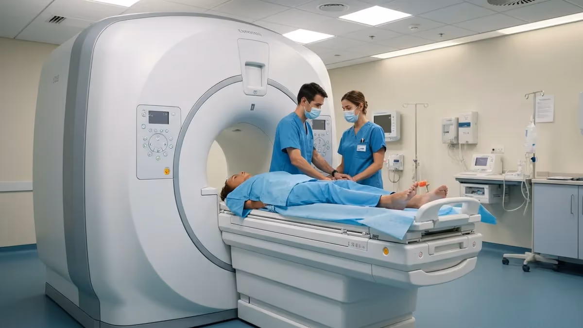
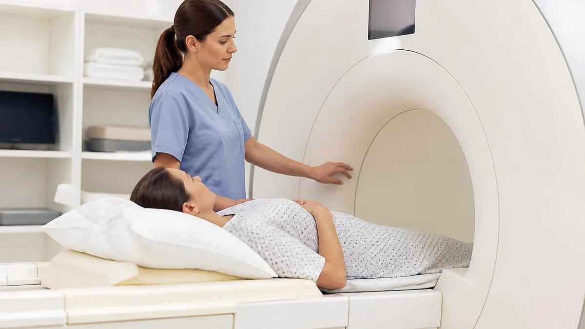
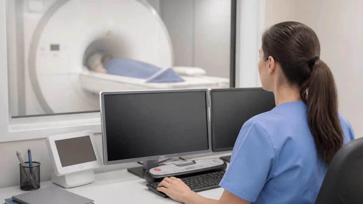
The watchman device MRI is one of the most frequently asked safety questions in modern MRI practice. The WATCHMAN Left Atrial Appendage Closure (LAAC) device, manufactured by Boston Scientific, is implanted in patients with non-valvular atrial fibrillation to reduce stroke risk without long-term anticoagulation therapy. As the device has grown in popularity — with hundreds of thousands implanted worldwide — MRI technologists and radiologists face an increasing number of patients who arrive at the scanner with this cardiac implant. Understanding the exact conditions under which MRI scanning is safe is not optional; it is a clinical and legal necessity.
Patients with the WATCHMAN device are not automatically excluded from MRI. In fact, current labeling from Boston Scientific designates the WATCHMAN FLX and legacy WATCHMAN 2.5 devices as MR Conditional, meaning a scan can proceed safely provided specific conditions are met. These conditions include field strength limits, spatial gradient restrictions, and specific absorption rate (SAR) ceilings. A technologist who scans a WATCHMAN patient without verifying these parameters against the device's Instructions for Use (IFU) exposes both the patient and the facility to serious risk.
This article covers everything MRI professionals and patients need to know about scanning with a WATCHMAN device in place. We will review the device's MR Conditional status, the precise scanning parameters that define safe conditions, how to screen patients effectively, and what protocols your department should have in writing before any WATCHMAN patient enters the magnet room. We also discuss how the WATCHMAN fits within the broader landscape of mri safety and devices, so that technologists can apply systematic thinking to all implanted devices, not just this one.
The WATCHMAN FLX, the current generation device approved by the FDA in 2020, is constructed from a nitinol frame with a polyethylene terephthalate (PET) fabric cover. Nitinol is a nickel-titanium alloy that is weakly paramagnetic, which means it does interact with magnetic fields but does not produce the gross deflection or heating seen with ferromagnetic materials. Understanding the material composition of any implant is the first step in assessing MRI compatibility, and nitinol's properties are why WATCHMAN devices can be scanned under controlled conditions rather than being absolutely contraindicated.
Both radiologists ordering studies and technologists performing them must be familiar with the post-implant timeline requirements. Boston Scientific's IFU specifies that MRI scanning of WATCHMAN patients should generally be deferred until adequate endothelialization has occurred — typically six weeks post-implantation. Before that window, the device is not yet fully anchored in tissue, and the combination of mechanical forces and radiofrequency heating creates an unacceptable risk profile. Exceptions exist in urgent clinical scenarios, and the ordering physician must document the medical necessity clearly in those cases.
Facility-level preparation is just as important as individual patient screening. Every MRI department should maintain an up-to-date binder or digital database of IFU documents for common cardiac devices, including both WATCHMAN generations. The MRI safety officer, typically a Level 2 MRI-trained technologist or radiologist, should audit this database at least annually. When a new device generation is released — as happened when the FLX replaced the 2.5 — the IFU parameters may differ significantly, and outdated protocols can lead to unsafe conditions even when staff believe they are following established procedure.
Staying current with the registry exam syllabus is equally important for MRI technologists preparing for ARRT credentialing. MRI safety, including implanted device management, constitutes a meaningful portion of the registry examination content outline. The concepts covered in this article — MR Conditional labeling, static field deflection testing, RF-induced heating, and spatial gradient forces — are all examinable topics that require both conceptual understanding and the ability to apply knowledge to clinical scenarios under time pressure.
WATCHMAN Device MRI Safety by the Numbers
WATCHMAN Device MR Conditional Status Explained
MR Conditional means an implant has been demonstrated to pose no known hazards in a specified MRI environment with specified conditions of use. This is distinct from MR Safe (no hazards in any MRI environment) and MR Unsafe (poses unacceptable risks in any MRI environment). The WATCHMAN FLX and 2.5 are both MR Conditional.
The FLX (approved 2020) and the legacy 2.5 device share MR Conditional status but may have different parameter limits in their respective IFUs. Always identify the exact device generation from the patient's implant card or operative report before checking conditions. Never assume both versions share identical restrictions.
MRI compatibility labeling follows ASTM International standard F2503. Under this framework, manufacturers perform bench testing for magnetically induced displacement, torque, RF heating, and image artifact before assigning a label. The WATCHMAN's nitinol construction contributes to its conditional rather than unsafe classification across field strengths up to 3 Tesla.
If your MRI system cannot achieve the conditions specified in the WATCHMAN IFU — for example, if the required SAR level cannot be achieved for the sequence needed for diagnosis — the scan should not proceed without explicit physician authorization documenting that the clinical benefit outweighs the risk.
Understanding the specific MRI scanning parameters allowed for WATCHMAN patients requires careful review of the device's Instructions for Use document, which is the definitive authority. For the WATCHMAN FLX, the primary conditions include a static magnetic field of 1.5 Tesla or 3.0 Tesla, a maximum spatial gradient magnetic field of 700 Gauss per centimeter (T/m is approximately 7 T/m), and a whole-body averaged SAR not exceeding 2.0 W/kg. The device has also been tested under specific peak spatial SAR conditions that technologists should verify with their MRI physicist when head coils are being used for other anatomical regions.
The spatial gradient field is a frequently misunderstood parameter. It refers to the rate of change of the static magnetic field with position — not the gradient fields used for imaging. High spatial gradients near the bore entrance exert translational forces on metallic implants.
For the WATCHMAN device, the nitinol frame is weakly paramagnetic and the force produced is low, but the IFU limit still exists because exceeding it has not been tested. Technologists positioning patients in or out of the bore should do so at the speeds prescribed by the IFU, particularly for patients scanned at 3.0T where spatial gradients are typically steeper.
Radiofrequency-induced heating is arguably the most important consideration for conductive implants. RF energy deposited in the body during MRI can be concentrated at the tips or edges of conductive structures, causing localized temperature increases. The WATCHMAN device's PET fabric cover and nitinol frame have been tested for RF-induced heating at both 1.5T and 3.0T under the SAR conditions listed in the IFU. Exceeding the SAR limit — for example, by using a high-resolution cardiac sequence with a high duty cycle — could produce heating that was not characterized during manufacturer testing.
The 45-day or 6-week post-implant waiting period deserves clinical scrutiny. During the first weeks after implantation, the WATCHMAN device is held in place primarily by its anchoring tines, which dig into the atrial appendage wall, and by the radial outward force of the nitinol frame. Over the following weeks, endothelial cells migrate across the device surface, eventually encasing it in tissue. Until that process is substantially complete, the device is more susceptible to both mechanical displacement forces and the effects of eddy-current heating in adjacent tissue that has not yet formed a stable interface with the nitinol.
Clinical urgency can override the waiting period in rare circumstances. If a patient with a recently implanted WATCHMAN device presents with an acute neurological emergency requiring brain MRI, or an acute aortic emergency requiring thoracic MRI, the risk-benefit analysis changes dramatically. In these cases, the ordering physician — typically in consultation with the implanting cardiologist — must document the medical necessity in the patient's chart. The MRI team should still follow all other applicable conditions from the IFU and should note the deviation from the post-implant timeline in the MRI report.
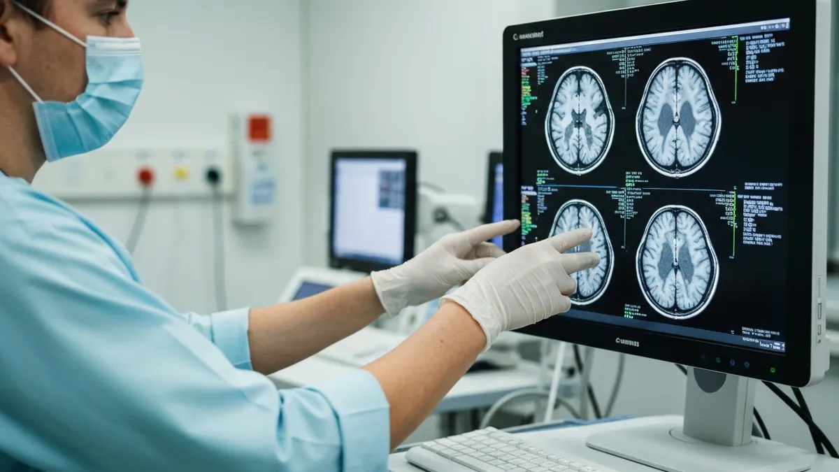
Image artifact from the WATCHMAN device is an additional practical consideration beyond safety. The nitinol frame produces a susceptibility artifact on MRI that can obscure adjacent cardiac structures. Sequences particularly vulnerable to susceptibility artifact, such as gradient echo sequences and EPI-based diffusion sequences, will show the most distortion near the left atrial appendage. When cardiac MRI is specifically ordered to evaluate the left atrium or pulmonary veins in a patient with a WATCHMAN device, the imaging team should adjust sequence selection and orient acquisition planes to minimize artifact overlap with the target anatomy.
Technologists should also document all scanning conditions used in the MRI report addendum. Recording the field strength, the maximum SAR achieved during the exam, the gradient mode used, and the scanning date relative to the implant date creates an auditable record that protects both the patient and the institution. Many facilities have begun incorporating WATCHMAN-specific fields into their MRI safety screening and reporting templates to standardize this documentation across all scanners and technologists.
MRI Safety Screening Protocols for Implanted Devices
Effective patient screening for WATCHMAN device MRI begins well before the patient arrives at the scanner. The referring physician's office should communicate implant details — including device model, serial number, implant date, and the implanting facility — at the time of ordering. Facilities should have a standardized intake questionnaire that specifically asks about left atrial appendage closure devices, cardiac occluders, and stroke prevention implants, since patients may not recognize the word "WATCHMAN" but will recall having a stroke prevention procedure.
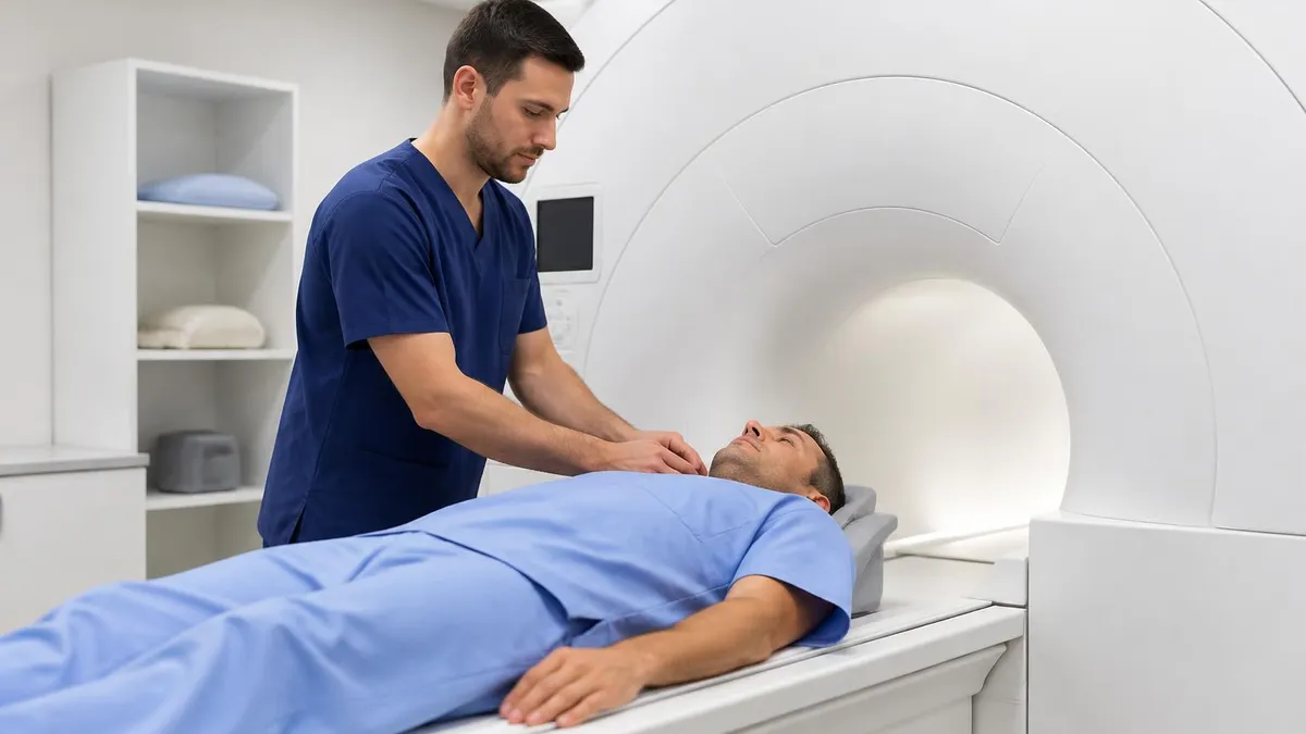
On the day of the scan, the MRI technologist must independently verify device information against an implant card or operative report. Verbal confirmation alone is insufficient. Once the device is confirmed, the technologist retrieves the current IFU from the manufacturer's MRI labeling database or the facility's implant management system. The technologist then compares the scanner's capabilities against each required condition in the IFU and documents this comparison in writing before proceeding. If any condition cannot be verified or met, the study is paused and the radiologist or MRI safety officer is notified immediately.
MR Conditional vs. MR Unsafe Implants: Key Differences for MRI Practice
- +MR Conditional devices like the WATCHMAN FLX can be scanned safely when IFU conditions are met
- +Clear parameter limits (SAR, field strength, gradient) allow standardized departmental protocols
- +Manufacturer testing data provides a documented safety basis that protects the institution
- +Most modern 1.5T and 3.0T scanners can achieve the conditions required for WATCHMAN scanning
- +MR Conditional labeling under ASTM F2503 is internationally recognized and legally defensible
- +Electronic IFU databases allow technologists to quickly verify current conditions without paper records
- −MR Conditional does not mean universally safe — every condition in the IFU must be verified individually
- −Different device generations (FLX vs. 2.5) may have different conditions, creating verification complexity
- −The 6-week post-implant waiting period can delay clinically important imaging in some patients
- −Nitinol susceptibility artifact near the left atrium can significantly degrade cardiac MRI image quality
- −Patients may not know their device model or implant date, requiring additional coordination with referring providers
- −Facilities without a formal MRI safety program may lack the protocols and training needed to scan these patients consistently
Pre-Scan Safety Checklist for WATCHMAN Device MRI Patients
- ✓Confirm device model and generation (FLX or 2.5) from implant card or operative report — do not rely on patient recall alone.
- ✓Verify implant date and confirm at least 45 days have elapsed since implantation for elective scans.
- ✓Retrieve the current IFU from the manufacturer's MRI labeling database or your facility's implant management system.
- ✓Confirm the scanner's static magnetic field strength is 1.5T or 3.0T — no other field strengths are covered by the WATCHMAN IFU.
- ✓Set the scanner's SAR limit to no more than 2.0 W/kg whole-body averaged and confirm the limit is active before scanning.
- ✓Verify the spatial gradient field at the patient's implant location does not exceed the IFU limit for the intended scanner.
- ✓Document the device information, scanner conditions, and post-implant timeline in the patient's MRI record before beginning the exam.
- ✓Notify the radiologist or MRI safety officer and obtain written approval before scanning any patient within the 45-day post-implant window.
- ✓Remove all extraneous metallic accessories from the patient and confirm compliance with standard MRI screening for all other implants.
- ✓After the exam, document the maximum SAR achieved, sequences used, and any artifact limiting diagnostic confidence in the report addendum.
MR Conditional Is Not a Green Light — It Is a Set of Required Conditions
Many patient safety incidents in MRI arise from misinterpreting "MR Conditional" as blanket permission to scan. The term means a device is safe only when every condition in the IFU is simultaneously satisfied. Verifying one parameter (field strength) while missing another (SAR limit) does not create a safe scan — it creates a partially checked scan that may still cause patient harm. Always work through every IFU condition systematically, document each one, and escalate to your safety officer when any condition cannot be confirmed.
The physics governing MRI safety for implanted devices like the WATCHMAN can be grouped into four distinct interaction mechanisms: static magnetic field effects, time-varying gradient field effects, radiofrequency electromagnetic field effects, and acoustic effects. Understanding these mechanisms in depth is essential for MRI technologists sitting for the registry exam and for those making real-time clinical decisions about whether and how to proceed with a scan involving a patient who has an implanted device.
Static magnetic field effects include magnetically induced displacement force and magnetically induced torque. Displacement force acts on paramagnetic and ferromagnetic materials, pulling them toward regions of higher field strength. Torque acts to align magnetic dipoles within the material along the field direction. For the WATCHMAN's nitinol frame, both forces are present but small because nitinol has a low magnetic susceptibility. ASTM F2052 bench testing quantifies these forces by suspending the device in a defined field gradient and measuring angular deflection; the WATCHMAN passes these tests at both 1.5T and 3.0T.
Time-varying gradient fields induce eddy currents in conductive implants. These eddy currents produce opposing magnetic fields (Lenz's law) and generate heat through resistive dissipation within the conductive material. For most implants, gradient-induced heating is far less significant than RF-induced heating, but it is not zero. The WATCHMAN's nitinol frame has finite resistance, and eddy-current heating during high-duty-cycle gradient sequences is a consideration that the manufacturer addresses in the IFU's sequence-specific guidance. Echo planar imaging sequences, used in diffusion and functional MRI, have particularly high gradient duty cycles and may require additional review.
Radiofrequency electromagnetic field effects are the most clinically significant heating concern for conductive implants. The RF transmit field (B1) oscillates at the Larmor frequency — approximately 64 MHz at 1.5T and 128 MHz at 3.0T. Conductive structures that are resonant at or near the Larmor frequency can act as antennas, concentrating RF energy and producing localized hotspots.
The WATCHMAN device's nitinol frame has a diameter of approximately 21 to 35 millimeters depending on device size, which is small relative to the RF wavelength in tissue even at 3.0T. This geometry limits resonance effects, which is reflected in the device's tested RF-induced temperature rise remaining below the 6-degree Celsius threshold used in ASTM F2182 testing.
The specific absorption rate is the metric used to regulate RF energy deposition in patients during MRI. SAR is measured in watts per kilogram (W/kg) and represents the rate of RF energy absorption per unit mass of tissue.
The IEC and FDA require that clinical MRI scanners operate within SAR limits for all patients, but implant-specific IFUs often impose stricter limits because implants can concentrate RF energy beyond what is predicted by body-averaged SAR calculations. For the WATCHMAN, the 2.0 W/kg whole-body limit is more restrictive than the 4.0 W/kg limit that applies to patients without implants, reflecting the additional heating risk from the conductive frame.
Acoustic effects — the loud banging and knocking sounds produced by gradient switching during MRI — are generally not a safety concern for implanted cardiac devices. The acoustic noise is produced by Lorentz forces on gradient coil windings, not by interactions with implants. However, in patients with cochlear implants or other auditory devices, acoustic considerations become primary. For WATCHMAN patients specifically, the implant itself presents no acoustic risk, but patient discomfort and anxiety from noise should still be managed with standard hearing protection and patient communication protocols used for all MRI examinations.
Understanding these four mechanisms also helps technologists reason about novel or less common implants when IFU documentation is unavailable or incomplete. By asking what material the implant is made of, what its geometry is, whether it contains leads or antennas, and whether it is electrically active, a Level 2 MRI-trained technologist can make a preliminary safety assessment that guides whether to proceed cautiously, defer the study, or escalate to the radiologist. This systematic reasoning framework is precisely what the ARRT registry exam tests in scenario-based safety questions.
IFU documents are updated by manufacturers when new safety data becomes available. A printed IFU in a facility binder may be outdated. Always verify the current version through the manufacturer's official MRI labeling website (Boston Scientific's MRI Labeling Resource Center for WATCHMAN devices) or through a validated implant management system. Using an outdated IFU could expose the patient to conditions that exceed currently recognized safety limits.
Preparing for the ARRT MRI registry examination requires a systematic approach to MRI safety content, and the WATCHMAN device is an excellent case study because it integrates multiple testable concepts: device classification terminology, specific MRI parameters, material science basics, and the clinical workflow for patient screening. Registry candidates who understand the WATCHMAN deeply will find that similar reasoning applies to cochlear implants, spinal cord stimulators, deep brain stimulators, and other complex implants that appear on the exam.
The registry content outline published by the ARRT allocates significant weight to patient care and MRI safety. Candidates should be able to define MR Safe, MR Conditional, and MR Unsafe under ASTM F2503; explain the physical basis of magnetically induced force and torque; describe how RF-induced heating is measured and regulated; and identify the role of the MRI safety officer in a clinical department. These are not obscure facts — they represent the foundational safety knowledge that every practicing MRI technologist must have.
When studying MRI safety, use active recall rather than passive review. Instead of rereading notes about the WATCHMAN, close this article and try to list from memory the five conditions in the WATCHMAN FLX IFU, the physical mechanism that makes RF heating dangerous for conductive implants, and the difference between MR Conditional and MR Safe labeling. Active retrieval strengthens long-term retention more effectively than any other study method, and the registry examination tests application, not just recognition.
Practice questions are the most efficient way to identify knowledge gaps in MRI safety. A student who answers a question about SAR limits incorrectly and then reads the explanation immediately consolidates the correct information in a way that simply reviewing the topic cannot replicate. Targeted practice on device safety scenarios — including questions about WATCHMAN, pacemakers, aneurysm clips, and retained metallic foreign bodies — will reveal which concepts need additional study time and which are already solidly understood.
Time management on the registry examination matters as much as content knowledge. The MRI registry includes 200 questions in a 3-hour window, giving candidates approximately 54 seconds per question. MRI safety questions are often scenario-based and involve reading a brief clinical situation before choosing the best course of action. Practicing under timed conditions builds the processing speed needed to read, reason, and answer within the allotted time without sacrificing accuracy on the safety content that is ethically and professionally non-negotiable.
Group study can be highly effective for MRI safety preparation when structured correctly. Discussing clinical scenarios out loud — explaining to a colleague why you would or would not scan a patient with a recently implanted WATCHMAN device — engages the same higher-order reasoning the registry tests. It also exposes gaps in understanding that self-study may miss, because explaining a concept to someone else requires a more complete and organized knowledge structure than simply recognizing the correct answer on a multiple-choice question.
After passing the registry, continuing education in MRI safety remains a professional obligation. The landscape of implanted devices changes rapidly as cardiology, neurology, and orthopedics adopt new technologies. New device approvals, updated IFUs, and changes to MRI scanner hardware all affect safe scanning conditions for specific patients. ARRT-approved continuing education in MRI safety helps technologists maintain currency with evolving standards and supports the kind of evidence-based practice that protects patients over an entire career. Resources like PracticeTestGeeks.com provide ongoing practice that reinforces safety knowledge long after initial certification.
Building a strong practical foundation in MRI safety requires going beyond textbook definitions and engaging with the clinical decision-making process that technologists face every day. When a patient arrives at your MRI suite holding an implant card that reads "WATCHMAN FLX," the sequence of actions you take in the next ten minutes will determine whether that patient receives a safe diagnostic study or is exposed to uncharacterized risk. Practicing that decision-making sequence mentally — or through simulation — before the first real patient encounter is the most effective form of safety preparation available to MRI students and early-career technologists.
Departmental policy is a critical safety layer that individual technologist competence alone cannot replace. Every MRI department should have a written policy for scanning patients with left atrial appendage closure devices, including the WATCHMAN, that specifies who has the authority to approve a scan outside of standard conditions, how IFU compliance is documented, and what the escalation pathway is when a scan cannot safely proceed. Policies should be reviewed and updated when new device generations are introduced and when the department's scanner hardware changes.
The relationship between the MRI team and the implanting cardiologist is particularly important for WATCHMAN patients. Cardiologists who perform LAAC procedures should be proactive about providing their patients with implant cards that include the device model, generation, and implant date. They should also be reachable for consultation when MRI teams have questions about scanning within the post-implant waiting period. MRI facilities that see high volumes of WATCHMAN patients may benefit from developing a formal communication protocol with the cardiology department or clinic to streamline information sharing before imaging appointments.
From the patient's perspective, understanding why MRI teams ask so many questions about implanted devices can reduce anxiety and improve cooperation. Patients should be encouraged to carry their implant card at all times and to proactively disclose the WATCHMAN to any provider who orders imaging. Patient education at the time of WATCHMAN implantation — including a clear explanation that MRI can still be performed safely under the right conditions — reduces the likelihood that patients will either conceal the device out of fear of being turned away or arrive without the documentation needed to proceed.
Technologists working in high-volume cardiac imaging centers may encounter WATCHMAN patients multiple times per week. Developing an efficient but thorough screening workflow — one that can be completed consistently in five to seven minutes without cutting corners — is both a productivity and a safety goal. Facilities should invest in electronic implant management systems that store IFU documents and patient implant records, generate automatic condition-checking checklists, and flag patients who fall within the post-implant waiting period at the time of appointment scheduling rather than on the day of the scan.
The WATCHMAN device represents one of the more straightforward MRI safety challenges in modern practice because its MR Conditional status is well-characterized, its conditions are clearly stated, and its clinical prevalence means that most MRI departments have at least some experience managing these patients. The skills developed in managing WATCHMAN patients — systematic IFU review, parameter verification, documentation, and escalation — apply directly to every other conditional device that will enter your MRI suite over the course of a career, including devices that are far less common or far more complex.
As MRI technology continues to advance — with ultra-high-field systems at 7T entering clinical use, new gradient and RF architectures being developed, and increasingly complex implants entering the market — the foundational approach to device safety will remain constant: identify the device precisely, find the current IFU, verify every condition, document your verification, and escalate when in doubt. That framework, applied consistently, is the best protection both for patients and for the professionals responsible for their safety inside the magnetic resonance environment.
MRI Questions and Answers
DWI MRI: Diffusion-Weighted Imaging Explained (2026 Guide)
Side Effects of Brain MRI: Understanding Risks, Safety, and What Every Patient Should Know
MRI Lumbar Spine Without Contrast: Complete Guide to the Scan, What It Shows, and How to Prepare
MRI History: From Nuclear Resonance to Modern Scanners
MRI of Cervical Spine: Complete Guide to Imaging, Anatomy, Pathology, and Patient Preparation
About the Author
Medical Laboratory Scientist & Clinical Certification Expert
Johns Hopkins UniversityDr. Sandra Kim holds a PhD in Clinical Laboratory Science from Johns Hopkins University and is certified as a Medical Technologist (MT) and Medical Laboratory Scientist (MLS) through ASCP. With 16 years of clinical laboratory experience spanning hematology, microbiology, and molecular diagnostics, she prepares candidates for ASCP board exams, MLT, MLS, and specialist certification tests.
Join the Discussion
Connect with other students preparing for this exam. Share tips, ask questions, and get advice from people who have been there.
View discussion (4 replies)