
MRI Sucked In: Real MRI Safety Accidents, Causes, and How Hospitals Prevent Them
MRI sucked in incidents explained — real accidents, projectile physics, Zone IV rules, screening failures, and how hospitals prevent magnet injuries. 📚

The phrase mri sucked in describes one of the most dramatic and dangerous accidents that can happen in modern medical imaging — a ferromagnetic object hurled across a scan room by a 1.5T or 3T magnetic field and slammed into the bore of an MRI machine, sometimes with a patient still inside. These incidents are rare but consistently catastrophic, and they have shaped nearly every MRI safety policy used in U.S. hospitals today. Understanding why they happen is essential for technologists, nurses, patients, and even visitors.
MRI scanners do not have an on/off switch for the static magnetic field. The superconducting magnet inside a clinical scanner remains active 24 hours a day, 7 days a week, even when no patient is being scanned and even during power outages. That is the central fact behind every projectile accident: a steel oxygen cylinder, a floor buffer, a wheelchair, a pair of trauma shears, or a police officer's sidearm becomes a missile the moment it crosses the 5-gauss line and the magnet's gradient field grips it.
The U.S. Food and Drug Administration's MAUDE database, the ACR Manual on MR Safety, and incident reports from The Joint Commission together document hundreds of serious projectile, burn, and quench events over the past two decades. Some have killed patients. Many have injured staff. Almost all of them trace back to a small number of recurring root causes: failed screening, untrained personnel entering Zone IV, or an exception made under time pressure during a code or trauma.
For radiologic technologists preparing for the ARRT MRI registry and for clinical staff who rotate through imaging, MRI safety is not optional knowledge — it is the single most heavily tested domain on the exam. Questions about the 5-gauss line, ferromagnetic detection, projectile risk, and quench procedures show up on every form of the test. If you want to see how these questions are framed, the MRI knowledge questions and answers set covers safety scenarios in detail.
This guide walks through real documented MRI accidents, the physics that drive them, the four-zone access control system the American College of Radiology requires, the screening process that should stop projectiles before they reach the magnet room, and the specific failures that allow incidents to slip through. We will also cover thermal injuries, gadolinium contrast reactions, and quench events — the three other major categories of MRI harm that get less media attention than projectile cases but injure far more patients each year.
If you work near an MRI scanner, or if a family member is preparing for a scan, the information here is meant to be practical, accurate, and grounded in published safety literature rather than viral news headlines. Some of the cases described are well known. Others are quieter near-misses that never made the news but appear in regulatory filings and peer-reviewed case reports. All of them illustrate the same lesson: the magnet is always on, and respect for that fact saves lives.
MRI Safety Accidents by the Numbers
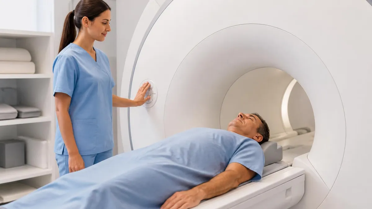
The Four ACR MRI Safety Zones
The general public can enter freely. This includes parking, lobbies, and hallways outside the imaging department. No magnetic field is present and no screening is required to be in this area.
Patients are greeted, registered, and begin screening here. Movement is supervised by MRI personnel. Metal objects may still be present, but no patient enters Zone III before screening is documented and confirmed.
Restricted to screened patients and trained staff. Physical barriers, locked doors, and ferromagnetic detectors guard the entry. Free access to Zone III by untrained personnel is the single most common cause of projectile accidents.
Contains the scanner itself and the 5-gauss field. Only Level 2 MR-trained personnel may bring patients or equipment inside. All items entering must be verified MR Safe or MR Conditional with documented limits.
To understand why an oxygen tank or a wheelchair can be ripped across a room, you have to start with the physics of the static magnetic field, often called B₀. A 1.5 Tesla MRI scanner produces a field roughly 30,000 times stronger than Earth's natural magnetic field, and a 3T scanner doubles that. But the projectile danger does not come from the static field alone — it comes from the spatial gradient of that field, the rate at which strength changes as you walk toward the bore.
Near the entrance to a magnet room, the field might be a few hundred gauss. Five feet closer, it can be several thousand. At the bore itself, you are inside a 15,000-gauss to 30,000-gauss environment. A ferromagnetic object dropped or carried into this gradient experiences a force proportional to both its mass and the steepness of that gradient. Once the pull exceeds the friction or grip holding the object in place, it accelerates rapidly toward the magnet's isocenter.
The result is what safety researchers call the projectile effect. A small object like a hairpin or paperclip will fly at relatively low energy. A 200-pound floor buffer or a steel oxygen cylinder, however, can accelerate to speeds that punch holes in fiberglass bore covers and crush anything in their path. The famous 2001 Michael Colombini case, in which a six-year-old patient at Westchester Medical Center died after a portable oxygen tank was carried into the scan room during a code, remains the textbook example.
Stainless steel is sometimes misunderstood by clinical staff. Many grades of stainless steel — including the 400-series used in some scissors, hemostats, and tool handles — are strongly ferromagnetic. Other grades, particularly 304 and 316 austenitic stainless used in surgical instruments, are weakly ferromagnetic or essentially non-magnetic. The label "stainless" alone tells you nothing about MRI safety. This is why each device must be tested and labeled as MR Safe, MR Conditional, or MR Unsafe under the ASTM F2503 standard.
Cell phones, hearing aids, credit cards, mechanical watches, and analog wristwatches are routinely damaged by the field even when they are not violently attracted. The magnetic strip on a credit card can be erased within Zone III, and mechanical watches can lose their timing permanently. These are minor losses compared to projectile injuries, but they remind us that the field interacts with everyday objects in ways that are invisible until they happen.
The bore of the scanner itself is also where many burns and pinch injuries occur, even without a projectile event. Patients touching the inside of the bore, looped ECG leads, conductive tattoo ink, and cabling laid against bare skin can all create radiofrequency hot spots that cause second-degree burns during the scan. If you want a refresher on the underlying physics, the MRI physics questions and answers resource covers the gradient, RF, and static field interactions in registry-style format.
None of these mechanisms require operator error in the moment — they require failures in the system surrounding the scanner. The magnet does exactly what the laws of physics dictate. Everything that protects patients and staff happens before the door to Zone IV opens.
MRI Practice Test Questions
Prepare for the MRI - Magnetic Resonance Imaging exam with our free practice test modules. Each quiz covers key topics to help you pass on your first try.
MRI Knowledge
MRI Exam Questions covering Knowledge. Master MRI Test concepts for certification prep.
MRI Physics
Free MRI Practice Test featuring Physics. Improve your MRI Exam score with mock test prep.
MRI Anatomy and Pathology
MRI Test Prep for MRI Anatomy and Pathology. Practice MRI Quiz questions and boost your score.
MRI Anatomy and Positioning
MRI Questions and Answers on MRI Anatomy and Positioning. Free MRI practice for exam readiness.
MRI Contrast Agents
Free MRI Quiz on MRI Contrast Agents. MRI Exam prep questions with detailed explanations.
MRI Patient Care and Positioning
MRI Practice Questions for MRI Patient Care and Positioning. Build confidence for your MRI certification exam.
Major Categories of MRI Sucked In and Related Accidents
Projectile accidents involve ferromagnetic objects being pulled into the bore. Documented cases include oxygen cylinders, IV poles, floor polishers, mop buckets, wheelchairs, firearms, sandbags with iron filings, and trauma shears. The injuries range from broken bores and torn safety covers to fatal head trauma. The U.S. case most often cited is the 2001 pediatric oxygen tank fatality, but FDA MAUDE filings show several projectile reports each year, including more recent events involving police weapons and contractor tools.
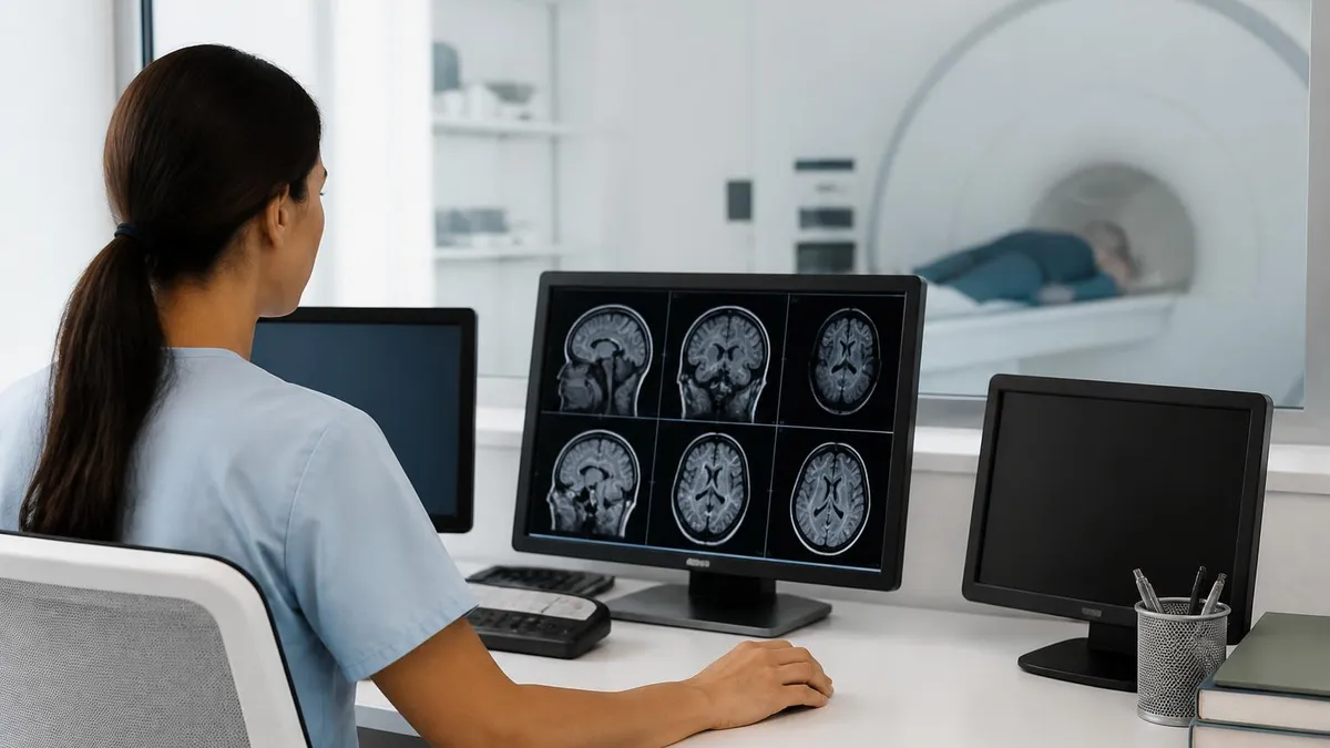
Every projectile event traces back to one of three failures: an unscreened person entering Zone IV, an item missed during screening, or a deliberate decision to bring equipment in during an emergency. Modern safety programs now require ferromagnetic detection systems at the Zone III–IV boundary, but these detectors are aids, not replacements for human screening protocols.
Ferromagnetic Detection Systems: Worth the Investment?
- +Catches small ferrous items missed in verbal screening
- +Reduces reliance on patient self-disclosure of implants
- +Provides documented audit trail for risk management
- +Lowers insurance premiums in many ACR-accredited sites
- +Detects items concealed in clothing or pockets
- +Required by Joint Commission expectations at new builds
- −Capital cost ranges from $15,000 to $80,000 per unit
- −False positives from non-ferrous metals slow throughput
- −Cannot replace trained MR personnel or written screening
- −Requires daily calibration and operator training
- −Will not detect implanted devices that are non-ferrous but still MR Unsafe
- −Detector placement still allows projectile risk if Zone III door is propped open
Pre-Scan MRI Safety Screening Checklist
- ✓Verify patient identity and confirm the ordered exam matches the body part
- ✓Complete written ferromagnetic screening form, signed by patient and technologist
- ✓Ask specifically about pacemakers, ICDs, neurostimulators, and cochlear implants
- ✓Document any aneurysm clips, stents, or surgical hardware with year and manufacturer
- ✓Confirm no shrapnel, BBs, or metallic foreign body history without prior orbit X-ray
- ✓Remove all jewelry, hairpins, piercings, hearing aids, dentures, and wigs
- ✓Check pockets for keys, coins, phones, USB drives, and credit cards
- ✓Pass the patient and any escort through the ferromagnetic detector
- ✓Verify all monitoring equipment entering Zone IV is MR Conditional with documented limits
- ✓Brief the patient on emergency squeeze ball, communication, and quench scenario
Even during a power outage, the static field remains at full strength.
Superconducting MRI magnets store enormous amounts of energy in their persistent current. They do not require continuous electricity to maintain B₀ — only cryogenic cooling. This is why projectile accidents have occurred during nights, weekends, fire drills, and outages. Any object brought into Zone IV at any hour is subject to the full magnetic pull.
Several MRI accidents have become reference cases in the safety literature, taught in every MR technologist program and cited in ACR documents. The 2001 death of six-year-old Michael Colombini at Westchester Medical Center in Valhalla, New York, is the most widely known. During a sedated brain MRI, an anesthesiologist responded to a desaturation event and an external oxygen tank — a portable steel cylinder — was carried into the scan room. The tank was pulled from the staff member's hands, flew into the bore, and struck the child in the head. He died two days later.
That single case drove the creation of the four-zone access control model, the formalization of MR personnel training levels, and the widespread adoption of aluminum and composite oxygen cylinders in imaging departments. Today, color-coded MR Conditional cylinders are standard equipment in every accredited MRI suite in the United States, and the practice of carrying a standard hospital oxygen tank into Zone IV is treated as a never event.
In 2014, a hospital in India saw an attendant pulled into a 1.5T scanner along with an oxygen cylinder he was carrying for a patient. He was pinned against the bore for hours until the magnet could be ramped down. He died from inhalation of leaking oxygen and the crush injuries he sustained. In 2018, another death was reported in Mumbai under similar circumstances, again involving a cylinder carried in during a chaotic patient handoff.
The United States has also documented projectile events involving floor buffers brought in by housekeeping, police officers entering with sidearms during emergency responses, contractors carrying tool belts into the bore for HVAC work, and even a sandbag whose interior contained iron-filled material rather than the expected silica sand. None of these required malice or recklessness — they required only a single moment in which the magnet's continuous, invisible presence was forgotten.
Burn cases are less dramatic in headlines but appear in larger numbers in the FDA MAUDE database. Patients have received second-degree burns from looped ECG leads, from pulse oximeter cables touching skin, from medication patches with metallic backing that were not disclosed during screening, and from conductive tattoo ink concentrated in dark designs. One published series documented dozens of burn cases linked to fentanyl patches alone before manufacturers updated labeling.
Gadolinium-based contrast reactions account for another category. Nephrogenic systemic fibrosis, linked to certain linear gadolinium agents in patients with severely impaired renal function, was identified in the mid-2000s and led to changes in screening for renal function before contrast administration. Modern macrocyclic agents have dramatically reduced this risk, but the safety lesson — screen kidney function, document GFR — became permanent.
Looking at the full body of accident data, a striking pattern emerges. The most dangerous projectile events happen during emergencies, not routine scans. Code blues, trauma resuscitations, contractor work, and night shifts produce a disproportionate share of incidents because the normal slow, deliberate screening process is short-circuited by time pressure. Every safety policy in MRI is essentially designed to keep that pressure from breaching the door to Zone IV.
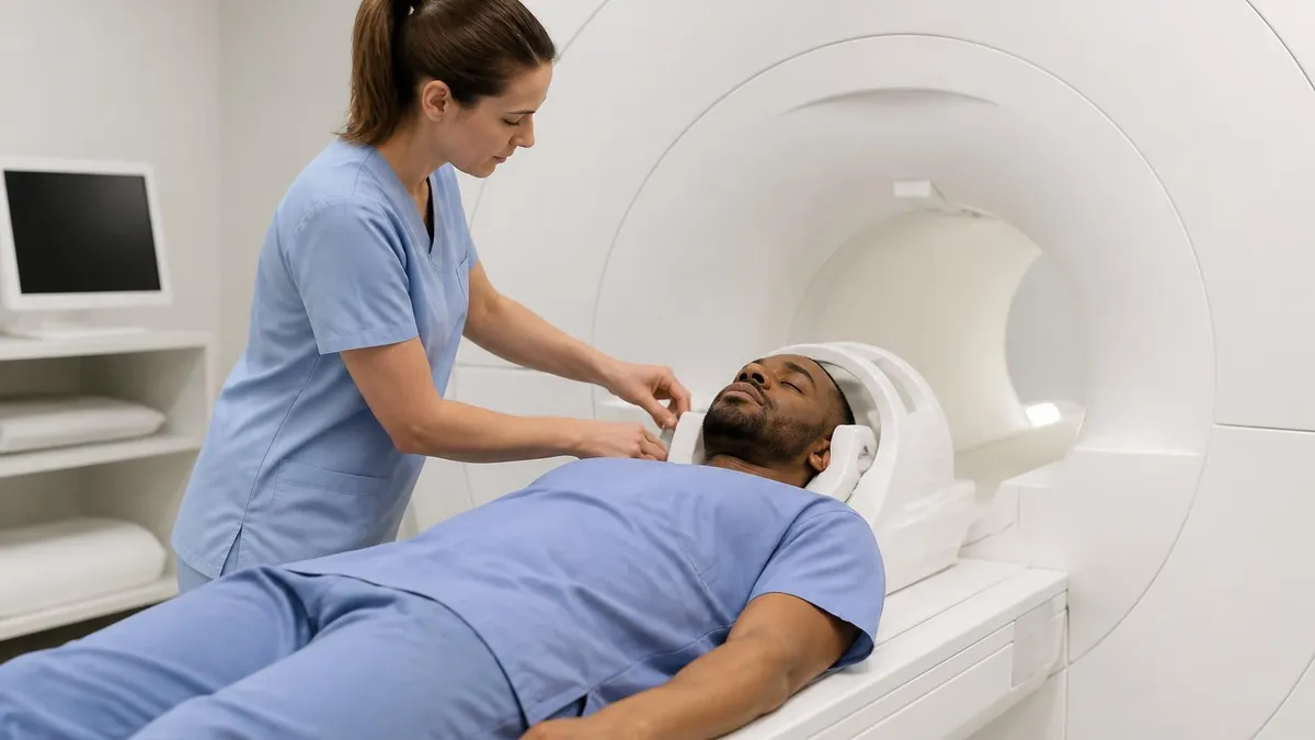
Standard steel oxygen cylinders are strongly ferromagnetic and have caused multiple fatalities in MRI suites worldwide. Only aluminum or composite MR Conditional cylinders, clearly color-coded and labeled, may enter the scan room. If an emergency requires resuscitation, the patient must be moved out of Zone IV first — never bring resuscitation equipment to the patient inside the bore.
Prevention of MRI sucked in accidents rests on layered defenses, none of which is sufficient alone. The first layer is environmental design. Scan rooms must be physically locked when not in use, with key control limited to MR personnel. Doors must not be propped open. Windows into Zone IV should provide line-of-sight from the control room so the technologist can see anyone approaching. Signage must clearly mark the 5-gauss line and the Zone III boundary in multiple languages.
The second layer is access control. The ACR defines Level 1 MR personnel as those who have completed basic safety education and may enter Zone III under supervision, and Level 2 personnel as those who have completed advanced training and may bring patients into Zone IV. Anesthesiologists, nurses, housekeeping, security, fire response teams, and contractors all need site-specific orientation before they ever step into Zone III. Most accidents involve people who would not pass a Level 2 quiz.
The third layer is screening. Written forms, verbal confirmation, and ferromagnetic detection together create redundancy. A patient who answers "no" to all questions but who triggers the detector still gets re-screened. A patient who declares an implant is researched in MRI safety databases — MRIsafety.com, the manufacturer's documentation, and the ACR Manual on MR Safe Practices — before the scan is approved. No assumption is made on the basis of an old chart note alone.
The fourth layer is equipment labeling under ASTM F2503. Every device entering Zone IV must carry one of the three labels: MR Safe (green square), MR Conditional (yellow triangle with conditions), or MR Unsafe (red circle with diagonal bar). MR Conditional devices have limits on field strength, gradient, SAR, and sometimes patient positioning. A pacemaker labeled MR Conditional at 1.5T may not be safe at 3T, and crossing those conditions can be fatal. For background on how modern scanners are designed, see what is an MRI test.
The fifth layer is emergency planning. Every MRI suite must have written protocols for code blue inside Zone IV (move patient out, do not bring equipment in), fire response (battery-powered MR Conditional extinguishers only), quench response (evacuate, ventilate, account for all personnel), and projectile recovery (do not attempt to remove the object, ramp down or quench depending on injury status). These drills should be practiced at least annually with all departments that may respond.
The sixth layer is continuous improvement. Near-misses must be reported and reviewed without blame. A staff member who realizes they almost walked into Zone IV with a clipboard containing a metal clip should feel safe reporting that event. Patterns in near-miss data — repeated screening failures for the same implant type, repeated breaches at the same door — reveal weaknesses that can be fixed before a serious accident occurs.
Taken together, these layers explain why serious projectile accidents are rare even though the underlying physics is unforgiving. The system works most of the time. The cases that do happen almost always involve breaches in two or more layers simultaneously, which is exactly the pattern predicted by accident-analysis frameworks like the Swiss Cheese Model and which is why redundancy matters more than any single control.
For technologists, students, and clinical staff preparing to work safely around MRI equipment, a few practical habits separate the careful from the lucky. Never assume an object is safe because it looks non-metallic — composite tools, plastic-coated scissors, and electronic devices all contain hidden ferromagnetic components. Verify the label, check the documentation, and when in doubt, do not bring it into Zone III. The cost of delaying a scan is always lower than the cost of a projectile event.
Treat verbal screening as a conversation, not a checklist. Patients often forget implants placed decades ago, or describe them in non-medical terms. Ask follow-up questions: "Have you ever had surgery on your heart or brain?" "Were any pieces of metal ever removed from your eye?" "Do you wear any patches, even for nicotine or pain?" Yes-answers should always trigger documentation rather than verbal reassurance. The patient's safety is more important than their schedule.
Set the tone in the control room. New staff, students, and visitors should be briefed before they cross the Zone II threshold. Make the words "the magnet is always on" part of the introduction. Walk people through the 5-gauss line and explain that nothing — phones, watches, ID badges with magnetic strips — crosses without verification. A culture that takes safety seriously is more protective than any single piece of equipment.
During emergencies, slow down. The instinct in a code is to rush, but every documented projectile fatality involves rushing. The correct sequence is always: stop the scan, move the patient to a designated resuscitation area outside Zone IV, then bring resuscitation equipment to the patient there. This may add 60 to 90 seconds to the response. Studies of MRI code outcomes show that this delay does not change patient survival, while the alternative repeatedly produces fatalities.
Document everything. Screening forms, contrast administration, vital signs, scan parameters, and any deviations from protocol all belong in the chart. If a contractor or fire-response team enters Zone III, log who entered, when, what they brought, and who supervised them. This paperwork seems excessive on normal days, but when something goes wrong, it is the only way to reconstruct what happened and prevent recurrence. Risk management and Joint Commission surveyors expect to see it.
For students preparing for the ARRT MRI registry, focus your safety study on three areas: identifying MR Safe versus MR Conditional versus MR Unsafe labels, recognizing the four-zone model and what activities belong in each zone, and understanding the physics that drive projectile force, RF burns, and quench events. Roughly a quarter of registry questions touch safety in some form, and they tend to use scenario wording — "A patient with X enters Zone Y, what is your next step?"
Finally, remember that MRI safety is a living field. The 2024 ACR Manual on MR Safe Practices differs in important ways from the 2007 version, and new implant designs, new contrast agents, and new field strengths up to 7T continue to reshape best practice. Subscribe to updates, attend annual safety refreshers, and treat the screening protocol you learned in school as a starting point rather than a finished body of knowledge.
MRI Questions and Answers
What Is an MRI Test? How Magnetic Resonance Imaging Scans Diagnose Disease in 2026
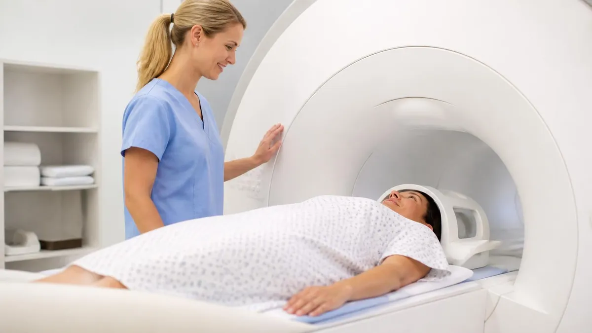
MRI Medical Abbreviation: What MRI Stands For and Why It Matters
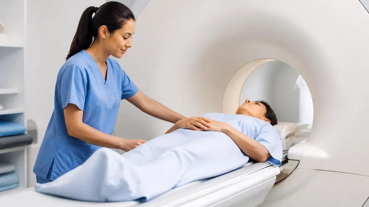
The History of MRI: From Discovery to Modern Medicine

Knee MRI Images: A Complete Guide to Reading, Understanding, and Interpreting Knee Scans
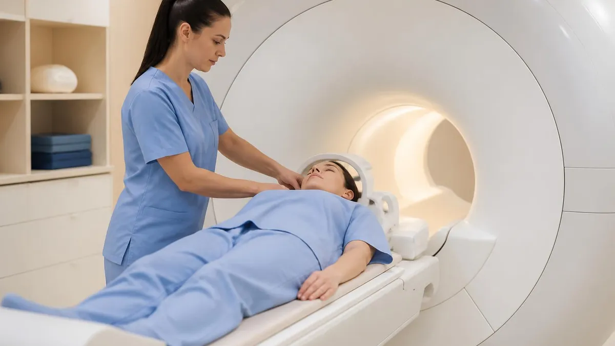
Noise of MRI Machine: Why MRI Scanners Are So Loud and What to Expect
About the Author

Medical Laboratory Scientist & Clinical Certification Expert
Johns Hopkins UniversityDr. Sandra Kim holds a PhD in Clinical Laboratory Science from Johns Hopkins University and is certified as a Medical Technologist (MT) and Medical Laboratory Scientist (MLS) through ASCP. With 16 years of clinical laboratory experience spanning hematology, microbiology, and molecular diagnostics, she prepares candidates for ASCP board exams, MLT, MLS, and specialist certification tests.
Join the Discussion
Connect with other students preparing for this exam. Share tips, ask questions, and get advice from people who have been there.
View discussion (8 replies)