Uterine Fibroid on MRI: Imaging, Diagnosis, and Treatment Planning
How a uterine fibroid on MRI appears, what sequences radiologists use, FIGO mapping, and what findings mean for treatment planning.
A uterine fibroid on MRI is one of the most accurately characterized pelvic findings in modern radiology, and for good reason. Magnetic resonance imaging maps the size, number, location, and internal composition of leiomyomas with a precision that ultrasound cannot match, especially when the uterus is enlarged, multiply fibroid, or distorted by prior surgery. For symptomatic women considering myomectomy, uterine artery embolization, or focused ultrasound, MRI is the imaging study that turns vague pelvic complaints into a clear surgical or interventional plan.
Fibroids are extraordinarily common. By age 50, an estimated 70 to 80 percent of women will have at least one leiomyoma, although only about a quarter become symptomatic enough to seek treatment. Symptoms typically include heavy menstrual bleeding, pelvic pressure, urinary frequency, constipation, dyspareunia, and infertility. Because these complaints overlap with adenomyosis, endometriosis, and ovarian pathology, accurate imaging is essential before any irreversible therapy is recommended.
Pelvic MRI excels here because it provides multiplanar, high-contrast images of the uterus, endometrium, junctional zone, and adjacent organs without ionizing radiation. A typical protocol combines T2-weighted sagittal, axial, and coronal sequences with T1-weighted imaging before and after intravenous gadolinium. Diffusion-weighted imaging and dynamic contrast enhancement add tissue characterization that helps separate benign degenerated fibroids from rare uterine sarcomas, a distinction that has major treatment implications.
Radiologists assess each lesion against the FIGO leiomyoma classification, which numbers fibroids 0 through 8 based on whether they are submucosal, intramural, subserosal, or pedunculated. This mapping drives almost every clinical decision that follows. A FIGO 0 pedunculated submucosal fibroid may be removed hysteroscopically in an outpatient setting, while a FIGO 7 pedunculated subserosal lesion on a thin stalk raises concern for torsion and usually requires laparoscopic resection.
MRI also identifies features that change management beyond simple location. Hyaline, cystic, myxoid, and red degeneration each produce a recognizable signal pattern, and the presence of restricted diffusion combined with necrosis or rapid growth raises the question of leiomyosarcoma. For patients planning MRI with and without contrast, the post-gadolinium phase is what allows the radiologist to confirm viable, perfused tissue versus avascular degeneration.
This guide explains how a uterine fibroid on MRI appears across sequences, how it is classified, how it is differentiated from look-alike lesions, and how radiology reports translate into treatment options. We will cover protocol design, FIGO mapping, degeneration patterns, the sarcoma question, embolization screening, focused ultrasound candidacy, and the practical patient experience from bowel prep to scan day. Whether you are a technologist, a referring clinician, or a patient preparing for your study, the goal is to leave you confident in what the MRI is showing and why it matters.
By the end, you should be able to recognize a typical leiomyoma on T2 imaging, understand why some fibroids are dark and others bright, and know which MRI features prompt a phone call to the gynecologic surgeon. We will also touch on post-treatment imaging after embolization or focused ultrasound, because follow-up MRI is how clinicians confirm that an intervention actually worked.
Uterine Fibroids on MRI by the Numbers
Standard Pelvic MRI Protocol for Fibroids
Sagittal, axial, and coronal T2 sequences are the workhorse. Fibroids appear as well-circumscribed, low-signal masses against the higher-signal myometrium, allowing precise mapping of size, number, and FIGO location.
Axial T1 imaging establishes baseline signal. Most fibroids are isointense to muscle, but hemorrhagic or red-degeneration fibroids show intrinsic T1 hyperintensity, which is important to recognize before contrast is given.
DWI with ADC mapping evaluates cellularity. Benign cellular fibroids can restrict, but markedly low ADC values combined with T2 heterogeneity raise concern for leiomyosarcoma and prompt further workup.
Gadolinium-enhanced sequences confirm tissue viability. Avascular areas suggest degeneration, while heterogeneous early enhancement with washout in a rapidly growing mass is a red flag for malignancy.
Some centers add cine imaging to assess junctional zone peristalsis and to distinguish adenomyosis from a fibroid, particularly when symptoms are atypical or the uterus is diffusely enlarged.
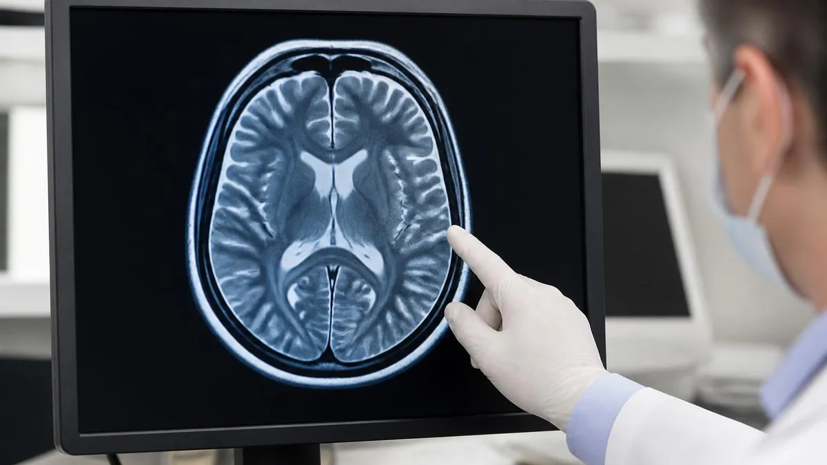
The classic uterine fibroid on MRI is a well-circumscribed, round or oval mass that appears markedly hypointense on T2-weighted images relative to the surrounding myometrium. This dark T2 signal reflects the dense, whorled bundles of smooth muscle and fibrous tissue that define a leiomyoma histologically. A pseudocapsule of compressed myometrium often outlines the fibroid, giving it a clean border that helps the radiologist count lesions and assign FIGO numbers accurately.
On T1-weighted imaging, most untreated fibroids are isointense to skeletal muscle and blend into the uterine background. Exceptions matter clinically. A fibroid with intrinsic T1 hyperintensity often represents red degeneration, an infarction-type change that occurs most commonly during pregnancy or after embolization. Recognizing this pattern is important because pregnant women presenting with acute pelvic pain and an enlarging fibroid frequently have red degeneration rather than torsion or abruption.
After gadolinium, a typical viable fibroid enhances heterogeneously but distinctly, confirming intact vascular supply. The dynamic phases show progressive enhancement that usually parallels or slightly lags the myometrium. Non-enhancing portions correspond to cystic, hyaline, or red degeneration. This distinction is essential before uterine artery embolization because pre-embolization viability predicts how much volume reduction the patient can expect after treatment.
Diffusion-weighted imaging adds another layer of characterization. Cellular leiomyomas can show high signal on high-b-value DWI with corresponding low ADC values, mimicking a malignant tumor. Radiologists therefore interpret DWI in context with T2 morphology, growth rate, and contrast pattern rather than in isolation. A benign cellular fibroid is smooth-bordered and homogeneously enhancing, while a sarcoma typically shows irregular margins, necrosis, and very low ADC.
Location drives both symptoms and treatment. Submucosal fibroids, even small ones, cause heavy menstrual bleeding because they distort the endometrial cavity. Intramural fibroids contribute to bulk symptoms and may impair fertility when they encroach on the cavity. Subserosal fibroids often grow large before causing pressure symptoms on the bladder or rectum. Pedunculated subserosal lesions can torse, and pedunculated submucosal lesions can prolapse through the cervix as so-called aborting fibroids.
Adenomyosis is the most important imaging differential. On T2 images, adenomyosis appears as diffuse or focal thickening of the junctional zone with embedded high-signal foci representing ectopic endometrial glands. Unlike a discrete fibroid, adenomyosis has indistinct margins and does not displace adjacent structures with a mass effect. Recognizing coexistent adenomyosis is critical because it changes counseling, particularly when reviewing the common MRI findings of pelvic pain in reproductive-age women.
Finally, the radiology report should describe the dominant fibroid by FIGO type, three-dimensional measurements, distance from the endometrium and serosa, and relationship to the cervix and ureters. This level of detail allows the gynecologist or interventional radiologist to choose the right procedure without re-imaging the patient.
FIGO Classification and Key MRI Sequences
FIGO type 0 is a pedunculated intracavitary fibroid attached to the endometrium by a stalk. Type 1 is submucosal with less than 50 percent intramural extension, and type 2 is submucosal with 50 percent or more intramural extension. On T2 sagittal MRI, these lesions distort the endometrial stripe and are the leading cause of heavy menstrual bleeding and infertility from fibroids.
Hysteroscopic myomectomy is the preferred treatment for FIGO 0 and most type 1 lesions, while type 2 may require a combined or staged approach. Accurate MRI measurement of the intramural depth is essential because resecting too deeply during hysteroscopy risks uterine perforation and thermal injury to the serosa or adjacent bowel.
MRI Versus Ultrasound for Fibroid Evaluation
- +Precise mapping of size, number, and FIGO location in complex uteri
- +Multiplanar imaging unaffected by body habitus or bowel gas
- +Identifies adenomyosis, endometriosis, and ovarian pathology in one study
- +Characterizes degeneration patterns guiding embolization decisions
- +Detects features suspicious for leiomyosarcoma with DWI and contrast
- +Provides a reproducible baseline for post-treatment follow-up
- +No ionizing radiation, safe for repeated imaging
- −Higher cost than transvaginal ultrasound
- −Longer scan times of 30 to 45 minutes
- −Contraindicated in some patients with implanted devices
- −Gadolinium requires adequate renal function
- −Claustrophobia limits tolerance in some patients
- −Less accessible in rural or low-resource settings
- −Requires specialized pelvic protocol and reader expertise
Pre-Scan Checklist for a Uterine Fibroid on MRI
- ✓Confirm the order specifies pelvic MRI with and without contrast
- ✓Screen for renal function with recent eGFR before gadolinium
- ✓Document last menstrual period and pregnancy status
- ✓Review prior imaging including ultrasound and any previous MRI
- ✓Complete the MRI safety questionnaire for implants and devices
- ✓Remove all metallic clothing, jewelry, and transdermal patches
- ✓Ask the patient to void approximately 30 minutes before scanning
- ✓Administer antiperistaltic agent if protocol calls for it
- ✓Position with appropriate pelvic phased-array coil placement
- ✓Counsel the patient about breath holds and contrast injection
- ✓Confirm IV access is patent in a vein appropriate for power injection
- ✓Verify the radiologist receives the gynecology referral question
T2 signal predicts treatment response
Fibroids that are markedly hypointense on T2 imaging tend to be densely fibrous and respond less robustly to uterine artery embolization than those with intermediate or heterogeneous T2 signal. Radiologists routinely flag this signal characteristic in the report because it directly influences whether a patient is offered embolization, focused ultrasound, or surgical myomectomy.
Degeneration is the term radiologists use for the secondary changes that occur when a fibroid outgrows its blood supply. Hyaline degeneration is the most common and produces a homogeneous, slightly higher T2 signal than a typical leiomyoma without true fluid content. It usually does not change management. Cystic degeneration appears as well-defined fluid-signal areas within the fibroid, very bright on T2 and dark on T1, and non-enhancing after contrast.
Myxoid degeneration shows very high T2 signal that can approach the brightness of simple fluid but with septations and gel-like enhancement. This pattern occasionally mimics a sarcoma or a malignant ovarian neoplasm, and careful tracing of the lesion back to the uterus is essential. Red, or carneous, degeneration is hemorrhagic infarction. It typically shows peripheral T1 hyperintensity with a dark rim from hemosiderin and is the classic cause of acute pain in a pregnant patient with a known fibroid.
The differential diagnosis radiologists worry about most is uterine leiomyosarcoma. Although rare, with an incidence well under one percent of suspected fibroids, leiomyosarcoma carries a poor prognosis when treated as a benign fibroid with morcellation. On MRI, features that should raise concern include rapid interval growth, irregular margins, central necrosis, very low ADC values on diffusion-weighted imaging, and heterogeneous early enhancement with central washout.
A widely used qualitative tool combines T2 heterogeneity, intratumoral T1 hyperintensity, and restricted diffusion to flag the small subset of masses that need additional workup, biopsy, or upfront hysterectomy rather than uterine-sparing intervention. No single finding is diagnostic. Radiologists describe the constellation and recommend correlation with serum LDH, clinical history, and gynecologic oncology consultation when the picture is concerning.
Lipoleiomyoma is a benign variant containing macroscopic fat that follows fat signal on all sequences, dropping out on fat-suppressed imaging. This is important to recognize because it can otherwise be mistaken for a teratoma or a fat-containing pelvic mass. Calcified fibroids show low signal voids on all sequences and are often seen in postmenopausal women.
Imaging after embolization or focused ultrasound also relies on MRI. A successful embolization shows complete devascularization of the targeted fibroids on post-contrast imaging, with shrinkage over three to six months. Persistent enhancement indicates residual perfusion and predicts symptom recurrence. After focused ultrasound, the non-perfused volume ratio measured on contrast MRI correlates with durable symptom relief.
Adenomyosis can coexist with fibroids in up to 20 percent of patients. When the junctional zone exceeds 12 millimeters or shows ill-defined T2 hyperintense foci, the report should explicitly call this out because residual adenomyosis after myomectomy is a major reason symptoms persist post-operatively.
Rapid interval growth in a postmenopausal woman, irregular margins, marked restricted diffusion with very low ADC, central necrosis, and elevated serum LDH together raise concern for leiomyosarcoma. These patients should not undergo morcellation or unmonitored uterine-sparing therapy and require prompt gynecologic oncology referral.
Translating a uterine fibroid on MRI into a treatment plan begins with three questions: where are the fibroids located, how many are there, and is the patient finished with childbearing. MRI answers the first two definitively, and the answer to the third determines whether the goal is symptom control, uterine preservation, or both. Submucosal fibroids causing bleeding favor hysteroscopic myomectomy. Multiple intramural fibroids causing bulk symptoms in a woman who has completed childbearing often favor uterine artery embolization.
For women planning future pregnancy, abdominal or laparoscopic myomectomy remains the gold standard for intramural and subserosal fibroids that distort the cavity or measure more than four to five centimeters. MRI guides the surgeon by showing exactly how many lesions to expect and where the dominant ones sit relative to the endometrial cavity and serosa, reducing operative time and missed fibroids.
Uterine artery embolization is appropriate for symptomatic women not pursuing future pregnancy when MRI confirms perfused fibroids of typical T2 signal. Pre-procedure MRI also screens for ovarian arterial supply, pedunculated subserosal fibroids on thin stalks, and large submucosal fibroids that may sluff and cause infection. The interventional radiologist relies on the radiology report to anticipate these risks and counsel the patient appropriately.
Magnetic resonance guided focused ultrasound, sometimes called MRgFUS, uses the same MRI scanner that diagnoses the fibroid to deliver thermal ablation. Candidacy depends on lesion accessibility, size under approximately 10 centimeters, T2 signal characteristics, and the absence of intervening bowel or scar along the beam path. MRI is the only imaging modality that can both plan and monitor this treatment in real time. Patient experience is similar to a standard MRI history of imaging session but longer.
Medical therapy with GnRH analogues, selective progesterone receptor modulators where available, or tranexamic acid for bleeding can serve as a bridge to surgery or as definitive management in perimenopausal women. MRI before initiating medical therapy establishes a baseline so that response can be measured objectively. A 30 to 50 percent volume reduction is typical with GnRH agonists over three to six months.
Hysterectomy remains the only definitive cure and is appropriate when symptoms are severe, fertility is not desired, and other modalities have failed or are contraindicated. Even in this scenario, pre-operative MRI helps the surgeon plan the approach, vaginal versus laparoscopic versus open, by clarifying uterine size, fibroid burden, and the relationship to the bladder, ureters, and rectum.
Follow-up MRI is recommended three to six months after embolization or focused ultrasound to confirm devascularization and volume reduction. For surgical myomectomy, follow-up imaging is reserved for recurrent symptoms because new fibroids develop in up to a third of patients within five years, especially in younger women with multiple lesions at baseline.
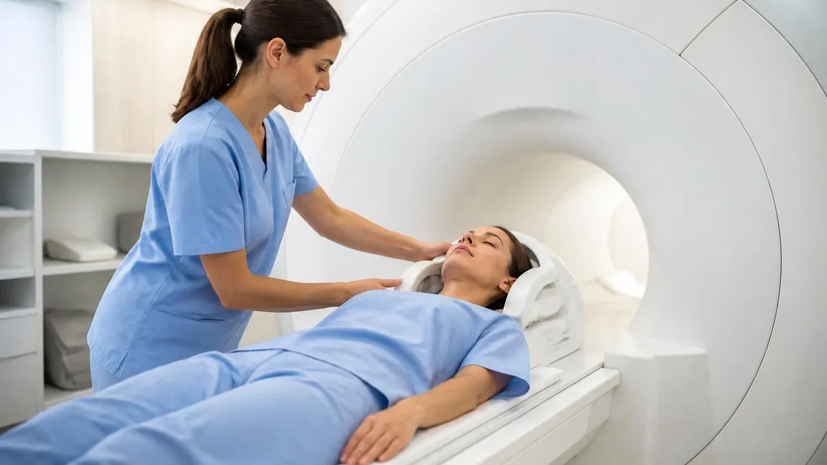
Practical preparation for a pelvic MRI is straightforward but worth getting right. Patients are typically asked to fast for four hours before the study to reduce bowel motion, although hydration with clear liquids is encouraged. A moderately full bladder helps stabilize the uterus and improves visualization of the bladder wall, but an overly distended bladder can introduce motion artifact and patient discomfort during a 45-minute scan.
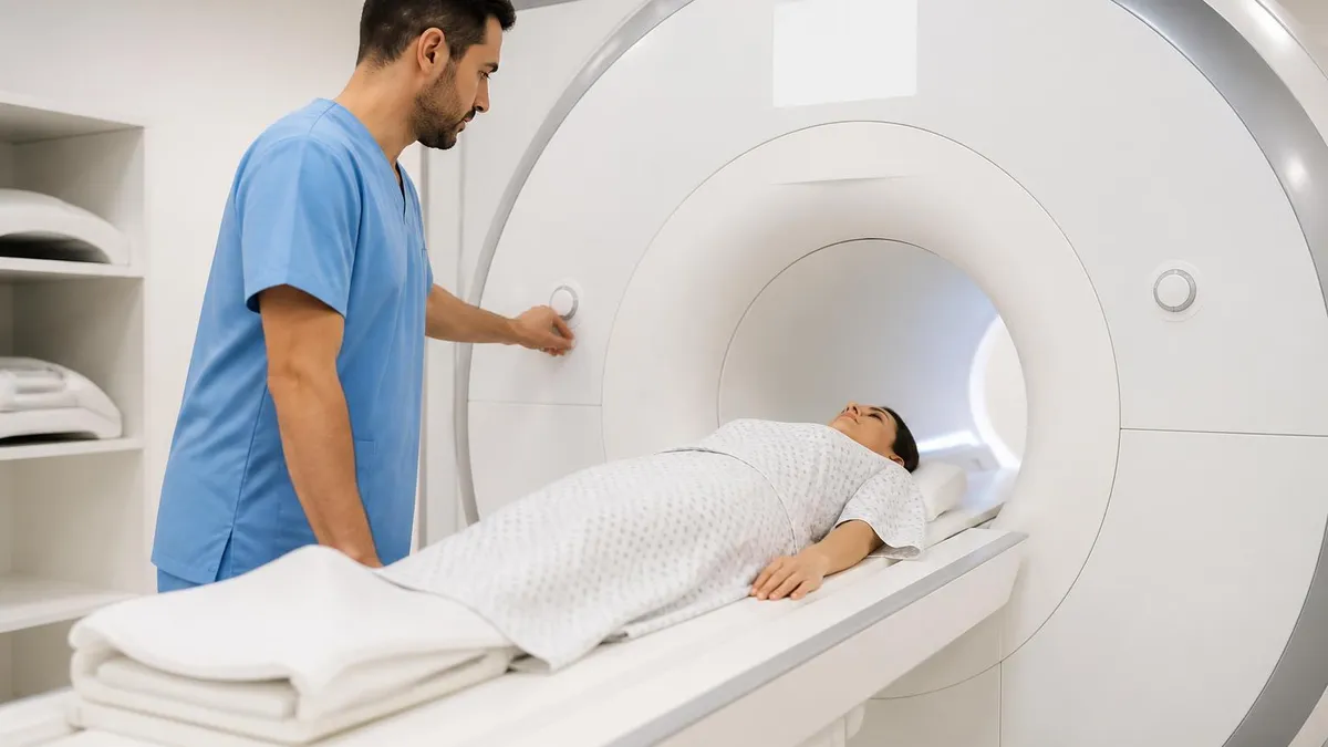
The technologist will position the patient supine with a pelvic phased-array coil draped over the lower abdomen. Many protocols include an antiperistaltic agent such as glucagon or hyoscine butylbromide given intramuscularly or intravenously to reduce bowel peristalsis artifact on T2 sequences. Breath-hold instructions are usually limited to the post-contrast dynamic acquisitions, which are short and well tolerated even by anxious patients.
Claustrophobia is the most common reason patients struggle with pelvic MRI. Wide-bore scanners, prone positioning when tolerated, eye masks, music, and oral anxiolytics prescribed by the referring clinician all help. Open MRI systems with lower field strength can be used when claustrophobia is severe, but image quality for fibroid mapping is meaningfully better at 1.5 or 3 Tesla closed-bore systems, and most centers prefer to accommodate the patient on a higher-field scanner.
Contrast administration deserves a clear conversation. Gadolinium-based contrast agents are generally safe with current macrocyclic formulations, and the older concerns about nephrogenic systemic fibrosis are largely confined to patients with severely impaired renal function. Pregnant patients do not receive gadolinium unless absolutely necessary, which is rarely the case for fibroid imaging because pregnancy itself usually defers elective evaluation.
Reading the radiology report is easier when patients know what to look for. The impression should state the number, location by FIGO type, dominant size, and any features of concern. Look for explicit mention of the endometrial cavity, the junctional zone, the ovaries, and the ureters. If your report is silent on adenomyosis or on the cavity in a patient with heavy bleeding, ask your gynecologist to clarify before proceeding with treatment.
Cost and access vary widely. In the United States, a pelvic MRI with and without contrast typically ranges from 1,000 to 3,500 dollars before insurance, depending on facility type and geography. Hospital outpatient departments tend to bill higher than freestanding imaging centers. Insurance coverage is generally reliable when the study is ordered for symptomatic fibroids and prior ultrasound has been performed, though prior authorization is common.
Finally, the value of a uterine fibroid on MRI is measured by how much it changes management. Studies repeatedly show that pre-treatment MRI alters the recommended therapy in roughly 20 to 30 percent of patients compared with ultrasound alone, most often by identifying coexistent adenomyosis, uncovering additional fibroids, or reclassifying a presumed fibroid as another pelvic mass. That diagnostic clarity is why MRI has become the standard of care before any uterine-sparing intervention.
MRI Questions and Answers
About the Author
Medical Laboratory Scientist & Clinical Certification Expert
Johns Hopkins UniversityDr. Sandra Kim holds a PhD in Clinical Laboratory Science from Johns Hopkins University and is certified as a Medical Technologist (MT) and Medical Laboratory Scientist (MLS) through ASCP. With 16 years of clinical laboratory experience spanning hematology, microbiology, and molecular diagnostics, she prepares candidates for ASCP board exams, MLT, MLS, and specialist certification tests.