Can an Aneurysm Be Seen on MRI? Detection Capabilities, Sensitivity, and Limitations Explained
Can aneurysm be seen on MRI? Learn how MRI and MRA detect brain, aortic, and peripheral aneurysms, sensitivity rates, contrast use, and key limitations.
The question can aneurysm be seen on MRI comes up constantly in neurology clinics, emergency departments, and primary care offices, and the short answer is yes, magnetic resonance imaging is one of the most powerful tools available for detecting aneurysms throughout the body. Whether the concern involves a cerebral aneurysm pressing on a cranial nerve, an aortic aneurysm expanding silently in the chest, or a peripheral aneurysm in the popliteal artery, MRI combined with magnetic resonance angiography (MRA) can identify, measure, and characterize these vascular lesions with remarkable accuracy.
MRI works without ionizing radiation, which makes it an attractive option for younger patients, pregnant women in their second and third trimesters, and individuals who need repeated surveillance imaging over many years. To understand why MRI excels at vascular imaging, it helps to review how does an MRI work at a fundamental level: powerful magnetic fields align hydrogen protons in tissue, radiofrequency pulses excite them, and the resulting signal differences between flowing blood and stationary vessel walls produce stunning anatomical detail.
For aneurysm detection specifically, radiologists rely on specialized sequences such as time-of-flight (TOF) MRA, phase-contrast MRA, and contrast-enhanced MRA. Each technique exploits the physics of flowing blood in a slightly different way. Time-of-flight imaging captures bright signal from inflowing unsaturated spins, while contrast-enhanced studies use gadolinium to highlight the vessel lumen against suppressed background tissue. Together these sequences allow detection of aneurysms as small as 2 to 3 millimeters in many cases.
Sensitivity for intracranial aneurysms larger than 5 millimeters approaches 95 percent on 3T MRA, and even 1.5T scanners reliably identify lesions in the 3 to 5 millimeter range. Below that threshold, sensitivity drops, and very small aneurysms near the skull base or adjacent to bone may require digital subtraction angiography for definitive evaluation. Still, MRI remains the first-line noninvasive screening tool for patients with a family history of subarachnoid hemorrhage, polycystic kidney disease, or connective tissue disorders.
Aortic aneurysms present a different challenge because the vessel is large, mobile with respiration, and surrounded by other moving structures. ECG-gated MRA and breath-hold sequences solve most of these problems, producing measurements that agree with CT angiography within 1 to 2 millimeters. For thoracic aortic aneurysms, MRI is often preferred for long-term surveillance because patients may need imaging every 6 to 12 months for decades, and avoiding cumulative radiation exposure becomes a major priority.
Peripheral aneurysms, including those in the popliteal, femoral, and splenic arteries, are also well visualized on MRA. These lesions often go undetected until they cause symptoms such as limb ischemia, embolic events, or palpable masses. Routine abdominal and lower extremity MRI studies frequently uncover incidental aneurysms, prompting further workup and sometimes elective repair before rupture or thrombosis occurs.
This guide walks through exactly what MRI can and cannot show, which sequences radiologists order, how contrast affects detection, and what patients and clinicians should expect from a typical aneurysm-focused MRI examination. Whether you are studying for a registry exam or trying to interpret a recent radiology report, the information here will help you understand the strengths and limitations of MRI for aneurysm detection.
MRI Aneurysm Detection by the Numbers

Key MRI Sequences Used for Aneurysm Detection
3D TOF MRA is the workhorse for intracranial aneurysm screening, generating bright vascular signal from inflowing unsaturated protons without requiring intravenous contrast. Ideal for arterial circulation evaluation.
Uses gadolinium to opacify the vascular lumen and is preferred for the aorta, neck vessels, and peripheral arteries. Provides high spatial resolution and minimizes flow-related artifacts seen with TOF.
Quantifies flow velocity and direction, useful for distinguishing slow-flow aneurysms from thrombus or characterizing complex hemodynamics in giant or fusiform aneurysms with turbulent flow patterns.
Standard sequences identify mass effect, perianeurysmal edema, and subarachnoid hemorrhage signs. FLAIR is particularly sensitive to small amounts of subarachnoid blood in subacute presentations.
Susceptibility-weighted imaging detects hemosiderin from prior microhemorrhages or sentinel bleeds. Helps identify previously ruptured aneurysms and small cortical microbleeds suggesting amyloid angiopathy.
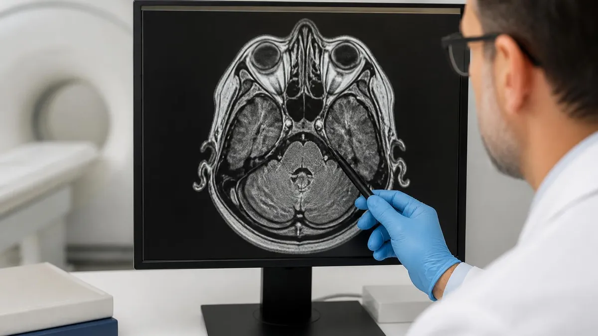
Cerebral aneurysms are the most clinically significant target for MRI evaluation because their rupture causes subarachnoid hemorrhage, a condition with 30 to 40 percent mortality and devastating long-term morbidity for survivors. Most cerebral aneurysms occur at branch points of the circle of Willis, with the anterior communicating artery, posterior communicating artery, and middle cerebral artery bifurcation accounting for roughly 85 percent of all saccular lesions. MRI and MRA can identify these locations reliably, even in patients who present with vague headaches or cranial nerve deficits.
For unruptured aneurysm screening, 3D time-of-flight MRA at 3 tesla has become the standard noninvasive test, with a published sensitivity of 92 to 97 percent for aneurysms 3 millimeters or larger. The technique requires no contrast, making it suitable for patients with renal impairment or gadolinium allergy. Source images and maximum intensity projection reconstructions are reviewed together because small aneurysms can be obscured by overlapping vessels on MIP images alone, a known interpretation pitfall.
When ruptured aneurysm is suspected based on a thunderclap headache, FLAIR sequences detect subarachnoid blood in the basal cisterns and sulci with sensitivity approaching that of non-contrast CT in the subacute phase. Beyond 48 to 72 hours, MRI actually outperforms CT for detecting prior subarachnoid hemorrhage because hemosiderin and methemoglobin remain visible on susceptibility-weighted imaging for weeks. This makes MRI invaluable in patients presenting late after a sentinel bleed.
Comparing MRI to other modalities is essential for understanding its role, and the differences between whats difference between mri and ct scan become particularly important in acute aneurysm presentations. CT angiography offers faster acquisition and is typically preferred in unstable patients, while MRI provides superior soft tissue characterization and avoids radiation in younger individuals undergoing repeated surveillance.
Giant aneurysms, defined as those exceeding 25 millimeters in diameter, present unique imaging features on MRI. They often contain mural thrombus that appears as concentric layers of varying signal intensity, reflecting blood products at different stages of degradation. The patent lumen shows flow void on T2-weighted imaging or bright signal on TOF MRA, while the thrombosed portion demonstrates intermediate signal with rim enhancement after gadolinium administration.
Mycotic and dissecting aneurysms have additional imaging characteristics that MRI captures particularly well. Mycotic lesions show perivascular inflammation and enhancement, while dissections demonstrate an intimal flap, intramural hematoma, and sometimes a double lumen. T1-weighted fat-suppressed sequences are especially useful for visualizing the crescentic intramural hematoma of arterial dissection in the cervical carotid and vertebral arteries.
Multidisciplinary teams use MRI findings to guide treatment decisions, including whether to observe a small unruptured aneurysm, proceed with endovascular coiling, or recommend surgical clipping. The morphology, location, neck width, and relationship to parent vessels are all evaluable on high-quality MRA, and many centers now plan interventions directly from MRI data without requiring catheter angiography in straightforward cases.
MRI Sensitivity by Aneurysm Location and Size
For intracranial aneurysms, MRI sensitivity varies dramatically with size. Lesions larger than 7 millimeters are detected with 98 percent sensitivity on 3T TOF MRA, while those between 3 and 5 millimeters show approximately 85 to 90 percent sensitivity. Below 3 millimeters, detection drops to 70 percent or lower, particularly for aneurysms adjacent to the cavernous sinus or skull base where susceptibility artifact degrades image quality.
Specificity is generally high at 95 percent or better, but false positives can occur from infundibular dilations at vessel origins, vascular loops mimicking saccular outpouchings, and motion artifact creating pseudoaneurysmal contours. Reading source images alongside MIPs and reviewing prior comparison studies dramatically reduces interpretive error rates in busy clinical practice.
MRI vs CT for Aneurysm Detection: Strengths and Weaknesses
- +No ionizing radiation, ideal for surveillance and younger patients
- +Superior soft tissue characterization including mural thrombus
- +Non-contrast TOF MRA option for patients with renal disease
- +Better visualization of associated edema and parenchymal changes
- +Detects chronic subarachnoid hemorrhage longer than CT
- +Can quantify flow velocity and direction with phase-contrast techniques
- −Longer acquisition time, typically 30 to 45 minutes per exam
- −Contraindicated with many cardiac devices and certain implants
- −Lower spatial resolution than digital subtraction angiography
- −Claustrophobic patients may not tolerate the exam
- −Motion artifact more problematic than with rapid CT acquisition
- −Less available in emergency settings than CT scanners
Patient Preparation Checklist for Aneurysm MRI
- ✓Screen thoroughly for ferromagnetic implants, pacemakers, and aneurysm clips placed before 1995
- ✓Verify renal function with recent eGFR before administering gadolinium contrast
- ✓Remove all metallic objects, jewelry, hairpins, and clothing with metal fasteners
- ✓Review prior imaging studies for direct comparison and measurement consistency
- ✓Confirm ability to lie flat and motionless for 30 to 45 minutes
- ✓Establish IV access in the antecubital fossa if contrast is planned
- ✓Discuss claustrophobia history and consider mild oral anxiolytic if needed
- ✓Document allergies to gadolinium-based contrast agents specifically
- ✓Provide hearing protection and explain the loud knocking noises in advance
- ✓Confirm pregnancy status in female patients of reproductive age
- ✓Hold metformin per institutional protocol if contrast is planned
- ✓Brief the patient on breath-holding instructions for aortic MRA sequences
Negative MRA does not always rule out aneurysm
While MRA has excellent sensitivity for aneurysms larger than 5 millimeters, very small lesions, blister aneurysms, and dissecting aneurysms at the skull base can be missed even on high-quality 3T studies. In patients with high pretest probability such as thunderclap headache with negative CT and lumbar puncture, digital subtraction angiography remains the gold standard. A negative MRA should be interpreted in the clinical context, not as definitive exclusion.
Despite its remarkable capabilities, MRI has well-documented limitations that radiologists, ordering clinicians, and patients should understand. The most important limitation is the inability to detect very small aneurysms, particularly those below 3 millimeters in diameter. These tiny lesions may be obscured by surrounding venous structures on TOF MRA, lost in the noise of source images, or hidden behind susceptibility artifact from adjacent bone. For patients with strong clinical suspicion despite a negative MRA, catheter angiography remains the definitive test.
Susceptibility artifact poses a particular challenge near the skull base, where the temporal bone, sphenoid sinus, and paranasal air create magnetic field inhomogeneities that distort MRA images. Aneurysms of the cavernous segment of the internal carotid artery, ophthalmic segment, and posterior communicating origin can be especially difficult to evaluate. Modern protocols using parallel imaging and shorter echo times help mitigate these artifacts but do not eliminate them entirely.
Motion artifact remains a persistent problem in patients who cannot lie still, are confused, or are in pain. Even small movements during a 5 to 7 minute MRA acquisition can blur vessels and create pseudoaneurysmal contours. Pediatric patients and those with dementia often require sedation, which adds cost, complexity, and risk to what would otherwise be a straightforward outpatient examination.
Contraindications limit MRI availability for a significant fraction of patients. Older pacemakers, cochlear implants, certain neurostimulators, and pre-1995 cerebral aneurysm clips made of ferromagnetic material remain absolute contraindications at most institutions. MRI-conditional devices have largely solved this problem for newly implanted hardware, but legacy devices in older patients still require careful screening and sometimes alternative imaging.
Gadolinium-based contrast agents, while generally safe, carry the small risk of nephrogenic systemic fibrosis in patients with severe renal impairment and a very rare risk of acute allergic reactions. Newer macrocyclic agents have markedly reduced these concerns, but patients with eGFR below 30 mL/min/1.73m² still require careful consideration before contrast administration. Non-contrast TOF MRA serves as an excellent alternative for intracranial vascular assessment in this population.
Interpretation pitfalls abound and include infundibular dilations mistaken for true aneurysms, vascular loops simulating saccular outpouchings, and slow-flow regions creating apparent filling defects. Experience matters substantially, and aneurysm detection accuracy improves significantly when studies are read by neuroradiologists with vascular subspecialty training rather than general radiologists working without dedicated workflow optimization for vascular imaging cases.
Finally, MRI cannot reliably predict rupture risk on a single examination. Size, location, morphology, and growth over time all contribute to risk stratification, but no imaging feature definitively predicts which aneurysm will rupture. The PHASES and UIATS scoring systems incorporate clinical and imaging factors to guide management decisions, but the inherent uncertainty means that shared decision-making with neurosurgeons, interventional neuroradiologists, and patients remains essential for every newly diagnosed unruptured aneurysm.
In the hyperacute phase of suspected subarachnoid hemorrhage, typically within 6 hours of headache onset, non-contrast CT remains the first-line imaging test with sensitivity exceeding 99 percent. MRI should not delay CT in unstable patients with thunderclap headache. After CT confirms or excludes acute blood, MRA or CTA can characterize the underlying aneurysm.
Clinical decision-making around aneurysm imaging requires balancing pretest probability, urgency, patient factors, and institutional resources. For asymptomatic screening in high-risk populations, MRA is generally preferred because of its excellent safety profile and absence of radiation. Patients with two or more first-degree relatives affected by intracranial aneurysm, autosomal dominant polycystic kidney disease, or specific connective tissue disorders benefit most from periodic noninvasive screening starting in their thirties or forties.
For acute presentations suggestive of ruptured aneurysm, CT and CTA remain the workhorses because of speed, availability in emergency departments, and high sensitivity in the first 24 hours after hemorrhage. MRI plays a complementary role for subacute presentations, recurrent symptoms after negative CT workup, and characterization of complex pathology such as giant or partially thrombosed aneurysms requiring detailed treatment planning.
Surveillance of known unruptured aneurysms is one of the most common indications for serial MRA. Small stable aneurysms below 5 millimeters in patients without high-risk features are typically imaged every 6 to 12 months initially, transitioning to annual or biennial intervals once stability is established. Any growth of 1 millimeter or more between studies typically prompts referral to neurovascular specialists for treatment discussion, including endovascular coiling or surgical clipping.
Post-treatment follow-up imaging is another major application. After endovascular coiling, MRA effectively detects residual neck filling, recurrence, and coil compaction without the streak artifact that limits CT angiography around metal coils. Time-of-flight sequences may underestimate residual flow due to dephasing near coil masses, so contrast-enhanced MRA is often preferred for post-coiling surveillance at 6 months, 1 year, and then annually depending on initial obliteration grade.
For surgically clipped aneurysms, MRI compatibility depends on clip material. Modern titanium and MP35N clips are MRI-conditional at 1.5T and 3T, allowing safe imaging within hours of surgery. However, local susceptibility artifact from clips obscures the immediate parent vessel and can mimic residual aneurysm. CT angiography or digital subtraction angiography may be needed to definitively evaluate clip placement and adjacent vasculature in the early postoperative period.
Decision-making is enhanced by reviewing the broader context of how MRI fits into modern imaging workflows, and understanding mri with or without contrast helps clinicians order the most appropriate study. For pure vascular questions, contrast typically improves sensitivity and specificity for aneurysms larger than 3 millimeters, while non-contrast TOF remains adequate for routine intracranial screening.
Ultimately, clinical questions drive protocol selection. A patient with suspected internal carotid dissection needs T1 fat-suppressed sequences in addition to MRA. A young woman with possible reversible cerebral vasoconstriction syndrome needs sequential MRA to document vasospasm resolution. A retiree under surveillance for a 4 millimeter middle cerebral artery aneurysm needs only repeat 3T TOF MRA. Tailoring the examination to the clinical question maximizes diagnostic yield while controlling costs and patient burden.
Practical preparation for an aneurysm-focused MRI starts long before the patient enters the scanner. Technologists and radiologists should review the clinical history, prior imaging, and specific question being asked. A patient referred for suspected unruptured aneurysm screening needs a different protocol than one being evaluated for postoperative clip residual or post-coiling follow-up. Standardized order sets within electronic health records help ensure the right sequences are obtained for each indication, reducing repeat examinations and missed findings.
Patient communication makes an enormous difference in image quality. Explaining the importance of stillness, demonstrating the breath-hold for aortic MRA, and offering reassurance about the noise and confined space reduces motion artifact dramatically. For pediatric patients, child-friendly scanners with cartoon themes, certified child life specialists, and parental presence in the room often eliminate the need for sedation in children old enough to follow instructions.
Coil selection matters more than many realize. Modern multichannel head and neck coils dramatically improve signal-to-noise ratio and enable parallel imaging that reduces scan time without sacrificing resolution. For aortic imaging, dedicated cardiac and torso coils with phased-array elements optimize coverage of the chest and abdomen. Ensuring the patient is centered properly in the coil and at the magnet isocenter is a small detail that significantly affects image quality.
Contrast timing for CE-MRA is critical and best handled with bolus tracking software that monitors arterial enhancement in real time and triggers acquisition at peak opacification. Test bolus methods using 1 to 2 milliliters of gadolinium provide an alternative approach for predicting transit time. Either way, the goal is arterial-phase imaging without venous contamination, which requires careful coordination between technologist, scanner software, and contrast injector.
Post-processing turns raw data into diagnostic images. Maximum intensity projections, multiplanar reformats, and volume-rendered images each contribute different perspectives on aneurysm morphology. Reviewing source images alongside reconstructions catches small lesions that might be obscured on MIPs by overlapping vessels or normal anatomic variation. Modern PACS workstations integrate these views and allow rapid measurements of aneurysm size, neck width, and dome-to-neck ratio.
Reporting standards continue to evolve. Structured reports for aneurysm imaging include location, size in three dimensions, morphology, neck characteristics, relationship to parent and branch vessels, presence of mural thrombus or calcification, and comparison with prior studies. Many centers now use anatomic templates and dictation macros to ensure consistent documentation, which improves both clinical care and research data quality across institutions.
Finally, ongoing education matters for everyone involved in aneurysm imaging. New sequences, higher field strengths, and emerging techniques like 4D flow MRI continue to expand what we can see and quantify. Technologists, radiologists, and ordering clinicians who stay current with the literature deliver better care to patients with cerebrovascular disease, and the field continues to advance toward earlier detection, better risk stratification, and improved outcomes for the millions of people living with known or suspected aneurysms.
MRI Questions and Answers
About the Author
Medical Laboratory Scientist & Clinical Certification Expert
Johns Hopkins UniversityDr. Sandra Kim holds a PhD in Clinical Laboratory Science from Johns Hopkins University and is certified as a Medical Technologist (MT) and Medical Laboratory Scientist (MLS) through ASCP. With 16 years of clinical laboratory experience spanning hematology, microbiology, and molecular diagnostics, she prepares candidates for ASCP board exams, MLT, MLS, and specialist certification tests.