MRI Breast Scan: The Complete 2026 July Guide to Breast MRI Imaging, Screening, and What Patients Should Expect
MRI breast scan explained: how breast MRI works, who needs it, preparation, contrast, costs, results, and what radiologists look for in 2026 July. 💯
An MRI breast scan is one of the most sensitive imaging tools available in modern medicine, capable of detecting cancers and tissue abnormalities that mammography and ultrasound can miss. Unlike X-ray-based imaging, breast MRI uses a powerful magnetic field, radiofrequency pulses, and a gadolinium-based contrast agent to produce detailed cross-sectional images of breast tissue, blood vessels, and lymph nodes. For high-risk patients, dense breast tissue, or post-surgical evaluation, an mri breast scan often becomes the deciding study in a complex diagnostic workup.
The procedure has expanded dramatically since the American Cancer Society first recommended supplemental MRI screening for women with greater than 20% lifetime breast cancer risk. In 2026, roughly 1.4 million breast MRI exams are performed annually in the United States alone, driven by genetic testing, denser screening guidelines, and growing demand from patients informed about their personal risk profile. Radiologists, MRI technologists, and ordering clinicians all need a working understanding of the modality.
Breast MRI is performed on either a 1.5T or 3.0T scanner using a dedicated bilateral breast coil. The patient lies prone with both breasts suspended into openings in the coil, allowing the tissue to hang freely away from the chest wall. This positioning minimizes motion, improves fat suppression quality, and provides the spatial resolution necessary to visualize sub-centimeter lesions. The exam takes 30–45 minutes and produces hundreds of high-resolution slices.
What makes breast MRI uniquely powerful is its ability to assess tissue physiology in addition to anatomy. After gadolinium contrast injection, malignant lesions typically demonstrate rapid early enhancement followed by washout, a kinetic pattern that helps radiologists differentiate cancer from benign findings such as fibroadenomas or cysts. Diffusion-weighted imaging (DWI) and apparent diffusion coefficient (ADC) mapping further refine specificity, reducing false positives that historically plagued early breast MRI protocols.
However, breast MRI is not a replacement for mammography. It is a supplemental study with its own indications, contraindications, and limitations. The exam is more expensive, requires intravenous contrast, and has a higher recall rate than mammography. Understanding when breast MRI is appropriate, how to prepare for it, and how results are interpreted is essential for anyone navigating a high-risk screening pathway, a new diagnosis, or a complex post-treatment surveillance plan.
This comprehensive guide walks through every aspect of the mri breast scan: how the machine and protocol work, who qualifies for screening MRI, what to expect on exam day, how contrast is used, what BI-RADS categories mean, typical costs and insurance coverage, and the most common questions patients ask their radiologists. Whether you are a patient scheduled for your first scan, a technologist preparing for the registry exam, or a clinician brushing up on indications, this article delivers the depth you need.
We will also examine 2026-specific developments, including abbreviated breast MRI (FAST MRI) protocols, AI-assisted lesion detection, contrast-enhanced mammography as an alternative, and updated supplemental screening recommendations from the American College of Radiology. Each section is grounded in current clinical practice and designed to translate dense radiology terminology into language a general reader, student, or patient can act on with confidence.
Breast MRI by the Numbers in 2026
Who Qualifies for a Breast MRI Screening?
Women with a calculated lifetime breast cancer risk of 20% or greater based on family history models like Tyrer-Cuzick or BRCAPRO qualify for annual screening MRI alongside mammography, typically starting at age 25–30.
Carriers of BRCA1, BRCA2, TP53, PALB2, CDH1, PTEN, and similar high-penetrance mutations are recommended for annual breast MRI starting at age 25, regardless of mammographic density or symptoms.
Patients who received mantle-field or chest radiation between ages 10–30 — typically for Hodgkin lymphoma — qualify for annual screening MRI starting 8 years after treatment or by age 25, whichever is later.
Breast MRI is often ordered at diagnosis to evaluate the extent of disease, search for multifocal or contralateral cancer, and inform surgical planning, particularly when considering breast-conserving therapy or oncoplastic reconstruction.
MRI is used when mammography and ultrasound findings are inconclusive, when there is suspicious nipple discharge with negative conventional imaging, or to evaluate implant integrity in patients with silicone implants.
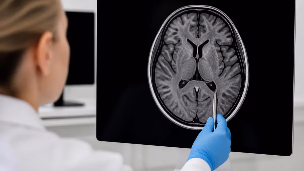
A breast MRI scan begins with the physics that make all MRI imaging possible: hydrogen protons in the body align with a strong external magnetic field, are excited by radiofrequency pulses, and then release detectable signal as they relax back toward equilibrium. In breast imaging, the scanner harvests this signal from fat, glandular tissue, water, and contrast-enhanced vessels to construct images with extraordinary soft-tissue contrast that no other modality can match for breast pathology.
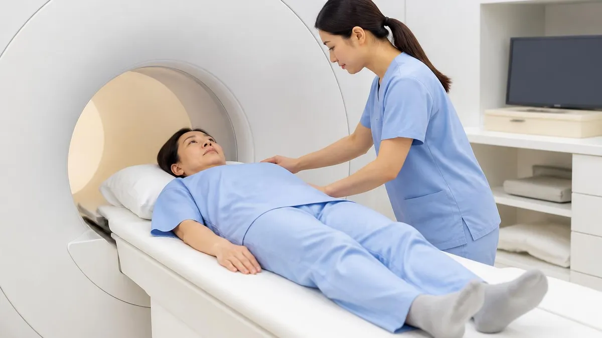
The patient is positioned prone on a specialized table with a dedicated bilateral breast coil. The breasts hang into two openings, suspended without compression, while the patient's arms are typically placed overhead or alongside the body to optimize coil sensitivity. Proper positioning is critical: poor centering causes wrap-around artifacts, while skin folds or nipple inversion mimic lesions. Most facilities use cushions and immobilizers to keep the patient comfortable for the full 30–45-minute exam.
Once positioning is confirmed, the technologist places an intravenous line — typically a 20- or 22-gauge catheter — for gadolinium-based contrast administration. The IV is connected to a power injector calibrated to deliver contrast at 2 mL/second, usually 0.1 mmol/kg of body weight, followed by a 20–30 mL saline flush. Timing of contrast delivery relative to imaging is choreographed precisely because the dynamic enhancement curve is the cornerstone of malignancy assessment.
The scan itself follows a standardized protocol. After localizer images orient the scanner to the anatomy, a T2-weighted sequence highlights fluid and edema, useful for identifying cysts, lymph nodes, and ductal dilatation. Then a series of T1-weighted fat-saturated dynamic sequences are acquired — one before contrast and at least three to five after contrast — at intervals of roughly 60–90 seconds. Diffusion-weighted imaging and post-contrast subtraction images round out the protocol.
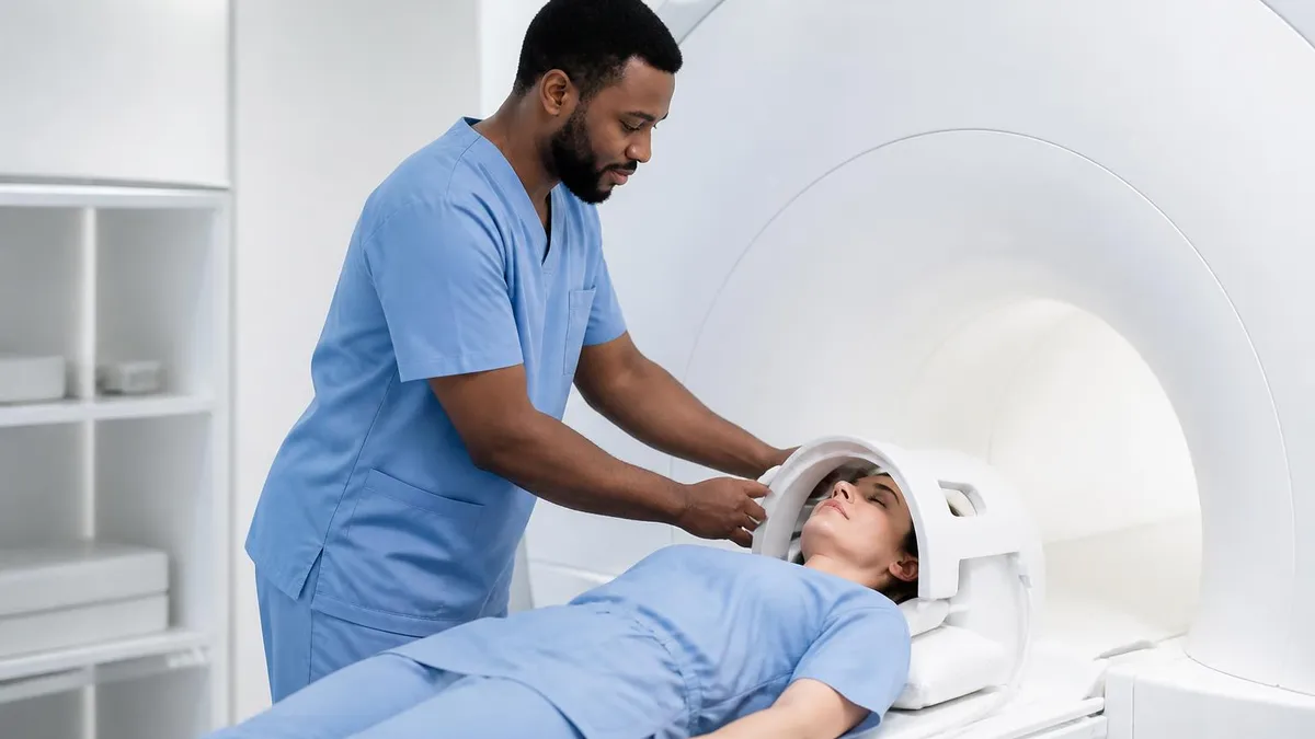
While inside the bore, patients hear loud knocking, humming, and buzzing as the gradient coils rapidly switch on and off. These sounds, which can exceed 110 decibels in some sequences, are completely normal and a fundamental part of MRI physics. Hearing protection is mandatory, and many centers provide patient-controlled music to reduce anxiety. A squeeze ball and intercom allow communication with the technologist throughout the exam.
Breast MRI is sensitive but not specific — meaning it detects almost every cancer but also picks up many benign findings. To improve specificity, modern protocols incorporate diffusion-weighted imaging, which measures the random motion of water molecules. Malignant tissue, with its dense cellularity, restricts diffusion and appears bright on DWI with low ADC values. Benign lesions typically show free diffusion. Combining morphology, kinetics, and DWI yields diagnostic accuracy approaching 95%.
If you've never sat inside an MRI scanner before, knowing about the loud knocking, claustrophobia mitigation strategies, and breathing technique can transform the experience. For a deeper look at scanner acoustics and why they sound the way they do, see our explainer on the mri machine noise environment. Patients who understand the physics tend to tolerate the exam significantly better than those who walk in cold.
MRI Practice Test Questions
Prepare for the MRI - Magnetic Resonance Imaging exam with our free practice test modules. Each quiz covers key topics to help you pass on your first try.
MRI Knowledge
MRI Exam Questions covering Knowledge. Master MRI Test concepts for certification prep.
MRI Physics
Free MRI Practice Test featuring Physics. Improve your MRI Exam score with mock test prep.
MRI Anatomy and Pathology
MRI Test Prep for MRI Anatomy and Pathology. Practice MRI Quiz questions and boost your score.
MRI Anatomy and Positioning
MRI Questions and Answers on MRI Anatomy and Positioning. Free MRI practice for exam readiness.
MRI Contrast Agents
Free MRI Quiz on MRI Contrast Agents. MRI Exam prep questions with detailed explanations.
MRI Patient Care and Positioning
MRI Practice Questions for MRI Patient Care and Positioning. Build confidence for your MRI certification exam.
Breast MRI Protocol Sequences Explained
The dynamic contrast-enhanced (DCE) T1-weighted fat-saturated sequence is the backbone of every breast MRI. One pre-contrast and multiple post-contrast acquisitions are obtained roughly every 60–90 seconds, creating a time-intensity curve for every voxel in the breast. Malignant lesions characteristically show rapid initial enhancement reaching peak intensity within 1–2 minutes after injection.
Radiologists analyze the delayed phase of the curve: persistent enhancement suggests benign tissue, plateau is indeterminate, and washout strongly suggests malignancy. Subtraction images — created by digitally subtracting the pre-contrast from post-contrast images — make enhancing lesions pop against the suppressed background. Maximum intensity projections (MIPs) help visualize overall enhancement patterns and vascular distribution across both breasts at a glance.

Pros and Cons of Breast MRI Screening
- +Highest sensitivity of any breast imaging modality, detecting up to 94% of invasive cancers in dense breast tissue
- +Detects multifocal, multicentric, and contralateral disease often missed on mammography or ultrasound
- +No ionizing radiation — safe for repeat annual screening in young high-risk patients
- +Excellent for evaluating silicone implant integrity and rupture without compression
- +Distinguishes post-surgical scar tissue from recurrent tumor more reliably than other modalities
- +Diffusion-weighted imaging adds physiologic data without additional contrast exposure
- −Higher recall and biopsy rate due to lower specificity compared to mammography
- −Requires intravenous gadolinium contrast, which carries small risk of allergic reaction or NSF
- −Significantly more expensive — often 5–10× the cost of a screening mammogram
- −Long scan time and prone positioning can be uncomfortable for patients with claustrophobia
- −Many implanted devices, including some pacemakers, may be contraindications
- −Limited availability in rural and underserved areas due to scanner and breast-coil requirements
How to Prepare for Your MRI Breast Scan
- ✓Schedule your exam between days 7–14 of your menstrual cycle to minimize hormonal background enhancement
- ✓Inform the scheduler about pregnancy, breastfeeding, or possible pregnancy before the exam
- ✓List all implants, devices, surgical clips, and tattoos for the MRI safety questionnaire
- ✓Stop nursing decisions: most guidelines now permit continued breastfeeding after gadolinium
- ✓Wear a two-piece outfit with no metal — you will change into a gown but valuables stay in a locker
- ✓Remove jewelry, hearing aids, dentures, hairpins, and any transdermal medication patches
- ✓Arrive 30 minutes early to complete the safety screening form and have your IV placed
- ✓Stay well-hydrated the day before and morning of your scan to ease contrast clearance
- ✓Bring prior imaging on CD if performed at a different facility — comparison studies prevent recalls
- ✓Tell the technologist immediately if you feel anxious so they can pause or coach you through it
Schedule between cycle days 7–14 for clearer images
Hormonal fluctuations cause normal breast tissue to enhance after gadolinium, which can mimic or mask cancer. Scanning during the second week of the menstrual cycle minimizes this background parenchymal enhancement. For postmenopausal women on hormone therapy, your radiologist may recommend pausing HRT for 4–6 weeks before the exam to improve image clarity and reduce false-positive findings.
When your radiologist interprets your mri breast scan, they follow the standardized Breast Imaging Reporting and Data System (BI-RADS) lexicon developed by the American College of Radiology. BI-RADS assigns every finding a category from 0 through 6, communicating the level of concern and the recommended next step. Understanding these categories is essential because they directly determine whether you need additional imaging, short-interval follow-up, or biopsy.
A BI-RADS 1 assessment means the study is completely negative — no masses, no abnormal enhancement, no suspicious findings. BI-RADS 2 indicates benign findings such as cysts, fibroadenomas with classic appearance, fat-containing lesions, or post-surgical scarring with no concerning features. Both categories return the patient to routine annual screening. The vast majority of high-risk screening MRIs land in one of these two reassuring categories.
BI-RADS 3 is reserved for probably benign findings with less than 2% likelihood of malignancy. These typically require short-interval follow-up MRI at six months to confirm stability. Common BI-RADS 3 findings include small foci of enhancement, non-mass enhancement with benign kinetics, or new but morphologically reassuring lesions. If stability is demonstrated over two to three years, the finding can be downgraded to BI-RADS 2.
BI-RADS 4 indicates suspicious findings warranting tissue diagnosis, subdivided into 4A (low suspicion, 2–10%), 4B (moderate, 10–50%), and 4C (high, 50–95%). BI-RADS 5 represents lesions with greater than 95% probability of malignancy — usually a mass with irregular shape, spiculated margins, washout kinetics, and restricted diffusion. Both BI-RADS 4 and 5 require biopsy, often performed under MRI guidance if the lesion is occult on mammogram and ultrasound.
BI-RADS 0 is a placeholder used when additional imaging is needed before a final assessment can be rendered. On MRI, this might mean the radiologist needs prior comparison studies, additional sequences, or a targeted ultrasound to evaluate a finding. BI-RADS 6 is reserved for biopsy-proven malignancies undergoing pre-treatment imaging to plan surgery, neoadjuvant chemotherapy, or radiation therapy.
Beyond the category, your report describes the breast composition (fibroglandular versus fatty), background parenchymal enhancement (minimal, mild, moderate, or marked), and any focal findings using a specific morphology lexicon. Masses are characterized by shape (oval, round, irregular), margin (circumscribed, irregular, spiculated), and internal enhancement pattern. Non-mass enhancement is described by distribution (focal, linear, segmental, regional, multiregional, diffuse) and internal pattern.
Kinetic curve analysis remains a central pillar of interpretation. The initial phase — within the first two minutes — is classified as slow, medium, or rapid. The delayed phase is described as persistent (continued enhancement), plateau (steady), or washout (decreasing signal). The most concerning combination is rapid initial uptake followed by washout, which carries malignancy probabilities exceeding 80% when combined with a morphologically suspicious mass.
Most patients tolerate gadolinium contrast without issue, but reactions can occur. Serious allergic reactions are rare (1 in 10,000), and nephrogenic systemic fibrosis (NSF) is now extremely uncommon with modern macrocyclic agents. Patients with eGFR below 30 mL/min should discuss risks with their radiologist. Always disclose pregnancy, prior contrast reactions, asthma, and severe allergies before your scan.
The cost of an mri breast scan in the United States varies dramatically depending on geography, facility type, insurance, and whether contrast is included. The national average sits around $1,084 in 2026, but the range stretches from roughly $500 at high-volume outpatient imaging centers to $3,500 or more at hospital-based academic radiology departments. Cash-pay patients can often negotiate significant discounts by requesting an itemized self-pay rate before scheduling.
Insurance coverage is generally strong when the exam meets medical-necessity criteria. The Affordable Care Act mandates coverage of screening mammography but not screening breast MRI; however, most commercial insurers cover annual screening MRI for women meeting the 20% lifetime-risk threshold, BRCA carriers, and patients with prior chest radiation. Pre-authorization is typically required, and denial rates remain higher than for mammography, so persistent advocacy with both the ordering provider and insurer pays off.
Diagnostic breast MRI — ordered to evaluate a known lesion, problem-solve mammographic findings, or stage a newly diagnosed cancer — is almost universally covered when the indication is documented. Coverage challenges most often arise around supplemental screening for women with dense breasts who do not otherwise meet high-risk criteria. Several states have passed laws expanding insurance mandates, and the federal landscape continues to evolve in 2026.
Access to high-quality breast MRI also varies. The American College of Radiology Breast MRI Accreditation Program certifies facilities that meet rigorous equipment, personnel, and image-quality standards. Choosing an accredited center significantly improves the odds of getting a high-resolution scan interpreted by a radiologist with subspecialty breast imaging training. Patients in rural areas may need to travel one to two hours to reach an accredited center, particularly for biopsy capability.
Beyond cost and access, the patient experience matters. Look for centers that offer wider-bore 70-cm scanners (more comfortable for claustrophobic or larger-bodied patients), patient-controlled music or video systems, and dedicated breast imaging navigators who help coordinate biopsies, MRI scheduling, and results delivery. Many cancer centers now provide same-day or next-day result review with a breast radiologist or nurse navigator, which dramatically reduces anxiety.
Emerging alternatives are reshaping the access conversation. Abbreviated breast MRI (also called fast breast MRI or FAST MRI) shortens the protocol to roughly 10 minutes by acquiring only one pre-contrast and one early post-contrast T1 sequence. Studies show sensitivity comparable to full-protocol MRI for invasive cancer detection, at substantially lower cost. Several centers now offer cash-pay abbreviated breast MRI for $250–$400 as a screening alternative for women with dense breasts.
Patients seeking a broader understanding of how MRI fits into the wider diagnostic landscape may benefit from reviewing what is mri test coverage, which compares MRI to CT, ultrasound, and X-ray across many clinical scenarios. A well-informed patient asks better questions, advocates more effectively for appropriate imaging, and arrives at the scanner less anxious — all of which translate into a smoother experience and a higher-quality study.
On the day of your mri breast scan, plan to arrive at least 30 minutes early. You will check in, verify insurance, and complete a detailed MRI safety screening questionnaire. The form asks about implanted devices, prior surgeries, metal exposure history (welding, shrapnel, embedded fragments), kidney function, pregnancy status, and allergies. Be thorough — even seemingly minor details like dental work, tattoos, or a 1980s aneurysm clip can change the safety plan.
You will change into a gown and store personal belongings in a locker. Bring nothing metallic into the magnet room: no jewelry, watches, hair clips, underwire bras, eyeglasses, hearing aids, or pocket items. Even credit cards can be demagnetized by the scanner. Many transdermal medication patches contain metal foil and must be removed beforehand. Tell the technologist if you have permanent eyeliner or eyebrow tattoos — they rarely cause issues but should be documented.
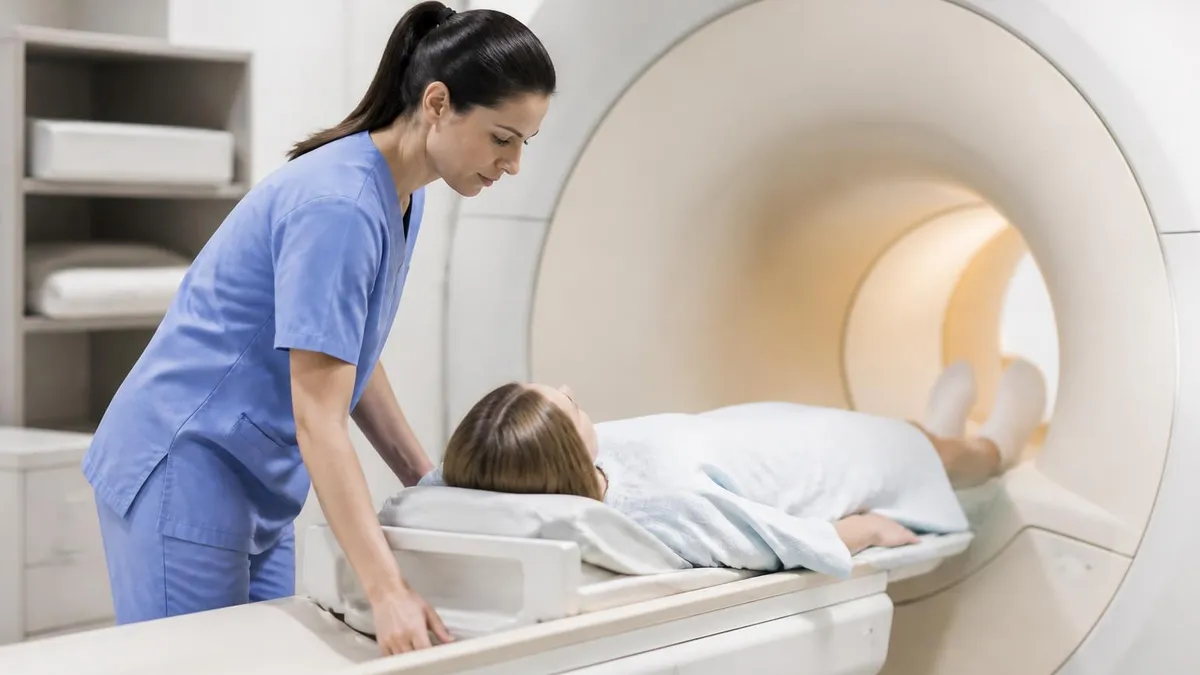
The technologist will start the IV, position you prone on the breast coil, and provide hearing protection — typically earplugs plus headphones. The table slides into the bore, and the exam begins with several minutes of localizers and T2 imaging. You will hear loud rhythmic knocking. Breathe normally and stay perfectly still; even small movements during the dynamic phase blur the images and can necessitate repeating sequences, extending your scan time.
The contrast injection itself usually causes no sensation, though some patients describe a brief cool feeling at the IV site or a metallic taste lasting 10–20 seconds. Mild nausea is uncommon. If you experience any unusual symptoms — itching, hives, difficulty breathing, chest tightness — squeeze the call button immediately. The technologist monitors you visually and via intercom throughout the exam and can stop the scan within seconds if needed.
After the scan, the IV is removed, you change back into your clothes, and you can resume normal activities immediately. Drink extra water for the next 24 hours to help your kidneys clear the gadolinium. There are no restrictions on driving, work, or exercise. If you are breastfeeding, current ACR guidelines confirm it is safe to continue nursing without interruption after a gadolinium dose, though some mothers choose to pump and dump for 24 hours as a personal preference.
Results are typically available within one to three business days. Your report will go to the ordering physician, and many centers also provide direct patient access through their portal. If the radiologist identifies a finding requiring biopsy, expect a phone call rather than a portal release. MRI-guided biopsy is scheduled separately, usually within one to two weeks, and is performed by a breast imaging radiologist using a dedicated biopsy grid attached to the breast coil.
If you have a strong family history but have never been formally assessed for breast cancer risk, ask your primary care provider for a referral to a genetic counselor or use a validated risk calculator such as the Tyrer-Cuzick model. Knowing your number is the single most important step in deciding whether annual screening MRI belongs in your care plan. Many women discover they qualify for supplemental MRI only after a relative is diagnosed — but earlier risk assessment changes outcomes.
MRI Questions and Answers
What Is an MRI Test? How Magnetic Resonance Imaging Scans Diagnose Disease in 2026

MRI Medical Abbreviation: What MRI Stands For and Why It Matters

The History of MRI: From Discovery to Modern Medicine

Knee MRI Images: A Complete Guide to Reading, Understanding, and Interpreting Knee Scans

Noise of MRI Machine: Why MRI Scanners Are So Loud and What to Expect
About the Author
Medical Laboratory Scientist & Clinical Certification Expert
Johns Hopkins UniversityDr. Sandra Kim holds a PhD in Clinical Laboratory Science from Johns Hopkins University and is certified as a Medical Technologist (MT) and Medical Laboratory Scientist (MLS) through ASCP. With 16 years of clinical laboratory experience spanning hematology, microbiology, and molecular diagnostics, she prepares candidates for ASCP board exams, MLT, MLS, and specialist certification tests.
Join the Discussion
Connect with other students preparing for this exam. Share tips, ask questions, and get advice from people who have been there.
View discussion (6 replies)