
MRI C-Spine: What Cervical Spine MRI Shows, How It Works, and Why It's Ordered
MRI c-spine guide: 🏆 what cervical spine MRI shows, how the scan works, why doctors order it, preparation, results interpretation, and what to expect.
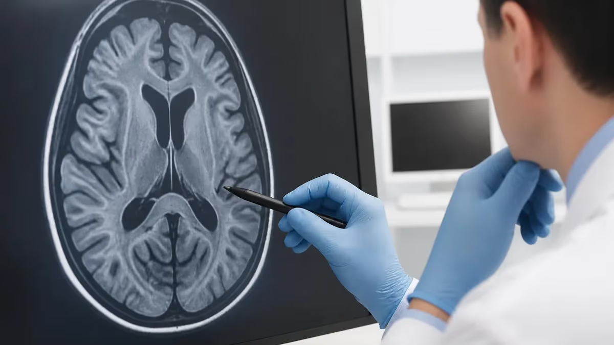
MRI of the cervical spine — often called an MRI c-spine — is the gold standard imaging study for diagnosing neck pain, radiating arm symptoms, and a range of conditions affecting the upper spine. Unlike X-rays which primarily show bone, an MRI shows soft tissues with remarkable detail: discs, nerves, spinal cord, ligaments, muscles, and fluid. When your doctor orders an MRI c-spine, they're looking for problems X-rays simply can't reveal.
This guide walks through what a cervical spine MRI actually shows, how the scan is performed, why it's ordered, what to expect during the appointment, and how to read your radiology report. We'll cover common findings like disc herniations, stenosis, and cord compression, as well as less common but important findings. Whether you're scheduled for an MRI c-spine or trying to understand a recent report, this guide gives you the foundation to engage with your healthcare team meaningfully.
Quick Overview
An MRI of the cervical spine captures detailed images of the seven cervical vertebrae (C1 through C7), the intervertebral discs between them, the spinal cord, nerve roots exiting the spine, surrounding ligaments and muscles, and the cerebrospinal fluid around the cord. The level of detail allows diagnosis of conditions ranging from minor disc bulges to spinal cord compression requiring urgent intervention.
Why Doctors Order MRI C-Spine
Neck pain that hasn't responded to conservative treatment over 4-6 weeks, especially when associated with arm symptoms or neurological signs.
Pain, numbness, tingling, or weakness in the arms — symptoms suggesting nerve root compression from a cervical disc or other spine problem.
After significant neck injury when X-rays are inconclusive or when clinical findings suggest soft tissue, disc, or cord injury.
Understanding what the cervical spine looks like helps make sense of MRI findings. Your neck contains seven cervical vertebrae stacked on top of each other, with intervertebral discs sandwiched between them. These discs act as shock absorbers and allow the neck to bend, twist, and extend. The spinal cord runs through the center of these vertebrae in a protective canal. At each level, pairs of spinal nerves exit through openings (foramina) on either side of the spine, traveling to your arms and other parts of your body.
The seven cervical vertebrae are numbered C1 through C7, with C1 (the atlas) at the top supporting the skull and C7 at the bottom transitioning to the thoracic spine. The discs are named by the vertebrae they sit between — the C5-C6 disc sits between vertebrae five and six. When you read an MRI report, levels are described by these numbers: 'C5-C6 disc herniation' tells you exactly which disc has a problem.
The cervical spine has both protective and functional roles. It protects the spinal cord — damage to which can cause permanent paralysis. It also allows the head to move freely, supporting the kind of three-dimensional awareness that's essential for daily life. The lower cervical levels (C5-C7) take the most stress from head movement and are the most common sites for age-related degenerative changes. The upper cervical levels (C1-C2) are designed for rotation and have unique anatomy that the MRI can clearly visualize.
MRI C-Spine By the Numbers
MRI Sequences Used for C-Spine
Shows anatomy with bright fat signal. Excellent for identifying tissue types — fat appears bright, water dark, healthy bone marrow bright. Standard sequence for initial anatomical overview.
What an MRI c-spine actually shows depends on what's being looked at. The most common findings are degenerative — disc dehydration (where discs lose water content and appear darker), disc bulges or herniations (where disc material extends beyond its normal border), bone spurs (osteophytes) that form as the body tries to stabilize aging joints, and facet joint changes. These age-related changes are extremely common and often unrelated to symptoms — many people with significant degenerative changes have no neck pain, and some with severe pain have minimal MRI findings.
Cervical disc herniations are a major focus when arm symptoms are present. The MRI shows whether disc material is pressing on a nerve root or the spinal cord itself. A herniation that compresses a nerve root produces radicular symptoms (pain, numbness, weakness in the arm following a specific nerve pattern). A herniation that compresses the cord can produce myelopathy (a more serious condition with gait problems, hand clumsiness, and sometimes bowel/bladder symptoms). The MRI clearly distinguishes these scenarios, which guides treatment.
Spinal stenosis — narrowing of the spinal canal — is another common finding, particularly in older adults. The narrowing can be central (affecting the cord) or foraminal (affecting nerve roots exiting the spine). MRI quantifies the severity of stenosis and identifies what's causing it — could be disc problems, bone spurs, facet joint hypertrophy, or thickening of ligaments. Severe stenosis with cord compression may require surgical decompression to prevent permanent neurological damage.
Many people have abnormal MRI findings without symptoms — and vice versa. Studies show 50%+ of people over 40 have disc bulges or other 'abnormalities' on MRI despite no neck pain. Your symptoms, physical exam, and history matter as much as the imaging. Don't make treatment decisions based on MRI findings alone. Discuss the full clinical picture with your doctor before deciding on next steps.
Common MRI C-Spine Findings
Mild extension of disc material beyond the vertebral edge. Very common with aging. Often asymptomatic and unrelated to symptoms.
More significant extrusion of disc material, sometimes pressing on nerves. May cause radiating arm pain matching specific nerve patterns.
Narrowing of the spinal canal or nerve root foramina. Can be mild and incidental or severe enough to require intervention.
Bright spots in the spinal cord on T2 imaging suggest cord injury or myelopathy. A more concerning finding requiring careful evaluation.
Preparing for an MRI c-spine is straightforward but worth knowing in advance. You'll be asked about metal in your body — implants, surgical clips, pacemakers, cochlear implants, or older shrapnel injuries. Most modern medical devices are MRI-compatible, but the technologist needs to verify safety. Pregnancy disclosure is also required, particularly in the first trimester. Bring a list of any prior surgeries, especially involving metal hardware.
On the day of the scan, wear comfortable clothing without metal — no zippers, snaps, underwire bras, or metal-embedded fabrics. Most facilities provide gowns to change into. Remove all jewelry, watches, hearing aids, and dentures with metal components. Empty your pockets — keys, coins, credit cards, and phones can't go into the scanner room.
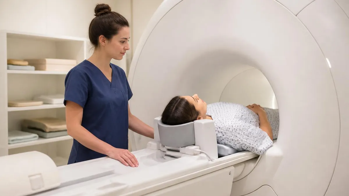
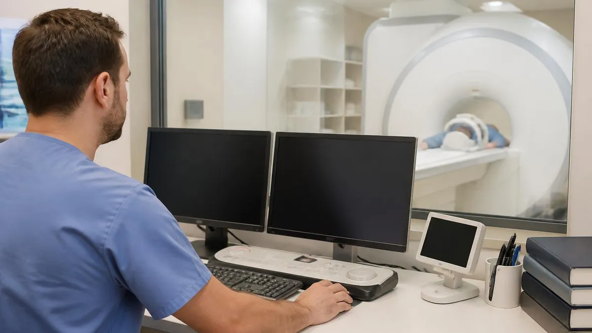
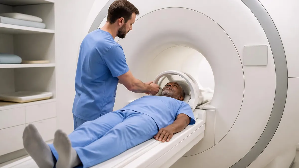
The scan itself involves lying still on a table that slides into the MRI machine. For cervical spine MRI, you lie on your back with your head and shoulders in the scanner. A coil (a specialized antenna) is placed around your neck to capture the signal. The machine makes loud knocking and beeping sounds during scanning — they'll give you earplugs or headphones with music. Most cervical spine scans take 30-45 minutes; complex protocols or contrast studies can extend to 60 minutes.
Conditions Diagnosed by MRI C-Spine
Nerve root irritation causing pain, numbness, or weakness in the arm following a specific nerve pattern. MRI identifies the specific level and cause — disc herniation, stenosis, or other compressive lesion.
Reading your MRI c-spine report can feel overwhelming. Reports follow a standard format: technique description, findings organized by anatomical region, and an impression summarizing the main diagnostic conclusions. The 'findings' section describes each anatomical level (alignment, disc spaces, cord, foramina) in detail. The 'impression' distills the most clinically relevant findings into a brief summary. Start with the impression for the overview, then look at findings for details.
Terminology can be intimidating. 'Posterior central disc protrusion' means disc material extends backward toward the spinal cord. 'Foraminal narrowing' means the openings where nerves exit are smaller than normal. 'Cord signal abnormality' means there's a bright or dark area in the spinal cord on certain sequences — usually concerning. 'Facet arthropathy' means the small joints in the back of the spine show degenerative changes. Don't panic at unfamiliar terms — most are descriptive and reflect common age-related changes.
The relationship between findings and symptoms is what your doctor will interpret. A C5-C6 disc herniation causing left arm pain in the C6 nerve distribution makes clinical sense — the imaging and symptoms align. A C5-C6 disc bulge in someone with right shoulder pain doesn't quite fit and suggests the bulge isn't the cause of the pain. Doctors integrate imaging with examination findings and symptom patterns to identify which findings actually matter.
Before Your MRI C-Spine Appointment
- ✓Tell the scheduler about any metal in your body during scheduling
- ✓Confirm whether contrast will be used and any contrast allergies
- ✓Wear comfortable, metal-free clothing or plan to change into a gown
- ✓Remove all jewelry, watches, and metal-containing items before the scan
- ✓Leave valuables at home or in a secured locker — not in the scanner room
- ✓Continue medications as usual unless told otherwise
- ✓Eat normally before the scan unless contrast is being used
- ✓Arrive 15-30 minutes early for paperwork and screening forms
- ✓Bring prior imaging studies if available (CDs or USB drives)
- ✓Plan to lie still for 30-60 minutes during the scan itself
Claustrophobia is a real concern for MRI scans, particularly traditional closed-bore machines where you're inside a narrow tube. Up to 15% of patients experience some level of anxiety during MRI scanning. If you know you struggle with enclosed spaces, discuss this with your doctor before scheduling. Options include mild oral sedation, headphones with music or guided meditation, eye covers to avoid visual claustrophobia triggers, and open or wide-bore MRI machines that reduce the tunnel sensation. For severe cases, open MRI or anesthesia-assisted scanning may be necessary.
Cost and insurance coverage vary widely. Hospital-based MRI scans tend to cost more than independent imaging centers. Insurance typically covers medically-necessary MRI but may require pre-authorization. Out-of-pocket costs can range from a few hundred to several thousand dollars depending on your insurance, the facility, and contrast use. Ask about pricing in advance — many facilities offer significant discounts for cash-pay patients compared to insurance-billed rates.
Contrast use is another consideration. Most cervical spine MRI scans don't require contrast. Contrast (gadolinium) is added selectively when looking for tumors, infections, or post-surgical changes. Gadolinium is generally safe but should be avoided or used cautiously in patients with significantly impaired kidney function due to a rare condition called nephrogenic systemic fibrosis. Your doctor will weigh benefits against any risks before ordering contrast.
Follow-up imaging is sometimes appropriate. If conservative treatment doesn't resolve symptoms over weeks to months, repeat imaging may help track changes. After surgical intervention, post-operative MRI assesses results and identifies any complications. Routine surveillance imaging isn't usually needed if symptoms are improving — imaging changes lag behind clinical improvement, and repeating scans without clinical indication adds cost without benefit.
Always request a copy of your MRI on disc or via patient portal. Imaging studies are part of your medical record, and having copies makes it easy to share with second-opinion physicians, surgeons, or specialists. Many facilities provide copies free or for a nominal fee. If you change doctors or seek a specialist opinion, having the images saves the cost and time of repeating the scan.
Alternatives to MRI exist and may be appropriate in specific situations. CT myelography combines CT scanning with contrast injected into the spinal fluid — useful when MRI is contraindicated (pacemakers, severe claustrophobia). Plain X-rays show bony alignment and can identify fractures, severe degenerative changes, or instability. Bone scans show areas of increased metabolic activity — useful for screening for tumors or infections. Each modality has strengths; MRI just happens to be best for most cervical spine questions involving soft tissues and the spinal cord.
The role of MRI in surgical planning deserves attention. Spine surgeons rely heavily on detailed MRI imaging when planning operations. The images show exactly where pathology lies, what structures must be navigated, and what tissues are at risk. Pre-operative MRI guides whether a single-level or multi-level operation is needed, whether anterior or posterior approach is best, and what specific structures need to be addressed. Surgical outcomes depend partly on accurate imaging.
Final notes on MRI c-spine: it's a powerful, non-invasive tool that has transformed evaluation of cervical spine problems. The detail it provides is remarkable, but findings must be interpreted in clinical context. Most age-related changes seen on MRI are common and not necessarily problematic. Acute, severe findings warrant urgent attention. The middle ground requires thoughtful conversation between you and your healthcare team about what the imaging means for your specific situation and what next steps make sense given the complete clinical picture.
MRI vs Other Spine Imaging
- +Excellent soft tissue detail — sees discs, nerves, cord, and ligaments clearly
- +No radiation exposure — uses magnetic fields and radio waves
- +Multiple sequences provide different views of the same anatomy
- +Highly sensitive for detecting most spine pathology
- +Gold standard for evaluating spinal cord and nerve root problems
- +Reproducible imaging that can be compared over time
- −Loud, lengthy scans (30-60 minutes typically)
- −Claustrophobia issues in traditional closed-bore machines
- −Contraindicated with certain implants (pacemakers, some clips)
- −Significant cost, especially without insurance coverage
- −Can show many incidental findings that aren't clinically significant
MRI is a tool that complements — not replaces — a thorough physical examination and clinical history. Your doctor combines all three to reach a diagnosis. A perfect MRI doesn't substitute for proper neurological examination. If your provider hasn't done a thorough exam alongside ordering imaging, ask for one before making treatment decisions based on imaging findings alone.
A few additional considerations help patients get the most from their cervical spine MRI experience. First, timing matters relative to symptoms. Imaging within the first few weeks of acute symptoms often shows clear pathology, while imaging months after an injury may show evolving findings as inflammation resolves and the body adapts. If your provider suggests waiting before imaging, it's often because conservative treatment is appropriate first and imaging is more useful if symptoms persist beyond the natural recovery window.
Second, getting second opinions on imaging is reasonable when major decisions hang on the results. If surgery is being recommended based on MRI findings, having another radiologist or spine specialist review the images can provide valuable confirmation. Many academic medical centers offer specialty image review services. The cost is modest relative to the magnitude of decisions being made. Some patients also find that telemedicine consultations with out-of-area specialists provide useful additional perspectives.
Third, understanding the difference between a radiologist's report and a treating physician's interpretation matters. Radiologists describe what they see anatomically. Treating physicians integrate those findings with your specific symptoms and examination. The radiology report may describe many findings, only some of which matter for your particular situation. When you discuss results with your doctor, focus on the findings that explain your symptoms rather than every abnormality listed in the report.
Fourth, advances in MRI technology continue to expand what's possible. Higher-field strength magnets (3T and stronger) provide better detail than older 1.5T machines. New sequences allow visualization of nerve fiber tracts (diffusion tensor imaging), cord function (functional MRI), and metabolic activity (MR spectroscopy). For most clinical questions, standard MRI sequences provide all the information needed. For unusual or complex cases, advanced sequences may add value.
Fifth, the patient experience varies significantly between facilities. Newer wide-bore and open MRI systems are much more comfortable than older closed-bore machines. Quieter sequences reduce the noise discomfort. Patient-friendly facilities provide music, climate control, and supportive staff. If you have flexibility about where to have your scan, asking about the facility's equipment age and patient amenities can meaningfully improve your experience.
Sixth, MRI safety has improved dramatically but remains worth taking seriously. The technologists screen every patient carefully before entering the scanner room. Don't ever attempt to enter the scanner room with anything metallic — even the smallest item can become a projectile in the magnetic field. People have been seriously injured by oxygen tanks, wheelchairs, and tools brought into MRI rooms accidentally. The screening process exists to protect you, and you should answer all questions honestly even when they feel repetitive.
Seventh, communication with your physician after the scan is just as important as the scan itself. Once results are available, schedule a dedicated appointment to discuss them — preferably in person or via video, not just over a portal message. Bring written questions. Ask which findings are most clinically relevant to your symptoms, what treatment options exist, what the natural course typically looks like, and what specifically would prompt a different approach. The conversation around imaging is often more valuable than the imaging itself.
Eighth, recovery and treatment planning depend on what the MRI shows. Conservative treatment with physical therapy, medications, and time resolves most cervical spine problems without surgery. Even significant disc herniations frequently improve over weeks to months without intervention. Surgery becomes appropriate when conservative treatment fails, when there's progressive neurological deficit, or when severe cord compression threatens permanent damage. The MRI helps establish where you stand on that spectrum.
Finally, remember that imaging is one piece of comprehensive care. Healthy outcomes come from a combination of accurate diagnosis, appropriate treatment matched to your specific situation, ongoing communication with your healthcare team, and active patient engagement in recovery. MRI provides exceptional diagnostic information — but the path to feeling better requires putting that information into action through whatever treatment plan emerges from the conversation with your provider team.
MRI Questions and Answers
About the Author

Medical Laboratory Scientist & Clinical Certification Expert
Johns Hopkins UniversityDr. Sandra Kim holds a PhD in Clinical Laboratory Science from Johns Hopkins University and is certified as a Medical Technologist (MT) and Medical Laboratory Scientist (MLS) through ASCP. With 16 years of clinical laboratory experience spanning hematology, microbiology, and molecular diagnostics, she prepares candidates for ASCP board exams, MLT, MLS, and specialist certification tests.
Join the Discussion
Connect with other students preparing for this exam. Share tips, ask questions, and get advice from people who have been there.
View discussion (8 replies)