IAC MRI: Complete Guide to Internal Auditory Canal MRI, Indications, Protocol, and Interpretation
Learn everything about IAC MRI — internal auditory canal imaging, protocol, indications, and how to interpret findings for hearing loss and tinnitus.
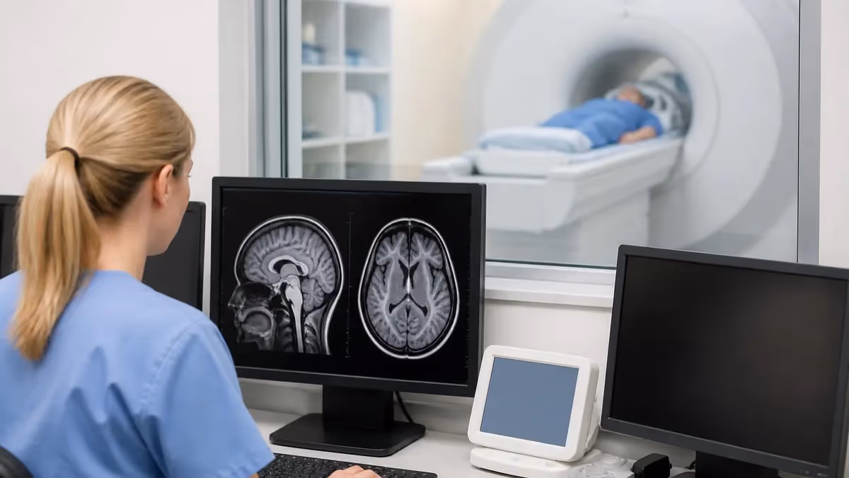
IAC MRI, or magnetic resonance imaging of the internal auditory canals, is one of the most clinically important specialized MRI protocols performed in modern radiology. The internal auditory canals are small bony tunnels within the temporal bone that house the facial nerve, the cochlear nerve, and the superior and inferior vestibular nerves. When patients present with unexplained sensorineural hearing loss, tinnitus, vertigo, or facial nerve dysfunction, IAC MRI is typically the first-line advanced imaging study ordered by otolaryngologists and neurologists. Understanding this protocol is essential for any MRI technologist or radiology student preparing for board examinations.
The anatomy covered by iac mri studies extends well beyond the canals themselves. A comprehensive IAC protocol images the entire posterior fossa, the cerebellopontine angle (CPA) cisterns, the cochlea, the semicircular canals, and the brainstem at the level of the pons. The CPA cistern is particularly important because it is the most common site for vestibular schwannomas, the benign nerve sheath tumors most frequently associated with progressive unilateral sensorineural hearing loss. These tumors can be as small as two millimeters in their intracanalicular portion, making high-resolution, thin-slice imaging with gadolinium contrast critical for reliable detection.
From a technical standpoint, IAC MRI protocols are demanding. They require dedicated high-resolution sequences including 3D heavily T2-weighted imaging such as FIESTA, CISS, or DRIVE depending on the MRI vendor, as well as post-contrast T1-weighted sequences with fat saturation. The 3D T2 sequences exploit the natural contrast between the fluid-filled labyrinth and membranous structures, generating what radiologists often call a cisternogram or MR cisternography image. These sequences can detect even tiny soft tissue filling defects within the fluid of the IAC and CPA cistern without the need for intravenous contrast in many cases.
Gadolinium contrast remains a cornerstone of the full IAC MRI protocol. When a vestibular schwannoma is present, it will enhance brightly and uniformly on post-contrast T1 images, making even intracanalicular tumors measuring just a few millimeters conspicuous. Meningiomas of the CPA also enhance avidly but tend to show a broad dural base and may calcify. Facial nerve neuromas, epidermoid cysts, arachnoid cysts, and vascular loops are among the other pathologies that can be differentiated with careful protocol selection and interpretation. The radiologist reading an IAC MRI must be systematic and thorough, evaluating each cranial nerve segment individually.
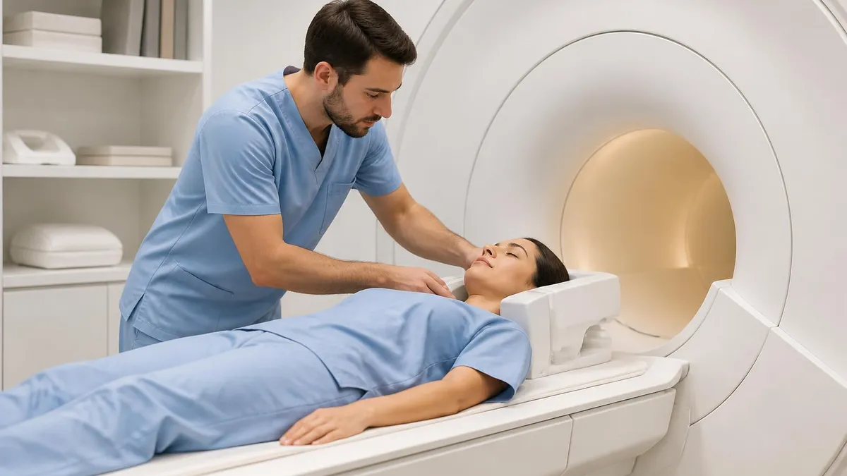
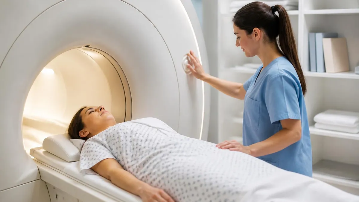
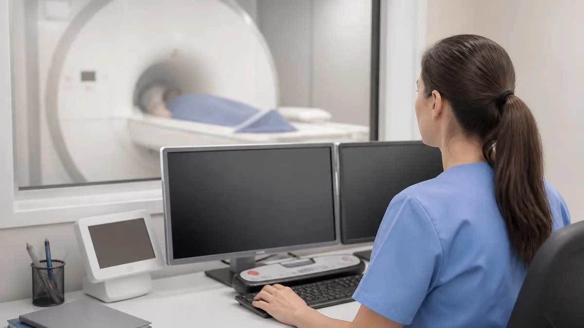
For MRI technologists, positioning and patient cooperation are critical to image quality. Patients must remain still for extended periods, often twenty to thirty minutes, while multiple high-resolution acquisitions are obtained. Even small amounts of motion can degrade the 3D T2 sequences significantly, potentially obscuring a small intracanalicular tumor. Using appropriate head coils, foam padding for immobilization, and clear patient education before scanning begins are all important steps. Technologists should also be prepared to troubleshoot wrap artifact, flow artifact from the jugular veins, and susceptibility artifacts from dental hardware.
The clinical impact of IAC MRI findings is substantial. A negative IAC MRI in a patient with sudden sensorineural hearing loss essentially rules out a retrocochlear mass lesion and redirects clinical management toward medical treatment. A positive finding of a vestibular schwannoma triggers a multidisciplinary discussion involving neurosurgery, radiation oncology, and otolaryngology to weigh observation, stereotactic radiosurgery, or microsurgical resection. Technologists who understand why the study is being performed and what radiologists are looking for will perform better scans and contribute meaningfully to patient care.
This guide covers everything you need to know about IAC MRI — from anatomy and clinical indications through imaging protocols, pathology interpretation, and practical preparation tips for registry and board examinations. Whether you are a student, a working technologist refreshing your knowledge, or a clinician seeking a deeper understanding of the imaging, the sections below provide comprehensive, accurate, and clinically relevant information designed to build real expertise.
IAC MRI by the Numbers
Anatomy of the Internal Auditory Canal
The internal auditory canal is a short bony tunnel approximately 8–10 mm long and 4–6 mm wide, running horizontally within the petrous portion of the temporal bone. It opens medially into the posterior cranial fossa at the porus acusticus and ends laterally at the fundus.
Four distinct nerve bundles travel through the IAC: the facial nerve (CN VII) anterosuperiorly, the cochlear nerve anteroinferiorly, and the superior and inferior vestibular nerves posteriorly. The transverse (falciform) crest and the vertical crest (Bill's bar) divide the fundus into four quadrants.
The CPA cistern is the CSF-filled space medial to the IAC where nerves enter from the brainstem. It is the most common location for vestibular schwannomas, meningiomas, and epidermoid cysts. The anterior inferior cerebellar artery (AICA) loops through this cistern and may contact or compress cranial nerves.
Just lateral to the fundus lies the membranous labyrinth, including the cochlea, the three semicircular canals, the utricle, and the saccule. These fluid-filled structures generate bright signal on 3D T2 sequences, making them exquisitely visible on IAC MRI and allowing detection of labyrinthine enhancement, fibrosis, or ossification.
The labyrinthine artery, a branch of the AICA, supplies the inner ear structures. Vascular loops of the AICA that indent into the IAC can mimic small tumors on imaging and are a well-recognized pitfall. Recognizing the pulsation artifact and the curved vascular morphology helps differentiate vascular loops from true neoplasms.
The clinical indications for ordering an IAC MRI are well-established and center on symptoms that suggest retrocochlear or posterior fossa pathology. Sudden sensorineural hearing loss (SSNHL) is perhaps the most common indication. While SSNHL most often has no identifiable structural cause and is treated empirically with steroids, a small but important percentage of cases result from a compressive CPA or intracanalicular mass. Guidelines from the American Academy of Otolaryngology recommend that all patients with sudden sensorineural hearing loss receive MRI evaluation to exclude an underlying neoplasm before attributing the hearing loss to idiopathic causes.
Asymmetric sensorineural hearing loss is another major indication for IAC MRI, even when the loss develops gradually over months or years. When audiometric testing reveals a difference of fifteen decibels or more at three consecutive frequencies between the two ears, or when speech discrimination scores differ significantly, clinicians are obligated to rule out a mass lesion. Many vestibular schwannomas present insidiously in this fashion rather than with sudden onset, and delays in diagnosis can allow tumors to grow to a size that complicates treatment options and risks facial nerve function during surgery.
Pulsatile and non-pulsatile tinnitus are both indications for posterior fossa imaging, though pulsatile tinnitus more often triggers vascular imaging as well. Non-pulsatile unilateral tinnitus, particularly when associated with any degree of hearing asymmetry, warrants IAC MRI to exclude an intracanalicular or CPA mass. Vertigo, particularly episodic vertigo that does not fit the typical pattern of benign paroxysmal positional vertigo, also prompts IAC MRI evaluation. Bilateral vestibular loss should raise suspicion for bilateral vestibular schwannomas associated with neurofibromatosis type 2, a genetic condition in which IAC MRI findings are pathognomonic.
Facial nerve palsy is a critically important indication for IAC MRI. While most cases of acute facial nerve palsy represent Bell's palsy, a clinical diagnosis of exclusion, the facial nerve's course through the IAC, the labyrinthine segment, the geniculate ganglion, and the parotid gland must all be evaluated when palsy is atypical, recurrent, or fails to recover within the expected timeframe. IAC MRI with gadolinium allows assessment of the intracanalicular facial nerve segment, which is not accessible by other imaging modalities and cannot be visualized on CT with the same soft tissue resolution.
Pre-operative and post-operative imaging for known CPA and IAC tumors is another major use case. Vestibular schwannomas are managed with active observation using serial IAC MRI at defined intervals, stereotactic radiosurgery such as Gamma Knife, or microsurgical resection. For observation patients, annual or biannual IAC MRI tracks tumor growth rate, which heavily influences the decision to intervene. Post-treatment imaging monitors for recurrence after surgery or for pseudoprogression versus true tumor growth after radiosurgery, which can be challenging to distinguish in the first two years after treatment.
Neurofibromatosis type 2 (NF2) is a specific indication that mandates regular surveillance IAC MRI beginning in childhood or adolescence. Patients with NF2 carry a mutation in the merlin tumor suppressor gene and invariably develop bilateral vestibular schwannomas along with meningiomas, ependymomas, and peripheral schwannomas. Surveillance imaging protocols for NF2 patients are rigorous, typically involving annual brain and spine MRI, and require technologists to be particularly careful about protocol consistency so that serial studies can be compared accurately and reliably over many years.
Other less common but important indications include evaluation for superior semicircular canal dehiscence, cochlear nerve aplasia or hypoplasia in pediatric patients with congenital hearing loss being considered for cochlear implantation, endolymphatic hydrops in Meniere's disease using intratympanic gadolinium protocols, and assessment of the internal auditory canal prior to auditory brainstem implantation. Each of these indications may require protocol modifications tailored to the specific clinical question, highlighting the importance of close communication between ordering clinicians and the MRI team.
MRI Practice Test Questions
Prepare for the MRI - Magnetic Resonance Imaging exam with our free practice test modules. Each quiz covers key topics to help you pass on your first try.
MRI Knowledge
MRI Exam Questions covering Knowledge. Master MRI Test concepts for certification prep.
MRI Physics
Free MRI Practice Test featuring Physics. Improve your MRI Exam score with mock test prep.
MRI Anatomy and Pathology
MRI Test Prep for MRI Anatomy and Pathology. Practice MRI Quiz questions and boost your score.
MRI Anatomy and Positioning
MRI Questions and Answers on MRI Anatomy and Positioning. Free MRI practice for exam readiness.
MRI Contrast Agents
Free MRI Quiz on MRI Contrast Agents. MRI Exam prep questions with detailed explanations.
MRI Patient Care and Positioning
MRI Practice Questions for MRI Patient Care and Positioning. Build confidence for your MRI certification exam.
IAC MRI Protocol: Sequences, Parameters, and Contrast
The 3D heavily T2-weighted sequence is the backbone of every IAC MRI protocol. Vendor-specific implementations include FIESTA (GE), CISS (Siemens), and DRIVE (Philips). These steady-state free precession sequences generate extremely bright fluid signal, creating a natural contrast between cerebrospinal fluid and the cranial nerves traversing the CPA cistern and IAC. Typical parameters include a 0.6–0.8 mm isotropic voxel size, a field of view of 180 mm centered on the posterior fossa, and a scan time of approximately eight to twelve minutes. Multiplanar reformations from the isotropic dataset allow the radiologist to trace each nerve segment in any plane without additional scanning.
A critical advantage of 3D T2 imaging is that it can detect intracanalicular tumors without gadolinium contrast by showing filling defects within the bright CSF of the canal. A vestibular schwannoma as small as two to three millimeters will appear as a small dark nodule against the bright background fluid. This makes 3D T2 sequences valuable for patients with renal impairment or gadolinium allergy who cannot receive contrast. However, the sequence is susceptible to pulsation artifact from nearby vascular structures, and image quality degrades significantly with even minor patient motion, requiring careful patient preparation and coaching before the scan begins.
IAC MRI: Benefits and Limitations Compared to Other Imaging
- +Highest soft tissue resolution for cranial nerve imaging — detects tumors as small as 2 mm
- +No ionizing radiation — safe for repeated surveillance imaging in NF2 patients
- +3D T2 sequences provide cisternogram effect without contrast in many cases
- +Directly visualizes all four nerves within the IAC simultaneously
- +Post-contrast sequences detect labyrinthine enhancement invisible on CT
- +Multiplanar reformatting from isotropic 3D datasets eliminates need for repeat acquisitions
- −Longer scan time (25–40 minutes) compared to CT of temporal bone (under 5 minutes)
- −Motion-sensitive — poor image quality if patient cannot hold still for 3D sequences
- −Gadolinium contraindicated in severe renal impairment (GFR under 30) and some allergy histories
- −More expensive than CT and requires MRI-compatible patient screening for metallic implants
- −Cannot assess bony detail of the otic capsule or ossicular chain as well as high-resolution CT
- −AICA vascular loops can mimic intracanalicular tumors, requiring careful correlation
Pre-Scan IAC MRI Checklist for MRI Technologists
- ✓Verify clinical indication and confirm correct protocol (IAC MRI with and without contrast vs. without only).
- ✓Complete thorough MRI safety screening for ferromagnetic implants, cochlear implants, and aneurysm clips.
- ✓Confirm IV access site and check renal function labs (eGFR) before drawing gadolinium contrast.
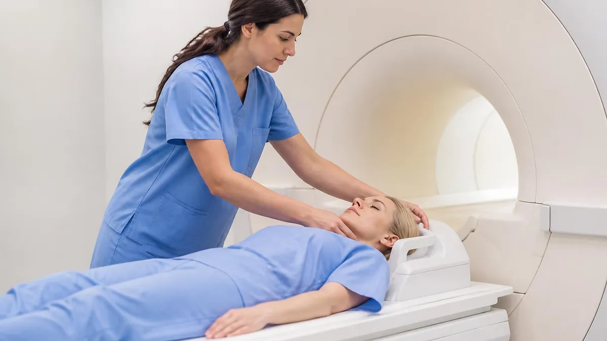
- ✓Educate the patient about scan duration, the importance of remaining still, and the noise level of 3D sequences.
- ✓Position the patient supine with the head centered in the head coil and immobilized with foam pads.
- ✓Ensure the field of view covers the full posterior fossa, brainstem, and both IACs symmetrically.
- ✓Acquire scout images and verify the 3D T2 volume is properly aligned over the petrous apices bilaterally.
- ✓Run the 3D T2 sequence first while the patient is freshest and best able to hold still.
- ✓Administer gadolinium contrast per institutional protocol after non-contrast sequences are complete.
- ✓Acquire post-contrast 3D T1 or 2D fat-saturated T1 sequences in axial and coronal planes.
Small Intracanalicular Tumors Require Both 3D T2 and Post-Contrast T1
Studies have shown that using 3D T2 sequences alone misses approximately 5–10% of small intracanalicular vestibular schwannomas because tiny enhancing nodules may not produce a visible filling defect in the fluid. Always acquire post-contrast T1 sequences in addition to the 3D T2 cisternogram to maximize sensitivity for lesions at the fundus of the IAC, where fluid volume is smallest and filling defect conspicuity is lowest.
Vestibular schwannoma, historically and incorrectly called acoustic neuroma, is by far the most common pathology detected on IAC MRI, accounting for approximately eighty to ninety percent of all CPA masses. These tumors arise from the Schwann cells of the vestibular nerve, almost always the superior vestibular division, and grow slowly within the IAC before extending medially into the CPA cistern.
On imaging, they are typically isointense to slightly hypointense to brain on T1-weighted sequences, heterogeneously bright on T2, and show avid, often homogeneous contrast enhancement. The Koos classification system grades these tumors from one to four based on their relationship to the IAC, CPA, and brainstem, guiding management decisions.
Meningioma is the second most common CPA mass, representing five to ten percent of cases. Unlike vestibular schwannomas, meningiomas typically arise from the dura of the posterior surface of the petrous bone or the porus acusticus and show a broad dural base rather than an epicenter within the IAC.
They tend to be homogeneously isointense to gray matter on both T1 and T2 sequences and enhance avidly. A dural tail sign — thickening and enhancement of the adjacent dura — is highly suggestive but not pathognomonic of meningioma. Calcification, seen as low signal on all MRI sequences, is common in meningiomas and can help differentiate them from schwannomas.
Epidermoid cysts are the third most common CPA lesion and represent a diagnostically important pitfall. They arise from ectodermal rests left during neural tube closure and grow by desquamation of keratin. On standard T1 and T2 sequences, they closely resemble arachnoid cysts — dark on T1 and bright on T2.
The critical distinguishing feature is diffusion-weighted imaging: epidermoids show bright signal on DWI and low signal on ADC maps due to restricted diffusion of the keratin contents, while arachnoid cysts follow CSF signal on DWI. Failure to obtain DWI on an IAC MRI protocol can result in this clinically significant misdiagnosis.
Facial nerve neuromas are rare but important CPA and IAC masses that can mimic vestibular schwannomas. They arise from the facial nerve rather than the vestibular nerve and may present with progressive facial weakness rather than hearing loss, though they can cause both.
On imaging, a mass that extends along the expected course of the facial nerve, particularly if it involves the labyrinthine segment or geniculate ganglion in addition to the IAC, should raise suspicion for a facial neuroma rather than a vestibular schwannoma. This distinction is critical because inadvertent surgical manipulation of a facial neuroma during what is believed to be vestibular schwannoma surgery can result in devastating facial paralysis.
Endolymphatic sac tumors are aggressive low-grade adenocarcinomas that arise from the endolymphatic sac on the posterior surface of the petrous bone. They are strongly associated with von Hippel-Lindau disease and should be suspected when imaging shows a destructive posterior petrous lesion with intratumoral hemorrhage.
On MRI, they characteristically show hyperintense foci on T1-weighted images due to blood products and cholesterol crystals. Early detection is important because small endolymphatic sac tumors can be resected with hearing preservation, while large tumors typically result in complete hearing loss and present significant surgical challenges due to their proximity to the jugular bulb and sigmoid sinus.
Labyrinthitis ossificans is a late complication of severe bacterial meningitis in which the membranous labyrinth is progressively replaced by fibrous tissue and ultimately bone. On early IAC MRI, the affected labyrinth shows abnormal enhancement on post-contrast sequences and loss of the normal bright 3D T2 fluid signal. In later stages, complete obliteration of the fluid spaces produces complete dark signal within the cochlea and semicircular canals on 3D T2 imaging. Detecting labyrinthitis ossificans before it reaches the cochlear implantation stage is clinically urgent because obliterated cochlear lumens require specialized surgical drilling and cannot receive standard cochlear implant electrode arrays.
Vascular loops represent an important normal variant and diagnostic pitfall on IAC MRI. The anterior inferior cerebellar artery and less commonly the posterior inferior cerebellar artery may loop into the IAC in normal individuals without producing symptoms. In some patients, however, vascular compression of cranial nerves VII and VIII within the IAC or at the root entry zone is believed to cause disabling tinnitus, hearing loss, or hemifacial spasm through a mechanism similar to trigeminal neuralgia.
Microvascular decompression surgery can be considered for carefully selected patients with neurovascular contact confirmed on dedicated MRI. Distinguishing a symptomatic vascular contact from an incidental loop requires clinical correlation and should never be based on imaging alone.
Many cochlear implants contain ferromagnetic magnets and were historically considered absolute MRI contraindications. Newer cochlear implant systems are conditionally MRI-safe at 1.5T with the internal magnet removed or with a head bandage applied to prevent magnet displacement, but policies vary by implant model and manufacturer. Always consult the manufacturer's MRI conditional guidelines and your institution's cochlear implant MRI protocol before scanning any patient with a cochlear implant. Failure to follow proper precautions can result in magnet displacement, pain, device damage, and serious patient injury.
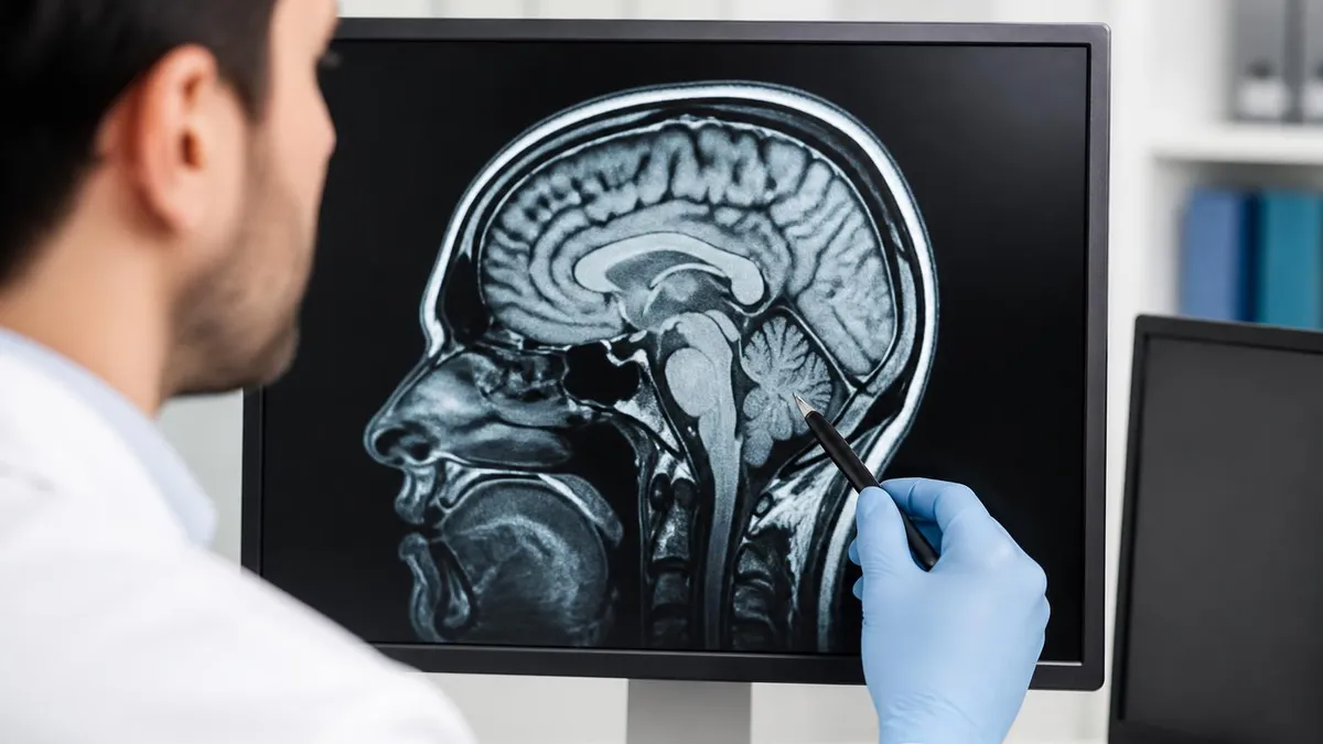
Interpreting an IAC MRI begins with a systematic review of the entire dataset rather than jumping immediately to look for masses. The radiologist or technologist reviewing images should first evaluate image quality, confirming that the 3D T2 sequence is free of motion artifact and that the post-contrast T1 sequences have adequate fat saturation.
Poor fat saturation in the post-contrast sequences can dramatically impair lesion detection by masking enhancing tissue against the bright background of fatty marrow. If fat saturation failure is identified, the sequence may need to be repeated or interpreted with significant caution, which should be communicated clearly in the report.
The systematic review of an IAC MRI should proceed anatomically from medial to lateral: begin at the brainstem and evaluate the root entry zones of cranial nerves VII and VIII, proceed through the CPA cistern evaluating for masses, arachnoid adhesions, and vascular contacts, then move to the porus acusticus and evaluate the proximal IAC, the mid-canal, and finally the fundus.
Each of these anatomic subregions is prone to different pathologies, and a region-by-region approach reduces the risk of overlooking a subtle finding. Compare the two sides carefully at each level — subtle asymmetry is often the first and only clue to an early intracanalicular lesion.
The size and location of any detected mass must be measured precisely and described in a standardized format. For vestibular schwannomas, the standard measurements include the maximum anteroposterior and transverse dimensions within the CPA cistern and the length of intracanalicular extension to the fundus.
The relationship of the mass to the fundus — whether there is a cap of CSF between the tumor and the fundus — is important to document because fundal involvement affects the choice of surgical approach. The Koos grade should be assigned based on the measurements, as this directly influences management discussions and allows consistent comparison on follow-up imaging.
Enhancement assessment requires careful comparison of pre- and post-contrast T1 sequences at identical slice positions. Subtle lesions, such as a small intracanalicular schwannoma measuring only a few millimeters, may show only modest enhancement that is easy to overlook if the reader is not systematically comparing pre- and post-contrast images side by side. Modern PACS workstations allow image subtraction, in which the pre-contrast T1 series is digitally subtracted from the post-contrast series, leaving only enhancing structures visible. Subtraction images are particularly useful for lesions at the fundus of the IAC, where they can dramatically improve conspicuity of small enhancing nodules.
The cochlea and labyrinth deserve specific attention on every IAC MRI, not just cases where labyrinthine pathology is suspected. Normal cochlear fluid should appear uniformly bright on the 3D T2 sequence with sharp, well-defined walls. Any asymmetry in signal intensity between the two cochleae, loss of the modiolus definition, or abnormal fluid-fluid levels within the cochlear turns should be noted and described.
On the post-contrast sequence, any enhancement within the cochlear turns, the semicircular canals, or the endolymphatic sac should be described in detail. These findings, even when subtle, can represent labyrinthitis, labyrinthine infarct, or early labyrinthitis ossificans and have immediate clinical management implications.
The facial nerve canal and its multiple segments deserve careful attention on IAC MRI, even when the primary clinical concern is hearing loss rather than facial weakness. The intracanalicular segment of the facial nerve travels in the anterosuperior compartment of the IAC, separated from the vestibular nerve by Bill's bar.
On the high-resolution 3D T2 sequence, the facial nerve should be visible as a thin dark line against the bright IAC fluid. Enhancement of the intracanalicular facial nerve on post-contrast T1 is abnormal in the intracanalicular segment but can be normal at the geniculate ganglion and in the tympanic segment, a common pitfall for less experienced readers.
After completing the targeted evaluation of the IAC and posterior fossa, the radiologist must perform a complete review of the entire imaging volume, which typically includes the entire posterior fossa, cerebellum, brainstem, and temporal lobes. Incidental findings such as cerebellar infarcts, demyelinating plaques, developmental venous anomalies, arachnoid cysts, and small meningiomas at sites remote from the IAC must be identified, characterized, and reported. The value of a comprehensive IAC MRI interpretation extends beyond just answering the primary clinical question and represents an important opportunity to detect clinically significant incidental pathology.
Preparing for MRI registry examinations and board certifications requires a solid understanding of IAC MRI because questions about cranial nerve anatomy, posterior fossa pathology, and specialized protocols appear consistently on the ARRT MRI examination and related certification tests.
Candidates who study IAC MRI systematically tend to perform well on anatomy-based questions because the IAC is a compact, well-defined anatomic region with a finite number of structures, relationships, and pathologies that can be learned thoroughly with targeted study. Begin your preparation by mastering the four cranial nerve subdivisions within the IAC and the anatomic landmarks that divide them — the falciform crest and Bill's bar — before moving on to pathology.
Understanding the signal characteristics of common CPA and IAC pathologies on T1, T2, and post-contrast sequences is essential for examination success. Create a systematic mental comparison table: vestibular schwannoma (iso T1, bright T2, avid enhancement), meningioma (iso T1, iso T2, avid enhancement with dural tail), epidermoid (dark T1, bright T2, restricted diffusion), and arachnoid cyst (dark T1, bright T2, follows CSF on all sequences including DWI). This type of systematic comparison not only helps on multiple-choice examinations but also builds the pattern recognition skills needed for clinical practice and practical examinations.
Sequence parameters for IAC MRI protocols are frequently tested on the ARRT registry. Know the target voxel size for 3D T2 sequences (0.6–0.8 mm isotropic), the standard gadolinium dose (0.1 mmol/kg), why fat saturation is required on post-contrast T1 sequences, and the specific advantages of CISS/FIESTA/DRIVE over standard T2 sequences for cranial nerve imaging. Understanding the physics behind steady-state free precession sequences, specifically why they generate such bright fluid signal with high signal-to-noise ratio at short repetition times, gives candidates a conceptual framework that helps answer not only protocol questions but also physics questions about related sequence families.
Artifact recognition is another high-yield area for examination preparation. Susceptibility artifact from dental amalgam, motion artifact degrading the 3D T2 sequence, chemical shift artifact at fat-water interfaces, and wrap (aliasing) artifact from inadequate field of view placement are all commonly depicted in registry examination images. For IAC MRI specifically, knowing that pulsation artifact from the jugular vein or sigmoid sinus can simulate signal abnormality in the posterior IAC, and that this artifact can be reduced with cardiac gating or modified phase-encode direction, demonstrates the clinical level of knowledge expected of registered technologists.
The ARRT examination also tests knowledge of patient safety considerations specific to the ear and posterior fossa region. Cochlear implants, as discussed above, are the most high-stakes safety consideration for IAC MRI. Know the general categories of cochlear implant MRI compatibility: older devices that are contraindicated at all field strengths, devices that are conditionally safe at 1.5T with magnet removal, and newer devices conditionally safe at 1.5T or 3T with a tight head bandage.
Auditory brainstem implants (ABIs), used in patients with NF2 who have lost both cochlear nerves, are generally considered MRI-unsafe and represent an absolute contraindication unless specific device documentation confirms otherwise.
Practice with imaging cases is the single most effective preparation strategy beyond reading. The ARRT MRI registry examination includes case-based questions that present images and ask candidates to identify findings, select the appropriate protocol modification, or choose the correct safety action. Seeking out IAC MRI cases through online case repositories, radiology training platforms, and the practice tests available on PracticeTestGeeks will dramatically accelerate your ability to recognize both classic presentations and subtle abnormalities under examination time pressure. Focus particularly on cases that compare the normal side to the abnormal side, since asymmetry recognition is fundamental to IAC MRI interpretation.
On examination day, read all IAC MRI questions carefully and look for key words that distinguish between protocols. Questions that specify gadolinium administration describe the full IAC protocol; questions that specify contraindication to contrast test your knowledge of the non-contrast protocol and its limitations.
Questions about posterior fossa anatomy in children may involve congenital inner ear malformations such as common cavity deformity, cochlear aplasia, or enlarged vestibular aqueduct syndrome, which present differently from the acquired pathologies most common in adults. Being aware of these age-specific considerations and knowing which imaging findings differentiate them ensures you are prepared for the full range of posterior fossa imaging questions the registry may present.
MRI Questions and Answers
MRI of Cervical Spine: Complete Guide to Imaging, Anatomy, Pathology, and Patient Preparation

Knee MRI Images: A Complete Guide to Reading, Understanding, and Interpreting Knee Scans
Ace Your Exam with MRI Practice Test 2026 Prep

MRI Tech School 2026 — Programs, Requirements, and What to Expect
Man Dies in MRI Machine: Understanding MRI Safety Incidents, Risks, and Prevention
About the Author
Medical Laboratory Scientist & Clinical Certification Expert
Johns Hopkins UniversityDr. Sandra Kim holds a PhD in Clinical Laboratory Science from Johns Hopkins University and is certified as a Medical Technologist (MT) and Medical Laboratory Scientist (MLS) through ASCP. With 16 years of clinical laboratory experience spanning hematology, microbiology, and molecular diagnostics, she prepares candidates for ASCP board exams, MLT, MLS, and specialist certification tests.
Join the Discussion
Connect with other students preparing for this exam. Share tips, ask questions, and get advice from people who have been there.
View discussion (4 replies)