Full Body MRI Scan Near Me: Manhattan Guide to Preventive Whole-Body Imaging
Full body MRI Manhattan guide: ✅ where to book whole-body scans near you, costs, what's detected, prep tips, and what results actually mean.
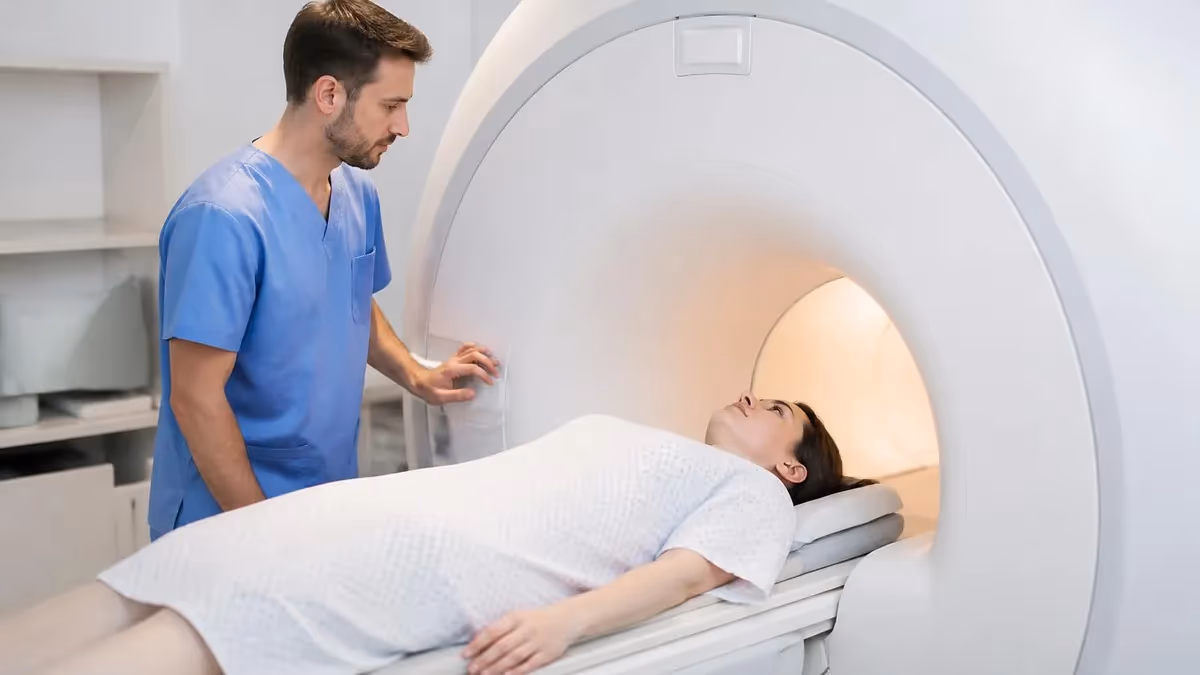
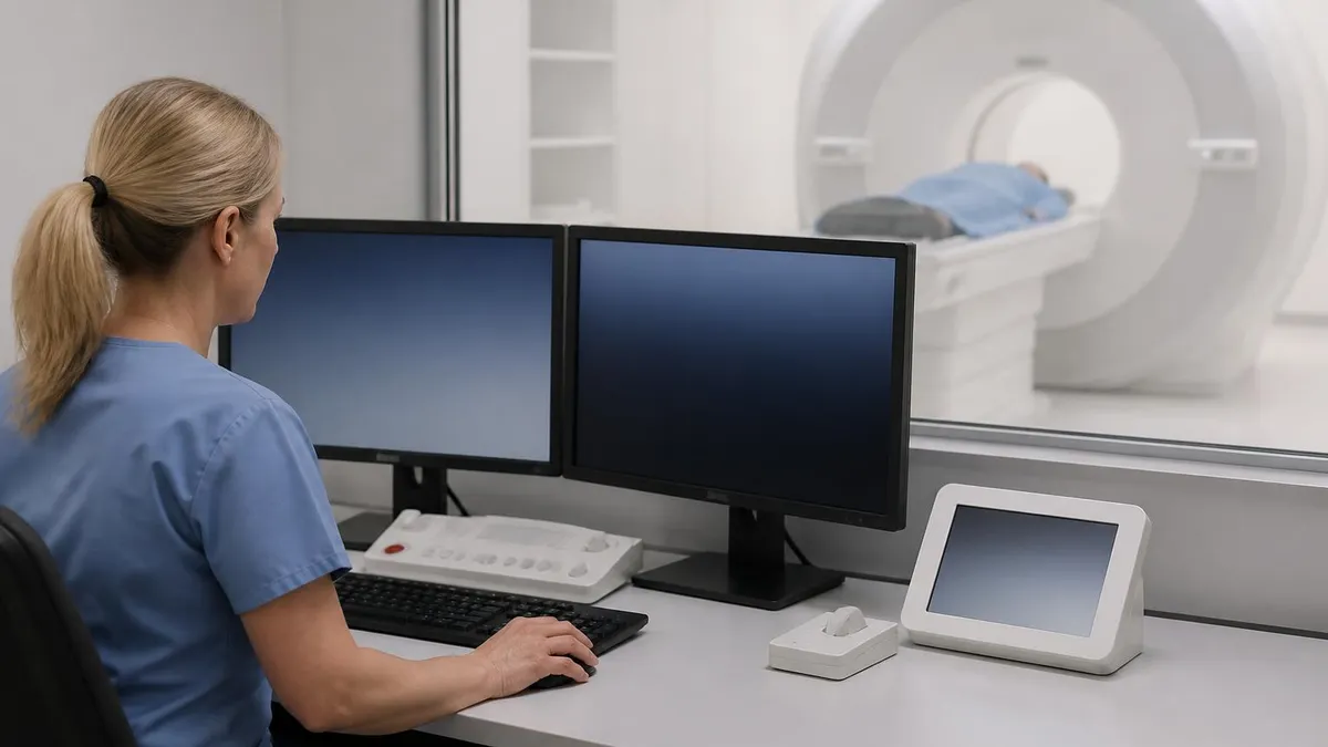
Searching for a full body mri manhattan provider has become one of the most common preventive health questions New Yorkers ask in 2026, fueled by celebrity endorsements, longevity clinics opening in Midtown, and rising consumer interest in catching disease early. A full body MRI is a non-contrast magnetic resonance scan that images the head, neck, chest, abdomen, pelvis, and sometimes the spine and extremities in a single 45 to 75 minute session, producing thousands of images without using ionizing radiation.
The appeal is obvious. Unlike CT or PET scans, MRI exposes you to zero radiation, making it suitable for repeat screening every one to three years. Manhattan now hosts more than a dozen dedicated preventive imaging centers, ranging from Prenuvo and Ezra in SoHo and the Flatiron District to hospital-based concierge programs at NYU Langone, Mount Sinai, and NewYork-Presbyterian. Pricing ranges from roughly $1,499 for a basic torso protocol to $2,999 for a comprehensive whole-body sequence with AI-assisted reporting.
Most people booking a scan want to answer one question: do I have anything serious growing inside me that I cannot feel yet? Full body MRI is genuinely good at detecting solid tumors larger than about 1 cm in the kidneys, liver, pancreas, ovaries, prostate, and brain, plus aneurysms, cysts, and structural abnormalities of the spine. It is not a substitute for mammography, colonoscopy, low-dose lung CT, or cardiac stress testing, all of which detect things MRI misses.
That nuance matters because the marketing rarely communicates it clearly. Patients sometimes assume the scan rules out cancer entirely, then skip their colonoscopy for five years and pay the price. A well-informed consumer understands the scan as one layer of surveillance, not a replacement for the rest. Reading the protocol document on any clinic's website before paying is the single best filter for separating serious providers from marketing-first operations.
Geographically, the densest cluster of providers sits below 42nd Street, with three options within a 10-minute walk of Union Square alone. Upper East Side residents have hospital-based programs within Lenox Hill and Weill Cornell, while Upper West Side patients tend to travel south or to Englewood, NJ. mri results wait times in early 2026 average two to four weeks for self-pay scans and six to eight weeks for hospital-affiliated programs that pre-screen for medical necessity.
This guide walks through what a full body scan actually captures, how Manhattan clinics compare on price and protocol, how to prepare on the day of your appointment, what your report will say and what it will leave unsaid, and how to think about follow-up if something incidental is found. It is written for the educated New Yorker who wants to make a smart decision rather than a marketing-driven one, and includes the questions you should ask before handing over your credit card.
If you are also studying MRI as a clinical professional rather than as a patient, the same principles apply in reverse: understanding what your patients have read online makes counseling them dramatically easier, and the registry concepts behind whole-body protocols come up regularly on board exams.
Full Body MRI in Manhattan by the Numbers
Top Full Body MRI Providers Near Manhattan
Located on Crosby Street, Prenuvo offers a 60-minute whole-body protocol with diffusion-weighted imaging and AI-assisted radiologist review. Self-pay pricing starts near $2,499 with optional brain MRA add-on for vascular screening.
Ezra runs a 30 to 60 minute scan in a 3T scanner near Madison Square Park, with reports delivered through a patient app within 7 to 10 business days. Tiers range from $1,495 torso-only to $2,395 full body plus prostate.
Hospital-affiliated whole-body imaging is available through NYU's executive health program with internist consultation included. Better suited for patients with family history or symptoms that justify medical necessity rather than pure screening.
Mount Sinai's preventive imaging combines whole-body MRI with genetic risk stratification, ideal for patients with strong family history of cancer. Pricing is bundled with a longevity consultation and bloodwork panel.
Several Lenox Hill Radiology outpatient centers across Manhattan accept self-referral for whole-body protocols on 1.5T and 3T magnets. Pricing is typically the lowest among brand-name options, though wait times can extend to four weeks.
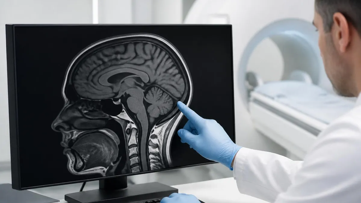
A full body MRI in 2026 is not a single sequence but a stitched mosaic of regional protocols, each tuned to the tissue beneath the coil. A typical Manhattan whole-body study includes T1-weighted and T2-weighted axial imaging from the skull base through the proximal thighs, fat-suppressed sequences across the abdomen and pelvis, diffusion-weighted imaging (DWI) of the trunk for brain tumor mri and a dedicated brain sequence with FLAIR and susceptibility weighted imaging for microbleeds and demyelinating lesions.
What this combination is genuinely good at finding is the category of solid organ lesions larger than roughly 1 centimeter. That includes renal cell carcinoma in the kidneys, hepatocellular carcinoma and metastases in the liver, pancreatic cysts and adenocarcinomas above about 1.5 cm, ovarian masses, uterine fibroids, prostate lesions on PI-RADS scoring when prostate sequences are added, and brain tumors, aneurysms larger than 3 mm, and old infarcts. Most spine pathology — disc herniations, stenosis, cord lesions — is also captured if the sagittal spine sequences are included in the package.
The list of things a full body MRI does not reliably catch is at least as important. It misses most early-stage colorectal cancers because the bowel is collapsed, gas-filled, and constantly moving. It misses lung cancers smaller than about 6 to 8 mm because air-filled lung tissue gives almost no MRI signal. It misses ductal carcinoma in situ of the breast unless dedicated breast MRI sequences are run separately with contrast. It cannot evaluate coronary artery calcification or functional cardiac ischemia, both of which require dedicated cardiac protocols.
Diffusion-weighted whole-body imaging is the technical advance that makes modern preventive scans worth their price. By measuring the restricted motion of water molecules in densely packed cellular tissue, DWI highlights malignant lesions as bright spots against a dark anatomic background, dramatically improving sensitivity for lymphoma, bone marrow metastases, and small soft-tissue tumors. Without DWI in the protocol, a so-called full body scan is dramatically less powerful, and patients should ask explicitly whether DWI is included before paying.
For comparison and context, learning how an MRI works at the physics level makes the protocol choices much more transparent. A 3T scanner delivers roughly twice the signal-to-noise of a 1.5T at equivalent scan time, which translates to higher spatial resolution or shorter exams. Most Manhattan preventive centers use 3T magnets; hospital scanners may be either field strength depending on the room.
One nuance worth understanding: most preventive scans are performed without intravenous contrast. Gadolinium contrast improves sensitivity for liver, pancreatic, and breast lesions but adds time, cost, and a small risk of allergic reaction or, in rare cases, nephrogenic systemic fibrosis in patients with poor kidney function. Skipping contrast is a deliberate choice for asymptomatic screening, where the population-level risk-benefit calculation favors a non-contrast protocol.
The takeaway: a full body MRI is a powerful but specific tool. Knowing what it captures and what it doesn't lets you slot it intelligently into a larger screening strategy that still includes age-appropriate colonoscopy, mammography, lung screening for smokers, and skin checks. It is not a single test that replaces all others, no matter how the marketing reads.
MRI Practice Test Questions
Prepare for the MRI - Magnetic Resonance Imaging exam with our free practice test modules. Each quiz covers key topics to help you pass on your first try.
MRI Knowledge
MRI Exam Questions covering Knowledge. Master MRI Test concepts for certification prep.
MRI Physics
Free MRI Practice Test featuring Physics. Improve your MRI Exam score with mock test prep.
MRI Anatomy and Pathology
MRI Test Prep for MRI Anatomy and Pathology. Practice MRI Quiz questions and boost your score.
MRI Anatomy and Positioning
MRI Questions and Answers on MRI Anatomy and Positioning. Free MRI practice for exam readiness.
MRI Contrast Agents
Free MRI Quiz on MRI Contrast Agents. MRI Exam prep questions with detailed explanations.
MRI Patient Care and Positioning
MRI Practice Questions for MRI Patient Care and Positioning. Build confidence for your MRI certification exam.
Comparing Full Body MRI Scan Manhattan Providers
Self-pay pricing in Manhattan as of mid-2026 clusters into three tiers. Entry-level scans at around $1,495 to $1,799 typically cover the torso from chest through pelvis on a 1.5T magnet, without brain imaging or DWI. These suit younger patients with no family history who want a basic look at solid organs.
Mid-tier scans at $1,999 to $2,499 add the brain, neck, and full diffusion-weighted imaging on a 3T magnet, with AI-assisted preliminary reporting. Premium tiers above $2,500 include spine sagittals, prostate or breast sub-protocols, optional cardiac structural imaging, and a one-on-one physician consultation to walk through findings. Pick the tier that matches your risk profile rather than your budget.
Is a Full Body MRI Worth It? Pros and Cons
- +Zero ionizing radiation, making repeat scans every one to three years biologically safe
- +Detects most solid organ tumors larger than 1 cm in kidneys, liver, pancreas, ovaries, and brain
- +Non-invasive, requires no fasting for most protocols, and uses no needles unless contrast is added
- +Provides a structural baseline that future scans can be compared against to detect change
- +Modern diffusion-weighted protocols are highly sensitive to lymphoma and bone marrow disease
- +Can identify aneurysms, structural heart abnormalities, and significant spine pathology incidentally
- −Misses early colorectal, lung, and breast cancers that require dedicated screening tests
- −Generates incidental findings in 30 to 40 percent of scans, many requiring follow-up imaging or biopsy
- −Not covered by most US insurance plans when ordered for asymptomatic screening purposes
- −False positives can drive anxiety, additional cost, and occasionally unnecessary invasive procedures
- −Limited evidence in long-term randomized trials that whole-body screening reduces mortality
- −Claustrophobic patients may struggle through a 60-plus minute scan even with sedation available
Pre-Scan Checklist Before Your Full Body MRI Appointment
- ✓Confirm in writing whether your scan includes diffusion-weighted imaging and brain sequences
- ✓Disclose every metal implant, pacemaker, cochlear device, surgical clip, and tattoo to the technologist
- ✓Remove all jewelry, piercings, hairpins, dentures, hearing aids, and underwire bras before entering the scan room
- ✓Wear loose clothing without metallic snaps or zippers, or plan to change into hospital scrubs on site
- ✓Eat lightly and avoid large amounts of caffeine within two hours of the appointment to reduce restlessness
- ✓Bring a list of prior imaging studies and previous radiology reports for the radiologist to compare against
- ✓If claustrophobic, request a wide-bore scanner and ask your primary care doctor for a single 0.5 mg lorazepam dose
- ✓Arrive 20 to 30 minutes early to complete the MRI safety screening questionnaire without rushing
- ✓Plan for a driver if you take any sedative, since you should not operate a car for at least four hours after
- ✓Have a follow-up plan ready for who will receive the report and which physician will counsel you on findings
Incidental findings are the norm, not the exception.
Roughly one in three asymptomatic patients who undergo a full body MRI in Manhattan will have at least one incidental finding flagged in the report, and most of these are benign cysts, lipomas, or vertebral hemangiomas that require no action. Before you book, decide in advance how you will react to ambiguous findings — both emotionally and financially — because follow-up scans, specialist consultations, and biopsies can add thousands of dollars to the original price tag.
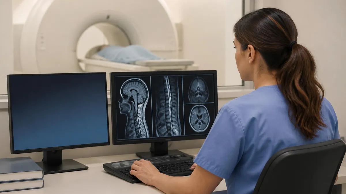
Your radiology report typically arrives one to two weeks after the scan as a 4 to 8 page PDF organized by anatomic region. Each section lists organs imaged, describes the appearance of normal structures, and then enumerates any abnormalities with their size, location, and a recommended follow-up. Modern preventive centers also attach a plain-language summary written for patients, often supplemented by annotated screenshots showing exactly where each finding sits.
The single most important sentence in the report is usually the impression at the bottom, where the radiologist condenses everything into a numbered list. Findings are flagged at one of several severity tiers: incidental and benign, requires routine follow-up imaging in 6 to 12 months, requires sub-specialist consultation, or requires urgent evaluation. Reading only the impression and ignoring the body of the report is a common mistake that misses important context about size, character, and uncertainty.
Common incidental findings include simple renal and hepatic cysts, vertebral hemangiomas, uterine fibroids, thyroid nodules, adrenal adenomas, and meningeal cysts. The overwhelming majority of these are biologically inert, and the appropriate response is documentation rather than action. The American College of Radiology publishes well-defined management guidelines (the ACR Incidental Findings white papers) that help radiologists decide which lesions need follow-up and which can simply be noted.
The trickier category is lesions that fall into the indeterminate range. A 1.2 cm pancreatic cystic lesion, for instance, sits in a zone where MRCP, EUS, or short-interval follow-up MRI may all be reasonable, depending on morphology and family history. This is where the quality of your radiologist matters enormously — board-certified abdominal sub-specialists generate dramatically more nuanced recommendations than general radiologists reading high volumes of screening studies.
If contrast was used, the report will also describe enhancement patterns of any masses, which often distinguishes benign from suspicious lesions. To understand the trade-offs of running contrast as part of your protocol, the breakdown of MRI with or without contrast walks through which clinical questions benefit from gadolinium and which do not, and why most asymptomatic screening protocols deliberately skip it.
The follow-up phase is where many patients underspend on planning. Booking a 30 to 60 minute consultation with the ordering physician or with an independent internist after the report arrives is the difference between a useful scan and a confusing one. Bring the full PDF, not just the impression, and prepare specific questions about each numbered finding before the appointment.
Document everything in a personal health folder so that any future scan can be compared directly against the baseline. Change over time is far more informative than any single snapshot, and the practical value of preventive imaging compounds with serial studies rather than living in a one-off result.
Patients with pacemakers, certain neurostimulators, cochlear implants, intraocular metal, or older aneurysm clips may be absolutely contraindicated for MRI, and entering the magnet room with an undisclosed implant has caused serious injuries. Always complete the safety screening questionnaire honestly, bring implant identification cards if available, and never assume a device is MRI-compatible without written documentation from the manufacturer or your surgeon.
Not everyone benefits equally from a full body MRI, and an honest provider will say so during the pre-scan consultation. The patients most likely to extract genuine value are those aged 40 to 70 with a meaningful family history of cancer, especially Lynch syndrome, BRCA mutations, Li-Fraumeni syndrome, or familial pancreatic cancer. For these patients, whole-body MRI surveillance has emerging evidence of mortality benefit and is sometimes recommended by national society guidelines.
A second clear-fit group includes patients with prior cancer diagnoses now in remission, who want a non-radiation modality for periodic surveillance between oncologist visits. Whole-body MRI can replace some CT-based follow-up imaging in this population and dramatically reduce cumulative lifetime radiation exposure. Coordination with the treating oncologist is essential so that the imaging informs rather than confuses the survivorship plan.
Conversely, low-risk adults under 35 with no family history typically derive little statistical benefit from screening MRI. The pre-test probability of finding a clinically significant lesion is so low that the rate of incidental false positives can actually generate net harm in the form of anxiety, additional imaging, and unnecessary procedures. For these patients, the money is usually better spent on lifestyle interventions, evidence-based screening already covered by insurance, and a continuing relationship with a thoughtful primary care doctor.
Patients with severe claustrophobia, morbid obesity exceeding scanner bore weight limits (typically 350 to 500 pounds), or non-MRI-compatible implants will face logistical barriers that may make the scan impractical or unsafe. For these individuals, low-dose CT chest screening, ultrasound of the abdomen, and dedicated organ-specific screening may be more realistic alternatives, and a frank conversation with an internist will surface the right strategy.
Pregnant patients should generally defer non-emergent imaging, and patients with severe renal disease should not receive gadolinium contrast unless absolutely necessary. Both populations can still consider non-contrast screening MRI, but the conversation with the radiologist about protocol modifications is more involved and should happen before payment.
If you are still deciding between modalities, the comparison of whats difference between mri and ct scan is essential reading because the two technologies excel at completely different things — CT for lung, bone, and acute trauma, MRI for soft tissue and central nervous system detail. They are complements, not substitutes, and a sophisticated screening strategy may pair them across years rather than choosing one and abandoning the other.
The bottom line is to match the test to the patient. Full body MRI is a powerful tool when slotted thoughtfully into a personalized prevention strategy, but it is neither a magic bullet nor a universal screening test. Talk to a physician who knows your full history before you book, and use the scan to ask better questions rather than to seek a single reassuring answer.
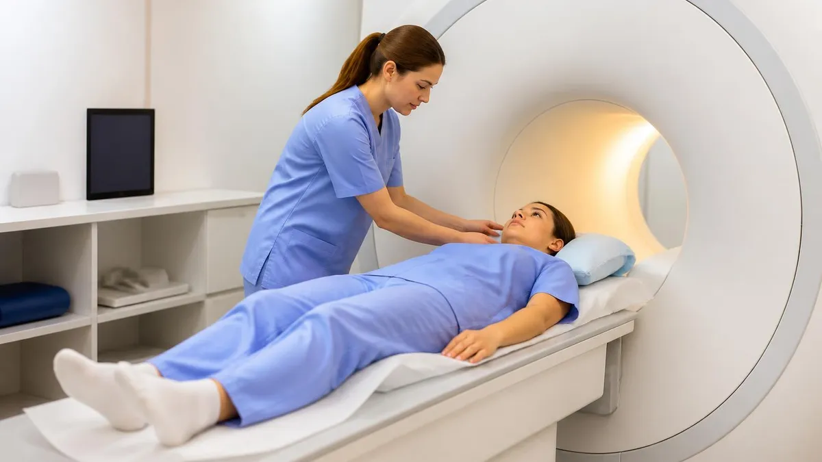
The day of the scan itself is logistically simpler than most patients expect. Arrive 20 to 30 minutes early so you have time to complete safety paperwork without rushing, change into scrubs if required, and store your belongings in a locker. Bring a photo ID and a credit card for the co-pay or self-pay balance, and confirm in advance whether your clinic emails the report or releases it through a patient portal.
Hydration is helpful in moderation. Drink a normal glass of water within two hours of the appointment so that abdominal sequences capture tissue with good contrast, but avoid downing a large bottle that will require a bathroom break mid-scan. Most protocols ask you to use the restroom immediately before the technologist positions you on the table to minimize discomfort during the longer sequences.
Once positioned, expect a coil placed over the body region being imaged, foam pads to keep you still, and headphones for music or noise reduction. The technologist will hand you a squeeze ball connected to the control room — squeeze it any time you need to pause, communicate, or stop the exam. Modern scanners include cameras and microphones, so you are never out of contact with the staff during sequences.
The noise is the part most patients underestimate. MRI gradients produce clicks, knocks, and rapid pulsing sounds that reach 100 to 120 decibels at the scanner bore, comparable to a chainsaw. The provided ear protection brings this down to safe levels, but earplugs plus over-ear headphones together are essential. Many centers stream music or even short video to a goggle display, which can make a 60-minute scan feel meaningfully shorter.
Movement is the single biggest threat to image quality. Even small shifts during a diffusion-weighted or T2-weighted sequence can blur the entire stack, occasionally requiring repeats. Practice slow nasal breathing in advance, focus on relaxing your shoulders and jaw, and resist the urge to swallow during breath-hold sequences when the technologist instructs. If you cough or sneeze, tell the staff so they can rerun that sequence rather than letting a corrupted dataset reach the radiologist.
After the scan, you can drive yourself home unless you took sedation, eat normally, and resume all activity immediately. There is no recovery period and no after-effects from the magnetic field. The waiting period for results is psychologically the hardest part for many patients — schedule something distracting during the 7 to 10 day window so you are not refreshing your patient portal hourly.
Finally, share your report with your primary care doctor regardless of whether the preventive imaging center offers follow-up. Continuity of care matters more than the scan itself. A baseline file in your physician's chart means that any future symptom, lab anomaly, or imaging study can be interpreted against your known anatomy — turning a one-time purchase into a durable medical asset that pays dividends over decades.
MRI Questions and Answers
MRI MARS Protocol: Complete Guide to Metal Artifact Reduction Sequences for Orthopedic Imaging
How Does an MRI Work: Magnetic Resonance Imaging Explained
MRI With or Without Contrast: What to Expect

The History of MRI: From Discovery to Modern Medicine
What's the Difference Between MRI and CT Scan? A Complete Comparison Guide
About the Author

Medical Laboratory Scientist & Clinical Certification Expert
Johns Hopkins UniversityDr. Sandra Kim holds a PhD in Clinical Laboratory Science from Johns Hopkins University and is certified as a Medical Technologist (MT) and Medical Laboratory Scientist (MLS) through ASCP. With 16 years of clinical laboratory experience spanning hematology, microbiology, and molecular diagnostics, she prepares candidates for ASCP board exams, MLT, MLS, and specialist certification tests.
Join the Discussion
Connect with other students preparing for this exam. Share tips, ask questions, and get advice from people who have been there.
View discussion (6 replies)