
CPT Code for Foot MRI: Complete Guide to 73718, 73719, 73720 and 73721–73723
💡 Find the right CPT code for foot MRI: 73718–73720 vs 73721–73723, contrast rules, modifiers, costs, and billing tips for patients and coders.
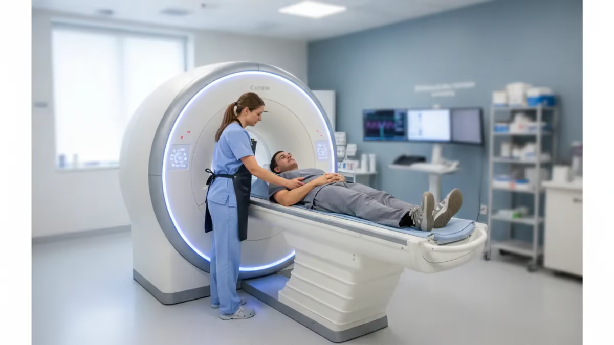
Finding the right cpt code for foot mri sounds simple until you realize the answer depends on which part of the foot was scanned, whether contrast was given, and how the ordering provider documented the clinical question. Most foot studies land in one of two code families: 73718 through 73720 for lower extremity exams that are not a joint, and 73721 through 73723 for a joint of the lower extremity. Choosing between them affects payment, denials, and patient bills.
This guide walks through each code in plain language so patients, schedulers, and billing staff can speak the same language. Our overview of the foot mri cpt code options connects the coding rules to real-world details such as contrast agents, laterality modifiers, and prior authorization. If you have ever received an explanation of benefits showing a charge you did not expect, understanding these five-digit numbers is the fastest way to ask informed questions and catch errors early.
Current Procedural Terminology, or CPT, is maintained by the American Medical Association and updated every year. Every imaging study receives a code describing the body region, the technique, and the contrast status. Payers use that code to decide coverage, set the allowed amount, and apply edits that flag mismatches with the diagnosis. A single digit difference, such as 73720 versus 73718, can change reimbursement by several hundred dollars at a hospital outpatient department.
Foot MRI is one of the more ambiguous areas for coders because the foot contains both joints and non-joint structures. The ankle and hindfoot are generally treated as a joint, while a study aimed at a metatarsal, a toe, or a plantar soft tissue mass may fit the non-joint codes. Radiology practices often publish internal rules, and the ordering provider's wording on the requisition usually settles the close calls.
These codes describe the technical and professional components together unless a modifier splits them. Hospitals typically bill the technical component, modifier TC, for the scanner, technologist, and facility overhead. The radiologist bills the professional component, modifier 26, for interpreting the images and writing the report. Freestanding imaging centers frequently bill both together as a global service with no modifier, which is why one patient sees two separate bills and another only one.
Patients benefit from knowing the code before the scan, not after. Calling the imaging center with the scheduled code lets your insurer confirm whether prior authorization is needed and what your deductible will cover. Many commercial plans require authorization for every outpatient MRI, and a mismatch between the authorized code and the performed code is a common reason claims are denied. A two-minute phone call can prevent weeks of appeals.
Below you will find a quick-reference breakdown of each code, billing modifiers, cost ranges, and a checklist for avoiding denials. The information is educational and reflects general US coding conventions. Always confirm details with your payer, your imaging facility, and a certified coder, because local coverage policies and annual CPT updates can change the specifics. Bookmark this page and revisit it before your next scheduled study.
Foot MRI Coding by the Numbers
Foot MRI CPT Codes at a Glance
Used for lower extremity MRI that is not a joint and uses no contrast. Typical examples include a forefoot bone concern, a toe, or a soft tissue mass assessed on pre-contrast images alone. It is the baseline code for many non-joint foot studies.
Reported when contrast is given and imaging occurs only after injection. It is uncommon because radiologists usually want pre-contrast comparison images, but it may apply to follow-up studies focused on enhancement of a known mass or collection.
Covers images before and after contrast for a non-joint lower extremity region. Radiologists choose it for suspected infection, tumor, or deep abscess where comparing both phases separates fluid from enhancing tissue and improves diagnostic confidence.
The most commonly used code for ankle and hindfoot studies, and for midfoot or forefoot exams centered on a joint. It suits tendon, ligament, cartilage, and bone marrow evaluation, and it is usually the first-line choice for chronic pain.
73722 is contrast only, while 73723 is without followed by with contrast. Both apply when a joint is imaged for synovitis, infection, or inflammatory arthritis, and both require documentation showing the agent, volume, and phases performed.
How to Choose Between Joint and Non-Joint Foot MRI Codes
The central question is whether the scan targets a joint. CPT does not publish a definitive list of joints for every body part, so radiology societies and coding references fill the gap. The ankle, subtalar joint, and tarsometatarsal joints are generally regarded as joints, which points to 73721 through 73723. A study of a heel pad mass or a long bone segment of the forefoot fits 73718 through 73720.
Consider a runner with suspected second metatarsophalangeal joint capsulitis. The clinical question centers on the joint capsule, plantar plate, and cartilage, so most coders select 73721. Now consider a patient with a palpable lump on the arch. The question is the soft tissue mass itself, not an articulation, so 73718 or 73719 is more defensible. The same anatomy can produce different codes depending on the indication documented on the order.
Ankle protocols complicate matters because patients frequently say foot MRI when the physician ordered an ankle study for heel pain or Achilles problems. The requisition says ankle, the technologist scans from the distal tibia through the hindfoot, and the code is 73721. If the patient later sees a bill describing a foot MRI, the wording differs while the code remains correct. Always compare the CPT code, not the casual description, when reviewing paperwork.
Forefoot studies use smaller coils and thinner slices, often 3 millimeters or less, to visualize the plantar plate, sesamoids, and interdigital nerves. Hindfoot and ankle studies cover larger fields of view to include tendons such as the posterior tibial, peroneal, and Achilles. These protocol differences do not change the code on their own, but they support the documentation that explains why a joint or non-joint code was chosen.
Bilateral studies add another wrinkle. If both feet are scanned in a single session, the facility reports the code twice, once with the RT modifier and once with LT, or uses modifier 50 when the payer prefers it. Multiple procedure reductions may apply to the second side. Patients ordering bilateral exams for conditions such as inflammatory arthritis should ask about the expected allowed amount before the appointment.
Trauma and post-surgical cases rely on the same logic. A suspected Lisfranc injury involves the midfoot joint complex, so 73721 fits. A stress fracture of the fifth metatarsal shaft is often scanned with the non-joint codes. When hardware is present, the scan may take longer and require metal artifact reduction sequences, but the CPT code does not change; only the time and technologist effort increase.
When the choice is truly ambiguous, the safest approach is to ask the radiologist or the coding lead at the facility before scheduling. Payers audit imaging claims where diagnosis codes do not support the selected procedure, so aligning the ICD-10 diagnosis, the clinical indication, and the CPT code protects everyone. Document the specific structure of concern, such as possible plantar plate tear at the second MTP joint, rather than the vague phrase foot pain.
CPT Code for Foot MRI: Without Contrast, With Contrast, or Both
Most foot MRI studies are performed without contrast, which means the cpt code for foot mri will be 73718 for non-joint exams or 73721 for joint exams. Non-contrast imaging is excellent for tendons, ligaments, bone marrow edema, stress fractures, and plantar fasciitis. Fluid-sensitive sequences such as STIR and proton density with fat saturation show inflammation clearly without any injection, which keeps cost, scan time, and risk lower for the patient.
Because no needle is involved, a non-contrast study usually takes 25 to 40 minutes and requires no kidney function screening. Insurers also tend to approve non-contrast requests faster. If your doctor is evaluating chronic heel pain, an Achilles tear, or a suspected Morton neuroma, the non-contrast code is typically the correct starting point, and contrast can be added later only if the images show something that remains unexplained.
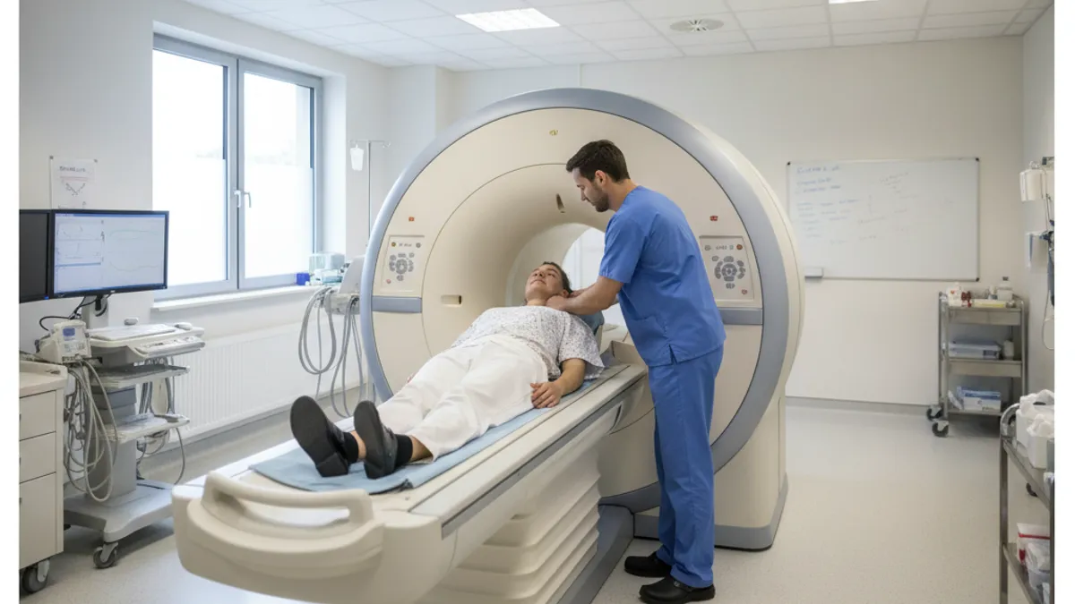
Is MRI Better Than X-ray or CT for Foot Problems?
- +Shows tendons, ligaments, cartilage, and nerves that X-ray cannot display
- +Detects bone marrow edema and stress fractures before they appear on radiographs
- +Uses no ionizing radiation, making it suitable for repeat imaging
- +Distinguishes infection, tumor, and inflammation with contrast-enhanced sequences
- +Provides multiplanar views that help surgeons plan procedures
- +Identifies plantar plate tears, neuromas, and Achilles pathology with high accuracy
- −Costs more than X-ray and often more than ultrasound
- −Takes 30 to 60 minutes compared with seconds for a radiograph
- −Metal implants and some devices may cause artifacts or safety concerns
- −Often requires prior authorization from the insurer
- −Contrast adds an IV, a separate drug charge, and a kidney function check for some patients
- −Fine cortical bone detail is usually better seen on CT
Checklist: Getting the CPT Code for Foot MRI Right
- ✓Confirm whether the ordered study targets a joint or a non-joint region.
- ✓Match the code to the contrast plan: without, with, or without then with.
- ✓Verify the requisition names the exact structure of concern.
- ✓Add the correct RT or LT laterality modifier for each foot scanned.
- ✓Check whether the payer requires prior authorization before scheduling.
- ✓Make sure the authorized CPT code matches the code performed on scan day.
- ✓Pair the CPT code with a specific ICD-10 diagnosis, not a vague symptom.
- ✓Document contrast agent, volume, and phases in the technologist notes.
- ✓Attach recent X-ray results and conservative treatment history to the request.
- ✓Request a written price estimate that lists the CPT code and facility fee.
The Ankle Is a Joint, and Many Foot Scans Use 73721
Many scans described as foot MRI are billed with 73721 because the ankle, hindfoot, and midfoot joints count as a joint of the lower extremity. Check the CPT number on your authorization and bill, not the casual study name, so the two always match.
Modifiers, Contrast Billing, and What a Foot MRI Costs
Modifiers tell the payer how and where the service was delivered. For foot MRI, the most common are RT and LT for laterality, TC and 26 for the technical and professional split, and 59 or XS when a distinct service must be separated from another procedure. Missing laterality modifiers is among the simplest errors to prevent, yet it triggers automatic rejections because the payer cannot tell which foot was imaged.
Contrast billing follows its own track. The scan code does not include the drug, so the facility adds an HCPCS supply code that matches the specific agent, along with units based on the volume administered. A typical foot study uses around 10 to 15 milliliters depending on patient weight. Auditors compare the units billed with the volume documented in the technologist notes, so accurate charting protects the claim from later recoupment.
Price varies enormously. Medicare pays hospital outpatient departments and freestanding centers different rates, and commercial insurers negotiate their own. A foot or ankle MRI without contrast may cost a self-pay patient at a freestanding center roughly $400 to $900, while the same scan at a hospital can be billed at $1,500 to $3,000 before insurance adjustments. Studies with and without contrast generally cost 20 to 40 percent more.
Patients with high-deductible plans pay the full allowed amount until the deductible is met, so choosing a freestanding imaging center can save hundreds of dollars. Ask for the CPT code, the facility fee, and the radiologist's fee before booking. Federal price transparency rules require hospitals to publish standard charges, and many centers quote a cash price lower than the insurance-billed amount. Comparing two or three facilities takes an hour and can save a significant sum.
Medicare coverage depends on medical necessity and the local coverage determination for the jurisdiction. Foot MRI is generally covered when conventional radiographs are inconclusive and the clinical picture suggests conditions such as occult fracture, osteomyelitis, tendon rupture, or tumor. Medicare Advantage plans may add prior authorization requirements, so beneficiaries should not assume original Medicare rules apply to their plan. Keep copies of the order and any authorization number.
Facilities must also consider bundling edits. National Correct Coding Initiative edits can bundle two MRI codes performed on the same body region and date. For example, billing a foot MRI and an ankle MRI in the same session may raise questions about overlap unless the documentation shows two distinct regions and separate clinical indications. Coders sometimes append modifier 59 with supporting notes to justify both studies.
Sedation, anesthesia, and special coils can generate additional charges outside the base CPT code. Patients who are claustrophobic may receive oral anxiolytics, which are billed separately if supervised by the facility. Foot studies are usually performed feet-first and many patients tolerate them without sedation because the head stays outside the bore. Still, ask whether any extras are planned so the final statement holds no surprises.
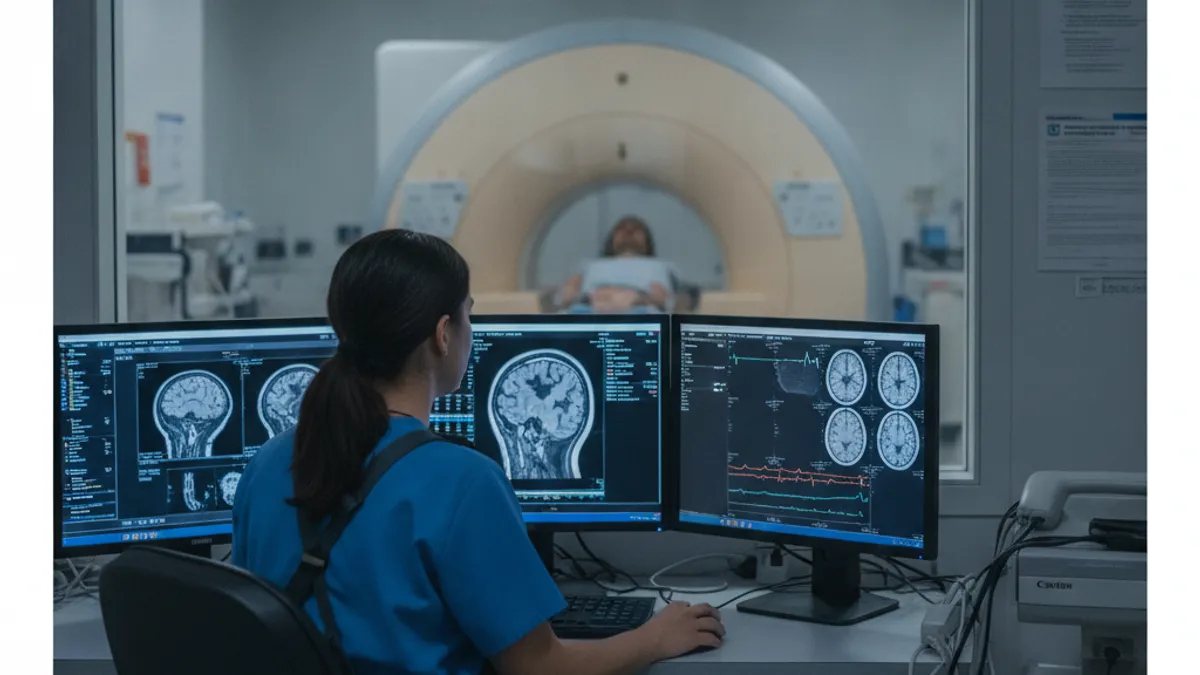
If the radiologist adds contrast on scan day, the performed code changes from 73721 to 73723, for example. Contact your insurer or the facility's authorization team first, because an unauthorized code change is a common cause of denied claims.
ICD-10 Diagnosis Pairing, Prior Authorization, and Denials
Every CPT code needs a diagnosis code to prove medical necessity. For foot MRI, common ICD-10 pairings include M72.2 for plantar fascial fibromatosis, M77.40 for metatarsalgia, M79.671 and M79.672 for pain in the right or left foot, and M25.571 or M25.572 for pain in the ankle and joints of the foot. Specific diagnoses outperform vague symptom codes when reviewers assess whether the scan was justified.
Payers typically expect conservative care first. Many medical policies require a documented period, often four to six weeks, of rest, bracing, anti-inflammatory medication, or physical therapy, plus a recent weight-bearing X-ray before approving MRI. Exceptions exist for red flags such as suspected infection, tumor, acute high-energy trauma, or neurological deficit. Referral offices should attach the clinic note that shows what was tried and why the images are needed.
Prior authorization is frequently handled by a radiology benefit manager such as eviCore or Carelon, which applies clinical criteria based on professional guidelines. The ordering office submits the requested CPT code, the diagnosis, and clinical history. Approval is tied to a specific code, facility, and date range, so a switch from 73721 to 73723 on the scan day may require an updated authorization before the contrast is given.
Denials usually come from a few predictable causes: no authorization, mismatched codes, missing laterality, a diagnosis that does not support the study, or duplicate imaging within a short window. Reading the denial reason code on the explanation of benefits tells you which problem to fix. Many denials are overturned on first appeal when the practice supplies the clinic notes and a letter of medical necessity from the ordering physician.
If you are appealing as a patient, act quickly. Most commercial plans give 180 days from the denial date for an internal appeal, and Medicare allows 120 days to request a redetermination after the initial decision. Request the full claim file, ask your doctor for a short supporting letter, and cite the specific clinical findings. If the internal appeal fails, the Affordable Care Act guarantees an external review by an independent organization for most plans.
Repeat imaging has its own rules. A follow-up foot MRI several weeks after surgery for a Charcot reconstruction or osteomyelitis treatment can be justified, but an identical study repeated without a change in symptoms may be denied. Record the new clinical question, such as persistent drainage or worsening pain, and cite what the first study showed. That context separates a necessary reassessment from a duplicate request.
Finally, remember that coding accuracy supports good care as well as payment. A correct code and indication help the radiologist tailor the protocol, for instance by adding fat-suppressed post-contrast sequences when infection is suspected. The scan answers the clinical question better, the report reaches the surgeon faster, and the patient avoids a repeat visit. Precision on paper translates directly into better imaging in the scanner.
Practical Tips Before You Book Your Foot MRI
Start with the order. Ask your provider which structure they are worried about, whether contrast is planned, and which CPT code they expect the facility to use. Having that information when you call to schedule lets the staff verify authorization correctly. If the answers are vague, ask the office to update the order with a specific indication, such as suspected stress fracture of the second metatarsal, before you book.
Compare facilities. Hospital outpatient departments often charge significantly more than freestanding imaging centers for the same CPT code, even when the same radiologists read the studies. Ask each location for the total patient responsibility, including the technical fee, professional fee, and any contrast charges. If you are paying cash, request a self-pay quote in writing, since discounts of 30 to 50 percent are sometimes available for prepayment.
Prepare for safety screening. Tell the technologist about pacemakers, cochlear implants, aneurysm clips, neurostimulators, metal fragments, or previous surgeries, and bring implant cards when you have them. Foot hardware such as screws and plates is usually MRI conditional, but the technologist needs details. Remove jewelry, belts, and clothing with metallic threads, and wear comfortable clothing without zippers so you may not need to change into a gown.
Plan for comfort. Foot scans usually place the feet in a dedicated coil with your head outside or near the scanner opening, which helps people who feel anxious in tight spaces. The machine is loud, so use the earplugs or headphones provided. Staying perfectly still matters because motion blurs small structures like the plantar plate. Ask for a pillow under your knees if lying flat hurts your back.
Bring what helps the radiologist. Prior X-rays, earlier MRI reports, surgical notes, and a list of medications make comparison easier. If you have kidney disease or diabetes and contrast is planned, bring recent lab results showing kidney function. Arrive 15 to 20 minutes early to complete screening forms, and confirm the CPT code printed on your paperwork matches the one on your authorization.
After the scan, review your bills. Compare the explanation of benefits with the facility statement, checking CPT code, laterality, date of service, and contrast line items. Call the billing office if you see two scanning charges for one foot or a code you did not expect. Many errors are corrected with a single phone call, and catching them within the first 30 days makes the process far smoother.
Use practice questions to cement what you learned, especially if you work in imaging or are preparing for a registry exam. Understanding how contrast, field strength, and pulse sequences affect the study helps you understand why codes are divided the way they are. Pairing coding knowledge with clinical and physics knowledge makes you a stronger technologist, scheduler, or informed patient advocate over the long term.
MRI Questions and Answers
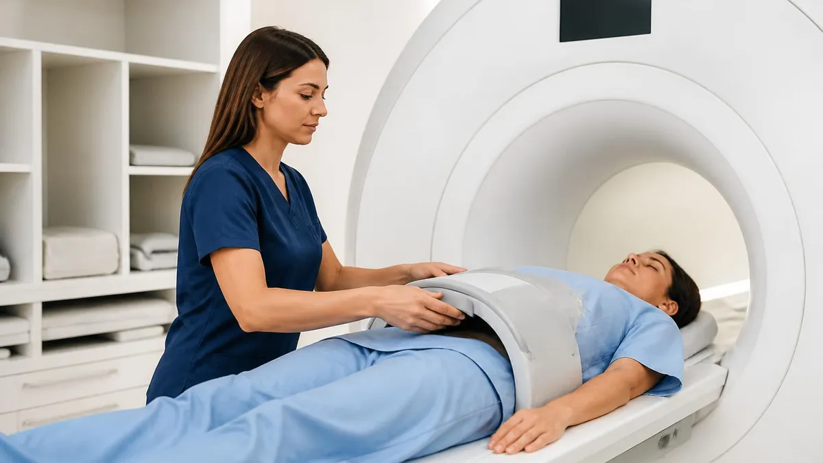
Contrast MRI: How It Works, Safety, Side Effects, and What to Expect

Can You Feel an MRI? What to Expect During Your MRI Procedure Experience
Multiple Sclerosis MRI: McDonald Criteria, Lesion Patterns and Protocols (2026 Guide)
Osteomyelitis MRI: Complete Guide to Imaging Findings, Protocols, Sequences, and Diagnostic Accuracy
Where to Get an MRI: Facilities, Costs, and How to Choose
About the Author

Medical Laboratory Scientist & Clinical Certification Expert
Johns Hopkins UniversityDr. Sandra Kim holds a PhD in Clinical Laboratory Science from Johns Hopkins University and is certified as a Medical Technologist (MT) and Medical Laboratory Scientist (MLS) through ASCP. With 16 years of clinical laboratory experience spanning hematology, microbiology, and molecular diagnostics, she prepares candidates for ASCP board exams, MLT, MLS, and specialist certification tests.
Join the Discussion
Connect with other students preparing for this exam. Share tips, ask questions, and get advice from people who have been there.
View discussion (8 replies)