
Bleed on MRI: How Brain Hemorrhages Appear on Magnetic Resonance Imaging 2026
🧠 Bleed on MRI explained — how hemorrhages appear on T1, T2, FLAIR, and GRE sequences, stages of blood breakdown, and clinical findings.
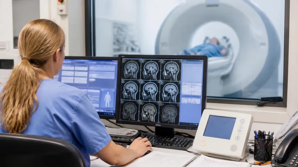
A bleed on MRI is one of the most clinically urgent findings a radiologist or MRI technologist can encounter, and understanding exactly how hemorrhage appears across different pulse sequences is essential knowledge for anyone working in neuroimaging. Unlike CT, which detects acute blood as a hyperdense white region due to protein content, MRI signal characteristics of hemorrhage change dramatically over time as hemoglobin undergoes a complex series of biochemical transformations. Recognizing these stages allows clinicians to date a bleed and guide treatment decisions. Practice with the free MRI - Magnetic Resonance Imaging practice test on PracticeTestGeeks (200+ questions with answer explanations).
When blood enters the brain parenchyma, the subarachnoid space, or the subdural and epidural compartments, it does not simply stay as whole blood. Oxyhemoglobin breaks down into deoxyhemoglobin, then methemoglobin, and finally hemosiderin, each molecule carrying distinct paramagnetic properties that interact with the MRI magnetic field in unique ways. This stepwise degradation is what creates the characteristic signal evolution radiologists rely on to stage intracranial hemorrhage precisely.
The appearance of brain bleed mri findings also depends heavily on which sequence is used. T1-weighted images, T2-weighted images, FLAIR, gradient echo (GRE), and susceptibility-weighted imaging (SWI) each highlight different aspects of hemorrhage. A lesion that is nearly invisible on one sequence may be strikingly obvious on another, which is why a comprehensive MRI brain protocol for suspected hemorrhage typically includes multiple sequence types acquired in specific orientations.
From a patient safety and clinical perspective, identifying a bleed on MRI quickly can be life-saving. Epidural hematomas can expand rapidly due to arterial bleeding and cause transtentorial herniation within minutes. Subdural hematomas, especially chronic ones in elderly or anticoagulated patients, may accumulate slowly but still demand urgent surgical evaluation. Parenchymal hemorrhages from hypertension, amyloid angiopathy, or underlying vascular malformations each carry distinct treatment pathways that imaging helps define.
For MRI students, registry candidates, and practicing technologists, mastering hemorrhage imaging is not merely an academic exercise. The ARRT MRI registry examination regularly tests knowledge of sequence-specific hemorrhage appearance, the role of hemoglobin oxidation states, and the indications for contrast administration in suspected vascular lesions. Understanding the physics behind why blood looks different at various stages also reinforces broader concepts in MRI physics such as T1 shortening, T2 shortening, and the effects of magnetic susceptibility.
This comprehensive guide covers the biochemical stages of hemorrhage, the MRI signal characteristics at each stage across multiple sequences, the major types of intracranial bleeds encountered clinically, and practical guidance for optimizing MRI protocols to detect even small microhemorrhages. Whether you are preparing for your registry exam or deepening your clinical knowledge, the information presented here provides a thorough, evidence-based foundation for understanding how MRI reveals bleeding in the brain with exceptional sensitivity and specificity compared to other imaging modalities.
MRI: upcoming dates and official changes
- Aug 17, 2026: ARRT launched a separate entry-level Imaging Assistant, Magnetic Resonance (I.A.) credential. Applications made Aug. 17–Oct. 14, 2026 paid a reduced $75 fee for testing Oct. 15–Nov. 30, 2026. Applicants after Oct. 14, 2026 pay $225 and can test on or after Jan. 1, 2027. The I.A. exam also requires a scaled section score of 7.5 or higher in MRI Screening and Safety.[4]
- The ARRT MRI postprimary exam has 220 questions: 200 scored and 20 unscored pilot items.[1]
- The ARRT MRI primary pathway exam has 230 questions: 200 scored and 30 unscored pilot items.[2]
- The ARRT MRI postprimary exam allows 210 minutes of test time within a 230-minute (3 hour 50 minute) appointment.[1]
- The ARRT MRI primary pathway exam allows 230 minutes of test time within a 250-minute (4 hour 10 minute) appointment.[2]
- ARRT exams, including MRI, are scored on a 1–99 scale, and candidates need a total scaled score of 75 to pass.[3]
- The ARRT MRI postprimary application fee is $225, or $450 for candidates using an NMTCB or ARDMS credential as the supporting category.[4]
- The ARRT MRI primary pathway application fee is $225.[4]
- ARRT MRI technologists holding an R.T. credential must complete Continuing Qualifications Requirements (CQR) every 10 years.[5]
- ARRT MRI credential holders renew every year for $65 and must complete 24 CE credits every two-year biennium.[5]
Numbers in brackets link to the official sources listed under Sources & last verified. Dates that have passed are removed automatically.
MRI Exam at a Glance
| Item | Detail | Source · Verified |
|---|---|---|
| Number of questions (Postprimary pathway) | 220 questions (200 scored + 20 pilot)[1] | assets-us-01.kc-usercontent.com · Oct 16, 2026 |
| Number of questions (Primary pathway) | 230 questions (200 scored + 30 pilot)[2] | assets-us-01.kc-usercontent.com · Oct 16, 2026 |
| Time limit (Postprimary pathway) | 210 minutes test time (230-minute appointment)[1] | assets-us-01.kc-usercontent.com · Oct 16, 2026 |
| Time limit (Primary pathway) | 230 minutes test time (250-minute appointment)[2] | assets-us-01.kc-usercontent.com · Oct 16, 2026 |
| Passing score | Scaled score of 75 (scale 1–99)[3] | The American Registry of Radiologic Technologists · Oct 16, 2026 |
| Exam fee (Postprimary pathway) | $225 application fee ($450 with an NMTCB or ARDMS supporting credential)[4] | The American Registry of Radiologic Technologists · Oct 16, 2026 |
| Exam fee (Primary pathway) | $225 application fee[4] | The American Registry of Radiologic Technologists · Oct 16, 2026 |
| Validity | Continuing Qualifications Requirements (CQR) every 10 years[5] | assets-us-01.kc-usercontent.com · Oct 16, 2026 |
| Renewal | Annual renewal, $65/year; 24 CE credits every 2 years[5] | assets-us-01.kc-usercontent.com · Oct 16, 2026 |
| Delivery | Computer-based at Pearson VUE test centers[2] | assets-us-01.kc-usercontent.com · Oct 16, 2026 |
| Eligibility (Postprimary pathway) | Supporting credential (ARRT, NMTCB or ARDMS) + structured education + clinical experience[1] | assets-us-01.kc-usercontent.com · Oct 16, 2026 |
| Eligibility (Primary pathway) | ARRT-verified MRI program + associate degree or higher + ethics[2] | assets-us-01.kc-usercontent.com · Oct 16, 2026 |
| Score release | Preliminary score at the test center; official results in about 10 business days[3] | The American Registry of Radiologic Technologists · Oct 16, 2026 |
| Testing window | One-year exam window per application[2] | assets-us-01.kc-usercontent.com · Oct 16, 2026 |
Free MRI - Magnetic Resonance Imaging Study Tools
Interactive MRI - Magnetic Resonance Imaging resources on PracticeTestGeeks, free to start:
- MRI - Magnetic Resonance Imaging practice test — 200+ questions with instant scoring and answer explanations
- MRI - Magnetic Resonance Imaging flashcards — review key terms and concepts card by card
- MRI - Magnetic Resonance Imaging study guide — topic-by-topic overview of what the exam covers
- MRI - Magnetic Resonance Imaging cheat sheet — one-page printable summary for last-minute review
Brain Hemorrhage and MRI — Key Numbers
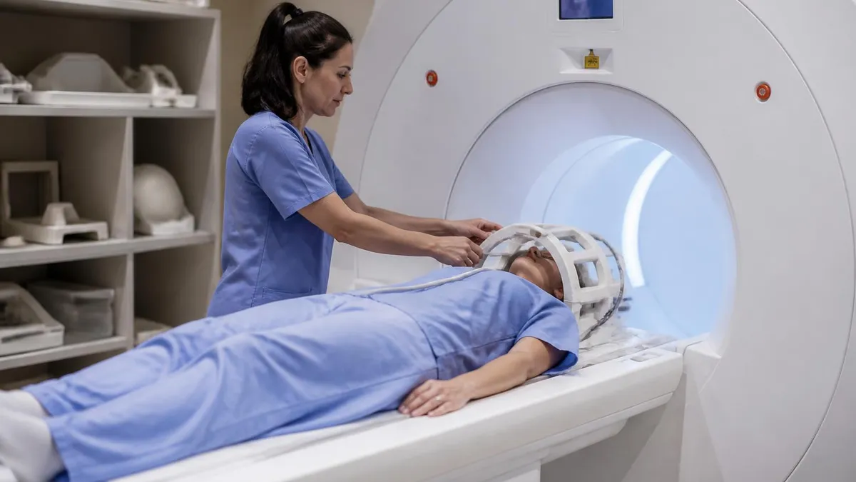
Stages of Brain Hemorrhage on MRI
Hyperacute (0–12 hours): Oxyhemoglobin
Acute (12–48 hours): Deoxyhemoglobin
Early Subacute (2–7 days): Intracellular Methemoglobin
Late Subacute (1–2 weeks): Extracellular Methemoglobin
Chronic (weeks to months): Hemosiderin
Understanding how each MRI sequence portrays a brain bleed requires grasping the fundamental physics of how paramagnetic molecules alter local magnetic fields. T1-weighted sequences are sensitive to longitudinal relaxation times: substances that shorten T1 appear bright. Methemoglobin dramatically shortens T1, which is why subacute hematomas glow white on T1 images. This property makes T1 the sequence of choice for confidently identifying subacute blood and distinguishing it from other T2 hyperintense lesions like edema, infarct, or demyelination.
T2-weighted imaging is sensitive to transverse relaxation. Deoxyhemoglobin and intracellular methemoglobin cause significant T2 shortening through a mechanism called susceptibility-induced dephasing, making acute and early subacute hematomas appear dark on T2. This is a crucial examination point: a lesion that is hypointense on T2 should always prompt consideration of hemorrhage, melanin, calcification, or high protein content. Correlating T2 hypointensity with clinical history and other sequences resolves most diagnostic uncertainty.
FLAIR (Fluid-Attenuated Inversion Recovery) suppresses free CSF signal and is particularly useful for detecting subarachnoid hemorrhage (SAH). In SAH, blood mixes with cerebrospinal fluid, which normally appears black on FLAIR. The protein and cellular content of blood prevents full suppression, making subarachnoid blood appear as sulcal hyperintensity on FLAIR. This sequence is more sensitive than non-contrast CT for SAH beyond 24 hours, an important clinical fact for triage decision-making in emergency settings.
Gradient echo (GRE) sequences are exquisitely sensitive to magnetic susceptibility effects. Because they lack a 180-degree refocusing pulse, dephasing from local field inhomogeneities accumulates rather than being corrected. Any hemoglobin breakdown product with paramagnetic or ferromagnetic properties — deoxyhemoglobin, intracellular methemoglobin, hemosiderin — produces a blooming artifact that makes the lesion appear far larger than its true anatomical size. This blooming effect is clinically valuable: it makes even tiny microhemorrhages visible that would be invisible on conventional spin-echo sequences.
Susceptibility-weighted imaging (SWI) takes GRE principles further by combining magnitude and phase information to create images with maximum sensitivity to magnetic susceptibility differences. SWI can detect microhemorrhages as small as 1 to 2 millimeters, making it the gold standard for evaluating cerebral amyloid angiopathy, diffuse axonal injury from trauma, cavernous malformations, and radiation-induced microbleeds. A dedicated SWI sequence should be included in any MRI brain protocol where hemorrhage is clinically suspected, even when conventional sequences appear normal.
Diffusion-weighted imaging (DWI) adds another dimension to hemorrhage evaluation. Hyperacute hematomas can restrict diffusion and appear bright on DWI due to the high viscosity and cellular packing of the clot, though this finding is not as consistent as in ischemic stroke. More importantly, DWI helps distinguish hemorrhagic transformation of an infarct from a primary hemorrhage: the surrounding penumbra of acute ischemia will show DWI restriction, while primary hypertensive hemorrhage typically lacks this surrounding diffusion abnormality. Combining DWI with the other sequences provides a comprehensive picture of the injury mechanism.
Contrast-enhanced MRI has a selective role in hemorrhage evaluation. In most cases of hypertensive or traumatic hemorrhage, gadolinium is not needed acutely. However, when there is clinical concern for an underlying lesion — tumor, vascular malformation, abscess, or metastasis — post-contrast T1 imaging is essential. Ring-enhancing lesions surrounded by hemorrhage raise concern for glioblastoma or metastatic disease. Arteriovenous malformations and dural arteriovenous fistulas may show enhancing vessels adjacent to the hematoma. MR angiography (MRA) or MR venography (MRV) can be added to evaluate for aneurysm or venous sinus thrombosis as the underlying etiology.
Types of Intracranial Hemorrhage on MRI
Intraparenchymal hemorrhage (IPH) occurs within the brain tissue itself and accounts for approximately 10 to 15 percent of all strokes in the United States. The most common cause is chronic hypertension, which damages small penetrating arteries in characteristic locations: the basal ganglia (especially the putamen), thalamus, pons, and cerebellum. On MRI, IPH follows the classic signal evolution from hyperacute through chronic stages described above, with surrounding vasogenic edema appearing as T2/FLAIR hyperintensity in the adjacent white matter.
Secondary causes of IPH include cerebral amyloid angiopathy, which preferentially causes lobar hemorrhages in elderly patients and is associated with multiple cortical microhemorrhages visible on SWI. Hemorrhagic transformation of ischemic infarct, coagulopathy, venous sinus thrombosis, and underlying tumors also produce parenchymal bleeds with distinct imaging signatures. Careful MRI sequence analysis combined with clinical context helps identify the etiology and guide the appropriate treatment pathway, whether medical management, surgical evacuation, or intervention on an underlying lesion.
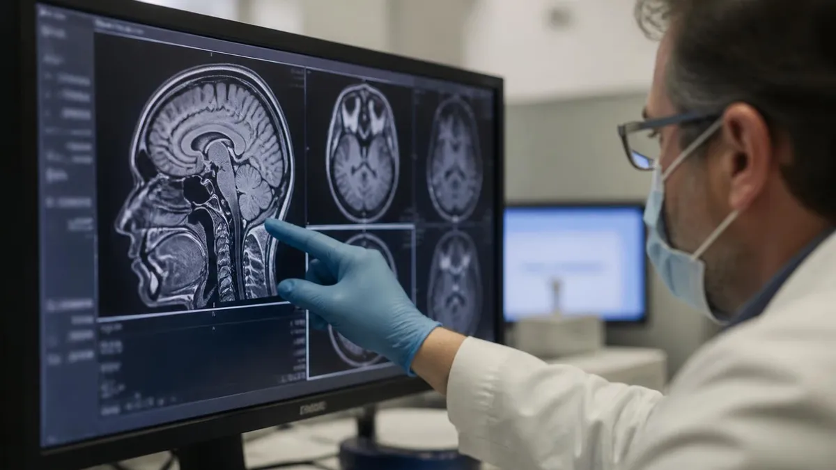
MRI vs. CT for Detecting Brain Bleeds: Key Comparisons
- +Superior sensitivity for subacute and chronic hemorrhage beyond 24–72 hours
- +SWI detects microhemorrhages invisible on CT and conventional MRI
- +FLAIR is more sensitive than CT for subarachnoid hemorrhage after 24 hours
- +Multi-sequence protocol can identify underlying cause (tumor, AVM, venous thrombosis)
- +No ionizing radiation — safer for repeated imaging in young or pregnant patients
- +Better soft tissue contrast reveals associated edema, herniation risk, and penumbra
- −Slower acquisition time (20–40 minutes vs. minutes for CT) — problematic in unstable patients
- −Less sensitive than CT for hyperacute hemorrhage in the first 6 hours
- −Contraindicated in patients with certain implants, pacemakers, or metallic foreign bodies
- −Patient cooperation required — motion degrades image quality and may require sedation
- −Limited availability in emergency and rural settings compared to CT scanners
- −Cost is significantly higher than CT, and insurance prior authorization may delay scanning
Pros and Cons at a Glance
| Pros | Cons |
|---|---|
| SWI detects microhemorrhages invisible on CT and conventional MRI | Contraindicated in patients with certain implants, pacemakers, or metallic foreign bodies |
| Multi-sequence protocol can identify underlying cause (tumor, AVM, venous thrombosis) | Patient cooperation required — motion degrades image quality and may require sedation |
| No ionizing radiation — safer for repeated imaging in young or pregnant patients | Limited availability in emergency and rural settings compared to CT scanners |
| Better soft tissue contrast reveals associated edema, herniation risk, and penumbra | Cost is significantly higher than CT, and insurance prior authorization may delay scanning |
MRI Brain Hemorrhage Protocol Checklist
- ✓Obtain complete clinical history including anticoagulant use, trauma, hypertension, and symptom onset time.
- ✓Screen for MRI contraindications: pacemakers, cochlear implants, metallic orbital foreign bodies, and aneurysm clips.
- ✓Include T1-weighted sequence to detect subacute methemoglobin as T1 hyperintensity.
- ✓Include T2-weighted sequence to identify edema, mass effect, and acute hemoglobin dephasing.
- ✓Acquire FLAIR sequence to evaluate for subarachnoid blood in sulci and periventricular lesions.
- ✓Add GRE or SWI sequence — mandatory for detecting microhemorrhages and confirming blood products.
- ✓Include DWI to distinguish hemorrhagic transformation of infarct from primary hemorrhage.
- ✓Consider post-contrast T1 if underlying lesion (tumor, AVM, abscess) is suspected clinically.
- ✓Add MRA (or CTA) if aneurysm, AVM, or vasculitis is in the differential diagnosis.
- ✓Document hematoma size, location, surrounding edema, midline shift, and ventricular involvement in the report.
The SWI Blooming Effect: Small Bleed, Big Signal
Susceptibility-weighted imaging (SWI) causes paramagnetic blood products to appear significantly larger than their true anatomical size — a phenomenon called blooming artifact. While this can occasionally complicate size measurements, it is clinically invaluable: it makes microhemorrhages smaller than 2 mm visible that would be completely missed on CT or conventional MRI. Always include SWI in any suspected hemorrhage protocol, especially when evaluating for cerebral amyloid angiopathy, traumatic diffuse axonal injury, or cavernous malformations.
The clinical significance of identifying a bleed on MRI extends far beyond simply confirming that blood is present. Location, size, associated findings, and the underlying etiology together determine urgency, surgical candidacy, and long-term prognosis. A hypertensive hemorrhage in the putamen of a patient who is awake and following commands is managed differently from a cerebellar hemorrhage causing obstructive hydrocephalus, which is a neurosurgical emergency regardless of clot volume. MRI provides the spatial resolution and soft tissue detail needed to make these distinctions accurately.
Mass effect and midline shift are critical measurements derived from MRI in hemorrhagic stroke management. When the brain shifts more than 5 millimeters across the midline, the risk of transtentorial herniation rises sharply. MRI beautifully demonstrates the anatomy of herniation: the uncus of the temporal lobe impinging on the brainstem, effacement of the ipsilateral perimesencephalic cistern, and compression of the posterior cerebral artery territory that can cause secondary occipital lobe infarction. These MRI findings directly inform neurosurgical decisions about craniotomy or craniectomy timing.
Cerebral venous sinus thrombosis (CVST) is an underdiagnosed cause of hemorrhagic venous infarction that MRI identifies with high accuracy. Unlike arterial infarcts, venous infarcts often present as cortical hemorrhages crossing arterial territories, associated with significant surrounding edema disproportionate to the apparent clot size. MR venography (MRV) reveals absence of normal flow signal in the affected dural sinus. The combination of cortical hemorrhage on standard sequences and sinus occlusion on MRV is diagnostic. Importantly, anticoagulation is the treatment of choice for CVST even in the presence of hemorrhage — a counterintuitive clinical decision that MRI helps justify.
Cavernous malformations (cavernomas) are vascular lesions that bleed repeatedly at low volume, producing the classic MRI appearance called a popcorn lesion: a mixed-signal core from blood products of different ages surrounded by a complete hemosiderin ring that appears intensely dark on GRE and SWI. They do not enhance on contrast and are not visible on conventional angiography (cryptic vascular malformations). Identifying cavernomas on MRI is clinically important because they carry a recurrent bleeding risk of approximately 3 to 6 percent per year per lesion, and surgical resection is considered for accessible symptomatic lesions.
Diffuse axonal injury (DAI) from high-velocity traumatic brain injury produces microscopic shearing hemorrhages at the gray-white junction, corpus callosum, and brainstem. These are invisible on CT and may not be apparent on standard T2 sequences, leading to underestimation of injury severity. SWI reveals dozens to hundreds of microhemorrhagic foci in severe DAI, providing a much more accurate picture of injury burden. Research shows that SWI lesion count correlates with Glasgow Outcome Scale scores, making it an imaging biomarker of prognosis — a rapidly growing area of clinical application for advanced MRI techniques in trauma.
Hemorrhagic metastases present a diagnostic challenge because the blood products can obscure the underlying tumor on routine sequences. Post-contrast T1 imaging is essential in this setting: tumor tissue enhances while the hemorrhagic component does not, revealing nodular or rim enhancement that marks the neoplastic origin. Certain tumor types are particularly prone to hemorrhage, including renal cell carcinoma, melanoma, thyroid carcinoma, choriocarcinoma, and lung carcinoma — a list worth memorizing for registry examination purposes. The presence of multiple hemorrhagic lesions in a patient with known malignancy essentially confirms metastatic disease.
Cerebral amyloid angiopathy (CAA) deserves special attention because its MRI diagnosis directly affects anticoagulation and thrombolysis decisions. CAA results from deposition of amyloid protein in the walls of cortical and leptomeningeal vessels, weakening them and predisposing to lobar hemorrhage. The Boston Criteria for CAA diagnosis rely on MRI findings: multiple lobar cortical microhemorrhages on SWI, cortical superficial siderosis (linear hemosiderin deposition along the cortical surface), and white matter hyperintensities in a posterior predominant distribution. A patient meeting modified Boston Criteria for probable CAA should generally not receive anticoagulation for atrial fibrillation without careful risk-benefit discussion.
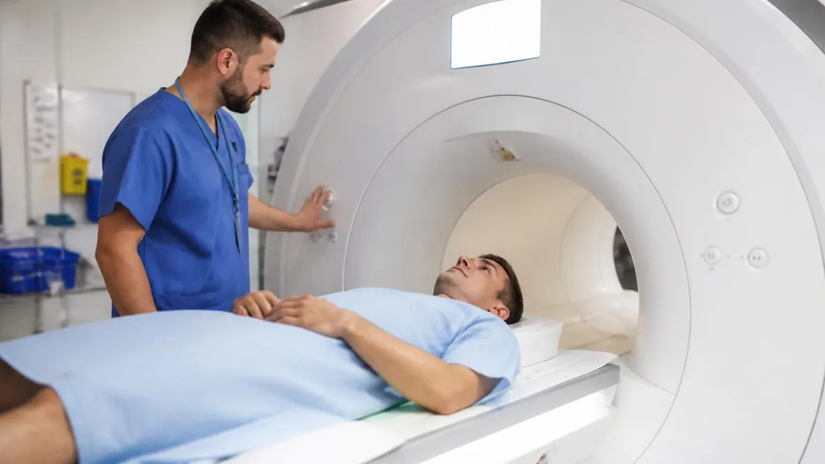
MRI should NOT replace CT as the first-line imaging study when acute intracranial hemorrhage is suspected in an unstable or rapidly deteriorating patient. CT is faster, more accessible, and highly sensitive for acute blood in the hyperacute window (0–6 hours). Reserve MRI for stable patients, subacute presentations, or when the underlying etiology needs further characterization. Always ensure MRI safety screening is completed before scanning any patient with suspected head trauma, as retained metallic foreign bodies may not be apparent without screening.
Registry examination questions about bleed on MRI focus heavily on matching the signal intensity pattern to the correct stage and explaining the underlying hemoglobin chemistry. The most commonly tested scenario involves identifying the acute stage: a hematoma that is isointense on T1 and hypointense on T2 corresponds to the deoxyhemoglobin phase, when intact red blood cells containing paramagnetic deoxyhemoglobin create T2 shortening through susceptibility-induced spin dephasing. Examinees must also know that this acute phase blooms on GRE — a classic exam distractor that rewards those who understand sequence physics rather than pattern memorization alone.
Another high-yield registry topic involves the distinction between intracellular and extracellular methemoglobin. Both stages produce T1 hyperintensity because methemoglobin is paramagnetic and shortens T1 regardless of its compartmental location. However, on T2, intracellular methemoglobin remains hypointense (because cell membranes maintain susceptibility gradients), while extracellular methemoglobin becomes hyperintense (because lysis removes the compartmental boundaries). This T2 signal flip from dark to bright, while T1 remains bright, is the signature of the transition from early to late subacute hemorrhage and is a reliable dating criterion that examiners test frequently.
The FLAIR sequence and subarachnoid hemorrhage represent another must-know registry pairing. When a patient presents with sudden severe headache (thunderclap headache) and CT is negative — a scenario that occurs in up to 15 percent of SAH cases within 6 hours of onset due to rapid dilution of blood — LP or MRI FLAIR becomes the next diagnostic step.
FLAIR shows sulcal hyperintensity where blood-tinged CSF has higher protein content and cellular elements that prevent the CSF signal from being fully suppressed by the inversion recovery pulse. Knowing this sequence-pathology relationship earns points on the registry and matters enormously in clinical practice.
SWI versus GRE is a comparison that appears in advanced registry questions. Both sequences detect blood products through susceptibility effects, but SWI is consistently more sensitive for microhemorrhages due to its use of both magnitude and phase information and its higher spatial resolution.
For clinical practice, the practical takeaway is that if your institution's MRI scanner has SWI capability, it should replace or supplement GRE in any brain hemorrhage protocol. For registry purposes, know that SWI creates its enhanced contrast through post-processing of the phase image multiplied into the magnitude image — a detail that tests physics understanding beyond simple sequence recognition.
Contrast administration in the context of hemorrhage is another area where registry candidates are frequently tested. Gadolinium-based contrast agents are generally safe to administer in hemorrhagic stroke when an underlying lesion is suspected, but the timing matters. In the acute phase, enhancement may be limited.
In the subacute phase, breakdown of the blood-brain barrier around the hematoma cavity produces rim enhancement that should not be mistaken for a ring-enhancing neoplasm. Knowing that inflammatory rim enhancement around a resolving hematoma peaks at approximately 3 to 6 weeks and resolves without treatment is a clinically important point that distinguishes it from tumoral enhancement, which persists or grows over time.
MR spectroscopy (MRS) and perfusion imaging have emerging roles in hemorrhage workup. MRS can detect elevated lipid and lactate peaks in and around acute hematomas, and the absence of N-acetyl aspartate (NAA) — a neuronal marker — within the hematoma core confirms non-viable tissue. Perfusion-weighted imaging assesses the penumbra surrounding a hemorrhage, identifying ischemic tissue at risk. These advanced techniques are increasingly included in research protocols and are beginning to appear in registry study materials as MRI technology becomes more sophisticated and accessible in clinical practice.
For students preparing for the ARRT MRI registry, a systematic approach to hemorrhage imaging pays dividends across multiple question categories. Start by memorizing the five stages of hemoglobin breakdown and their T1/T2 signatures as a table. Then learn which sequences are most sensitive for each stage and why, grounding the pattern in basic MRI physics.
Finally, understand the clinical context: where the bleed is, what caused it, and how imaging findings change management. This three-layer framework — biochemistry, physics, clinical application — covers the range of hemorrhage questions that appear on the registry and builds durable knowledge that serves throughout a career in MRI.
When preparing for registry exams or clinical practice in MRI, developing a systematic mental framework for evaluating any potential brain hemorrhage on MRI will serve you far better than rote memorization of individual facts. The approach begins the moment the order arrives: review the clinical indication carefully. A patient with sudden-onset severe headache requires SAH evaluation with FLAIR as a priority.
A patient with hypertension and acute focal deficit needs sequences that can distinguish acute hemorrhagic stroke from ischemic stroke, including DWI to look for coexisting infarction. A patient with known malignancy and new neurological symptoms needs post-contrast imaging to evaluate for hemorrhagic metastases.
Protocol optimization for hemorrhage detection requires collaboration between technologists, radiologists, and clinical teams. At minimum, a brain hemorrhage MRI protocol should include T1, T2 or T2*, FLAIR, DWI, and SWI or GRE. Adding post-contrast T1 and MRA when clinically indicated transforms a diagnostic study into a comprehensive vascular workup.
Field strength matters: 3T MRI provides significantly better sensitivity for microhemorrhages on SWI compared to 1.5T due to the greater susceptibility effects at higher field strength. If patients are imaged at 1.5T and SWI findings are equivocal, repeat imaging at 3T may be warranted in high-stakes clinical scenarios such as suspected amyloid angiopathy.
Patient preparation for a brain hemorrhage MRI study involves both technical and communication components. Technologists should explain to patients and families why the scan is being performed without creating alarm, since many patients are already frightened by their symptoms. Motion is the enemy of MRI quality: even small movements during SWI acquisition create ghost artifacts that can mimic or obscure microhemorrhages.
Coaching patients to remain still, using foam ear inserts to reduce scanner noise anxiety, and positioning the head securely in the coil with appropriate padding all contribute to diagnostic-quality images. Sedation may be necessary in agitated or confused patients, requiring coordination with nursing or anesthesia staff.
Reporting and communication of hemorrhage findings require a structured approach. Radiologists and technologists alike should be familiar with the key measurements: hematoma maximum diameter in three planes, estimated volume using the ABC/2 method (A × B × C / 2), degree of midline shift in millimeters, presence or absence of intraventricular extension, and status of the cisterns as a proxy for herniation risk.
These measurements should be communicated immediately to the clinical team when a new or expanding hemorrhage is identified. Most institutions have stroke alert or neurosurgery notification protocols that technologists participate in by escalating urgent findings through proper channels without delay.
Emerging MRI techniques are expanding the capabilities of hemorrhage detection and characterization. Quantitative susceptibility mapping (QSM) converts SWI phase data into a quantitative map of magnetic susceptibility, allowing measurement of iron concentration within a hematoma over time. This has potential applications in monitoring hematoma resolution, predicting rebleeding risk, and differentiating calcification from hemorrhage — two entities that are both dark on GRE/SWI but have opposite susceptibility values on QSM. Ultra-high-field 7T MRI offers extraordinary spatial resolution for depicting tiny vascular structures adjacent to a hematoma, though clinical applications remain primarily in research settings for now.
Artificial intelligence and machine learning are increasingly being applied to hemorrhage detection on MRI. Deep learning algorithms trained on large labeled datasets can now detect intracranial hemorrhage with sensitivity and specificity comparable to trained radiologists, and FDA-cleared commercial tools are being integrated into clinical workflows as triage aids.
These AI tools flag studies containing potential hemorrhage for priority review, reducing the time from scan completion to radiologist interpretation in busy emergency settings. For MRI technologists, understanding the role of AI in the diagnostic workflow is becoming part of professional competency, and registry content is beginning to reflect this evolving landscape of clinical practice.
The intersection of clinical decision-making and advanced MRI technology creates a dynamic and intellectually rewarding specialty area within radiology and radiologic technology. Whether you are a student encountering hemorrhage imaging for the first time or an experienced technologist seeking to deepen your understanding of advanced sequences, the field of brain hemorrhage MRI continues to evolve with new techniques, new clinical indications, and new levels of diagnostic precision. Investing time in mastering the fundamentals of hemoglobin biochemistry, pulse sequence physics, and clinical application creates a durable foundation that supports lifelong learning and career-long contributions to patient care in neuroimaging.
If you want more targeted practice, the mri abdomen and pelvis cpt code page covers the same material with additional questions and explanations.
If you want more targeted practice, the open mri near me page covers the same material with additional questions and explanations.
Sample MRI - Magnetic Resonance Imaging Practice Questions
Try these questions from our free MRI - Magnetic Resonance Imaging practice tests. The correct answer and an explanation follow each question.
MR spectroscopy (MRS) of the brain measures metabolite concentrations. Which metabolite, reduced in neuronal loss and gliosis, is considered a marker of neuronal integrity?
- A. Choline (Cho)
- B. Creatine (Cr)
- C. N-acetylaspartate (NAA)
- D. Lactate (Lac)
Answer: C. N-acetylaspartate (NAA)
NAA (N-acetylaspartate) is located predominantly in neurons; its reduction indicates neuronal loss or dysfunction, making it a key spectroscopic marker of neuronal integrity.
The landmark for centering an MRI brain exam using a head coil is located at:
- A. Top of the skull
- B. Nasion or bridge of the nose
- C. External auditory meatus
- D. Occiput
Answer: B. Nasion or bridge of the nose
The nasion (bridge of the nose) is the standard external landmark used to center the isocenter of the MRI system for brain imaging.
Which anatomical landmark is used to prescribe the standard axial plane for brain MRI?
- A. Frankfurt plane
- B. Anterior commissure–posterior commissure (AC-PC) line
- C. Hard palate line
- D. Orbitomeatal baseline
Answer: B. Anterior commissure–posterior commissure (AC-PC) line
Axial brain MRI slices are typically aligned parallel to the AC-PC line for standardized anatomical coverage and comparison.
The magnetic field's intensity is varied from head to foot by this gradient coil.
- A. Z
- B. X
- C. Y
Answer: A. Z
In most MRI systems, the main magnetic field (B0) is oriented along the Z-axis, which typically runs from the patient's head to foot. The Z-gradient coil is specifically designed to create a linear variation in the magnetic field strength along this Z-axis. This allows for precise slice selection and spatial encoding in the head-to-foot dimension.
Take the full MRI - Magnetic Resonance Imaging practice test
MRI Questions and Answers
Sources & last verified
- assets-us-01.kc-usercontent.com — number of questions, time limit, eligibility (official source). “MRI Scored Items 200 Pilot Items 20 Total Items 220 Test Time 210 min” Verified Oct 16, 2026.
- assets-us-01.kc-usercontent.com — number of questions, time limit, delivery, eligibility (official source). “MRI Scored Items 200 Pilot Items 30 Total Items 230 Test Time 230 min” Verified Oct 16, 2026.
- The American Registry of Radiologic Technologists — passing score, score release (official source). “Our exam scoring scale ranges from 1 to 99, and you’ll need a total score of 75 to pass the exam.” Verified Oct 16, 2026.
- The American Registry of Radiologic Technologists — exam fee, other (official source). “Postprimary (M, CT, MR, BD, CI, VI, VS, BS, R.R.A.): $225 Postprimary (CT, MR, BD, or VS—using NMTCB as supporting category): $450 Postprimary (MR, VS,…” Verified Oct 16, 2026.
- assets-us-01.kc-usercontent.com — validity, renewal (official source). “Completing your Continuing Qualifications Requirements (CQR) every 10 years (if you have an R.T. or R.R.A. credential)” Verified Oct 16, 2026 · source checked Oct 16, 2026.
How we verify: editorial & fact-checking policy. Report an error: contact us.
DWI MRI: Diffusion-Weighted Imaging Explained (2026 Guide)
Side Effects of Brain MRI: Understanding Risks, Safety, and What Every Patient Should Know
MRI Lumbar Spine Without Contrast: Complete Guide to the Scan, What It Shows, and How to Prepare
MRI History: From Nuclear Resonance to Modern Scanners

MRI of Cervical Spine: Complete Guide to Imaging, Anatomy, Pathology, and Patient Preparation
About the Author

Medical Laboratory Scientist & Clinical Certification Expert
Johns Hopkins UniversityDr. Sandra Kim holds a PhD in Clinical Laboratory Science from Johns Hopkins University and is certified as a Medical Technologist (MT) and Medical Laboratory Scientist (MLS) through ASCP. With 16 years of clinical laboratory experience spanning hematology, microbiology, and molecular diagnostics, she prepares candidates for ASCP board exams, MLT, MLS, and specialist certification tests.