ACL MRI: How Magnetic Resonance Imaging Diagnoses Anterior Cruciate Ligament Tears in 2026 July
ACL MRI guide covering protocols, sequences, findings, 💯 and accuracy for diagnosing anterior cruciate ligament tears and associated knee injuries.
An ACL MRI is the gold-standard imaging study for evaluating the anterior cruciate ligament, the critical stabilizer that prevents the tibia from sliding forward beneath the femur. When a patient hears a pop, feels the knee give way, and develops rapid swelling after a pivoting injury, magnetic resonance imaging becomes the single most important diagnostic tool. Unlike X-ray or ultrasound, MRI visualizes the ligament directly, alongside menisci, cartilage, and bone marrow edema patterns that confirm the mechanism of injury and guide surgical decision-making.
Radiologists rely on a combination of proton density, T2-weighted, and fat-suppressed sequences in sagittal, coronal, and axial planes to construct a complete picture of the ligament. A normal ACL appears as a taut, dark band of fibers running from the lateral femoral condyle to the anterior tibial spine, parallel to the roof of the intercondylar notch. When that fiber pattern becomes wavy, discontinuous, or replaced by fluid signal, the radiologist begins building a case for partial or complete rupture with high diagnostic confidence.
The clinical importance of an accurate ACL MRI cannot be overstated. Studies published in radiology journals consistently report sensitivity above 92% and specificity above 95% for complete ACL tears when modern 1.5T or 3T scanners are used with dedicated knee coils. That accuracy directly influences whether a patient receives conservative rehabilitation, ACL reconstruction with autograft, or staged surgery to address combined ligament and meniscus pathology before chronic instability sets in.
Beyond the ligament itself, an ACL MRI captures the so-called terrible triad and its variants, including medial collateral ligament injuries, medial or lateral meniscal tears, and the classic kissing contusion pattern at the lateral femoral condyle and posterolateral tibial plateau. These secondary findings often tell the radiologist exactly what happened, even when the patient cannot recall the precise moment of injury. The bone marrow edema footprint is essentially a fingerprint of the pivot-shift mechanism.
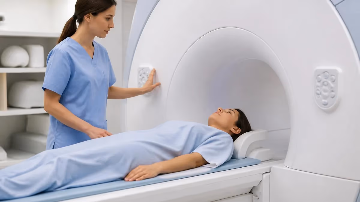
For technologists, ACL MRI presents a series of practical challenges. The patient must remain still for 20 to 30 minutes despite knee pain, and the knee should be slightly externally rotated by 10 to 15 degrees to align the ACL with the sagittal plane. Slice thickness, field of view, and the choice between 2D fast spin echo and 3D isotropic acquisitions all influence whether a small partial tear is captured or missed. Quality positioning is non-negotiable for diagnostic accuracy.
Whether you are a radiology resident, an MRI technologist preparing for the ARRT advanced registry, or a patient researching your own scan, understanding how ACL MRI works empowers better clinical conversations. If you are new to the broader topic, the what is an MRI test primer offers a useful foundation before diving into ligament-specific protocols. This guide walks through every layer of the exam, from sequence selection to grading criteria, with the depth a working technologist or trainee needs.
By the end of this article you will be able to identify normal and abnormal ACL signal, recognize the secondary signs that boost diagnostic confidence, troubleshoot common artifacts, and counsel patients on what to expect during the exam. We will also review the most recent ACR Appropriateness Criteria, current 3T protocols used in major sports medicine centers, and the practice questions you should expect on the ARRT MRI registry exam covering musculoskeletal anatomy and pathology.
ACL MRI by the Numbers
ACL Anatomy and Injury Mechanism
Tightens in flexion and primarily resists anterior tibial translation when the knee is bent. This bundle is the more commonly torn portion in pivoting injuries and is the focus of single-bundle reconstruction techniques used in most US sports medicine centers.
Tightens in extension and contributes to rotational stability, especially during the pivot-shift maneuver. Isolated posterolateral bundle tears are uncommon but can produce subtle rotational instability that is missed on standard physical exam yet visible on high-resolution MRI.
The ACL inserts on the anterior intercondylar area of the tibia between the medial and lateral tibial spines. Avulsion fractures here are more common in skeletally immature patients and require careful evaluation on sagittal and coronal MRI sequences.
The ligament originates on the posteromedial aspect of the lateral femoral condyle. Femoral-side avulsions and proximal stump tears are common and influence whether a primary repair, augmentation, or full reconstruction is performed surgically.
The standard ACL MRI protocol in 2026 combines high-resolution two-dimensional fast spin echo sequences with selective three-dimensional isotropic acquisitions that allow multiplanar reformatting. A typical exam includes sagittal proton density with fat saturation, sagittal T2 fat-saturated, coronal proton density, coronal T2 fat-saturated, and axial proton density fat-saturated sequences. Some institutions add a 3D PD CUBE or VISTA sequence to reconstruct oblique sagittal images aligned with the long axis of the ligament, which dramatically improves visualization of partial tears.
Sagittal imaging is the workhorse for ACL evaluation because the ligament runs roughly parallel to that plane. A slice thickness of 3 to 3.5 millimeters with no interslice gap is standard at 1.5T, while 3T scanners can push that to 2.5 millimeters without sacrificing signal-to-noise ratio. Field of view should be tight, typically 14 to 16 centimeters, with a matrix of at least 320 by 256 to preserve the spatial resolution required to resolve individual ligament fibers and small chondral defects on adjacent surfaces.
Positioning is one of the most underappreciated variables in ACL MRI quality. The patient is placed supine with the knee in 5 to 15 degrees of external rotation, a maneuver that aligns the obliquely oriented ACL with the sagittal plane and reduces the volume averaging that hides partial tears. Padding under the knee for slight flexion of 10 to 15 degrees relaxes the joint, improves patient comfort over the scan duration, and reduces motion artifact, which is the single most common cause of repeat sequences in busy outpatient centers.
Coil selection matters enormously. A dedicated 8-channel or 16-channel transmit-receive knee coil maximizes signal-to-noise and allows aggressive parallel imaging factors that shorten scan time without degrading image quality. Older quadrature coils still produce diagnostic images but lack the resolution needed for cartilage assessment and small ligament fiber detail. Technologists should confirm the coil is centered on the joint line, not the patella, before starting localizer sequences to avoid wrap artifacts that compromise the lateral structures.
Contrast is rarely needed for routine ACL evaluation. Direct MR arthrography with intra-articular gadolinium injection is reserved for postoperative graft assessment when imaging is ambiguous, or for evaluating chronic instability with suspected meniscal root tears. Indirect arthrography with intravenous contrast plays a smaller role and is generally limited to inflammatory or infectious workups. Most acute and chronic ACL injuries are diagnosed confidently on non-contrast studies when the protocol is executed correctly. The MRI medical abbreviation guide explains how exam codes communicate these protocol variations across electronic health record systems.
Scan time discipline keeps the exam under 30 minutes, which improves patient compliance and throughput. Modern compressed sensing and deep learning reconstruction allow protocols that previously took 35 minutes to complete in 18 to 22 minutes with equal or better image quality. These accelerated sequences are now FDA cleared on most major platforms and are increasingly standard in sports medicine programs handling high volumes of ACL evaluations across collegiate and professional athletics during the fall and winter seasons.
Finally, the MRI technologist must screen for contraindications before bringing any patient into the scanner room. Implanted devices, retained metallic foreign bodies, and certain neurostimulators require careful evaluation of MR conditional labeling. Renal function screening is unnecessary for non-contrast ACL exams but becomes relevant if direct arthrography or contrast-enhanced cartilage assessment is planned. Patient safety screening remains the foundation of every MRI workflow regardless of the body part being imaged or the urgency of the clinical question.
MRI Practice Test Questions
Prepare for the MRI - Magnetic Resonance Imaging exam with our free practice test modules. Each quiz covers key topics to help you pass on your first try.
MRI Knowledge
MRI Exam Questions covering Knowledge. Master MRI Test concepts for certification prep.
MRI Physics
Free MRI Practice Test featuring Physics. Improve your MRI Exam score with mock test prep.
MRI Anatomy and Pathology
MRI Test Prep for MRI Anatomy and Pathology. Practice MRI Quiz questions and boost your score.
MRI Anatomy and Positioning
MRI Questions and Answers on MRI Anatomy and Positioning. Free MRI practice for exam readiness.
MRI Contrast Agents
Free MRI Quiz on MRI Contrast Agents. MRI Exam prep questions with detailed explanations.
MRI Patient Care and Positioning
MRI Practice Questions for MRI Patient Care and Positioning. Build confidence for your MRI certification exam.
Reading the ACL MRI Scan
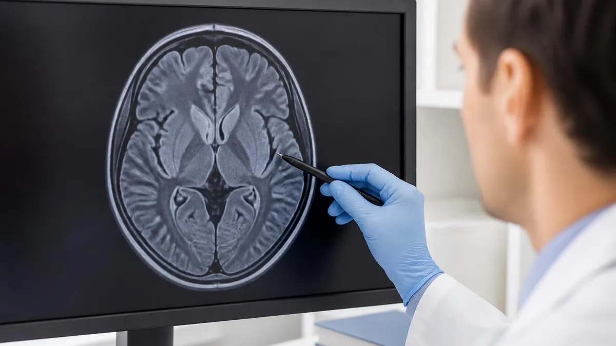
A normal ACL appears on sagittal images as a straight, taut, low-signal band running from the posteromedial aspect of the lateral femoral condyle to the anterior intercondylar area of the tibia. The fibers should be parallel to Blumensaat line, the roof of the intercondylar notch. Mild striation between the anteromedial and posterolateral bundles is normal and should not be mistaken for a tear.
On coronal imaging, the ligament is visible as a continuous structure with uniform low signal on proton density and T2 sequences. Axial images show the ligament as an ovoid low-signal structure in the intercondylar notch. The footprint, fiber bundle pattern, and absence of fluid signal collectively define a normal exam and serve as the baseline against which all pathology is compared on subsequent reads.
MRI vs Clinical Exam Alone for ACL Diagnosis
- +Direct visualization of the ligament fibers and tear morphology
- +Detects concomitant meniscal, cartilage, and collateral ligament injuries
- +Identifies bone marrow edema patterns confirming injury mechanism
- +Sensitivity above 92 percent for complete tears at 1.5T or 3T
- +Provides surgical roadmap before reconstruction is scheduled
- +Documents preexisting degenerative changes that affect prognosis
- −Higher cost than physical exam, ranging from $400 to $2,500
- −Scan time of 20 to 30 minutes can be uncomfortable for acute injuries
- −Contraindicated in some patients with implanted devices
- −Partial tear sensitivity is significantly lower than complete tear sensitivity
- −Image quality is operator and protocol dependent
- −Acute hemarthrosis can obscure subtle findings in the first 48 hours
ACL MRI Technologist Checklist
- ✓Complete MRI safety screening including implants and foreign bodies
- ✓Verify renal function only if contrast arthrography is planned
- ✓Position patient supine with knee in 5 to 15 degrees external rotation
- ✓Pad behind the knee for 10 to 15 degrees of comfortable flexion
- ✓Center the dedicated knee coil over the joint line, not the patella
- ✓Set field of view to 14 to 16 centimeters with matrix at least 320 by 256
- ✓Acquire sagittal PD, sagittal T2 FS, coronal PD, coronal T2 FS, axial PD FS
- ✓Use slice thickness of 3 to 3.5 mm at 1.5T or 2.5 mm at 3T
- ✓Add oblique sagittal 3D sequence aligned with ACL axis if partial tear suspected
- ✓Review images before releasing patient to confirm diagnostic quality
The Lateral Notch Sign
A deepening of the lateral femoral condylar sulcus greater than 2 millimeters, known as the lateral notch sign, is highly specific for ACL injury even when the ligament itself is partially obscured by hemarthrosis. Always check the sagittal lateral compartment slice closely before signing out the study.
Grading ACL injuries on MRI follows a practical three-tier system that aligns with surgical decision-making. Grade I represents a sprain with intact fibers and mild edema, grade II represents a partial tear with some fiber disruption but overall continuity, and grade III represents a complete rupture. While orthopedic surgeons may use additional descriptors based on examination findings such as the pivot-shift grade, the radiology report should always specify the location and extent of fiber discontinuity to facilitate surgical planning.
Secondary signs dramatically increase diagnostic confidence when the primary ligament appearance is ambiguous due to hemarthrosis or partial volume averaging. The most reliable secondary sign is the kissing contusion pattern, which shows bone marrow edema at the central lateral femoral condyle and the posterolateral aspect of the lateral tibial plateau. This pattern reflects the impaction that occurs as the tibia subluxes anteriorly during the pivot-shift mechanism that ruptures the ligament in the first place.
Anterior tibial translation greater than 5 to 7 millimeters relative to the femur on sagittal images is another strong secondary sign of complete ACL deficiency. It is measured by drawing a line tangent to the posterior femoral condyle and assessing how far the posterior tibial cortex sits behind that line. A second sagittal measurement, the posterior cruciate ligament angle, becomes more obtuse when the ACL is torn because the PCL buckles into a more vertical configuration as the tibia drifts anteriorly under gravity.
The posterior horn of the lateral meniscus normally sits covered by the posterior overhang of the lateral tibial plateau. When the tibia translates forward in an ACL-deficient knee, the meniscus becomes uncovered, a sign known as the uncovered lateral meniscus sign. Quantitatively, more than 3.5 millimeters of meniscus exposed beyond the tibial cortex is considered abnormal. This finding is highly specific and is one of the easiest to recognize on a single well-positioned sagittal slice through the lateral compartment.
Beyond the ACL itself, the radiologist must systematically evaluate the menisci, collateral ligaments, articular cartilage, extensor mechanism, and bone marrow. The medial meniscus posterior horn is the most commonly injured concomitant structure in acute ACL tears, while the lateral meniscus posterior horn root is more commonly torn in chronic ACL deficiency. Documentation of these associated injuries is essential because they often determine whether the surgeon performs a single-stage combined reconstruction and meniscal repair or a staged approach.
Cartilage assessment has become increasingly important in the era of joint preservation. High-resolution 3T imaging with dedicated cartilage sequences can identify chondral fissures, delamination, and full-thickness defects that may benefit from microfracture, osteochondral transplantation, or matrix-induced chondrocyte implantation performed concurrently with ACL reconstruction. Documenting cartilage status preoperatively also sets realistic expectations for postoperative recovery and long-term joint health, which is particularly relevant in younger athletes facing decades of joint loading.
Finally, postoperative ACL graft evaluation requires familiarity with normal graft appearance over time. A fresh graft is taut and intermediate signal, transitions through a ligamentization phase with heterogeneous signal at 6 to 12 months, and eventually returns toward more uniform low signal as it matures. Distinguishing normal ligamentization from graft retear, graft impingement, or cyclops lesion at the tibial tunnel requires careful comparison with operative reports and prior imaging when available.
In the first 48 to 72 hours after injury, large joint effusions and hemarthrosis can obscure subtle ligament fiber discontinuity. If clinical suspicion is high but MRI appears equivocal, recommend follow-up imaging in 2 to 3 weeks once the effusion resorbs. Never rule out an ACL tear based on a single ambiguous acute scan.
The clinical workflow from ACL injury to definitive treatment increasingly integrates MRI at the front end rather than as a confirmatory step. Primary care sports physicians, emergency departments, and orthopedic urgent care clinics now routinely order knee MRI within days of presentation when history and physical examination suggest ACL injury. The Lachman test, anterior drawer, and pivot-shift maneuver remain important, but MRI confirms the diagnosis and identifies the concomitant injuries that influence surgical planning and timing.
Surgical timing is informed directly by MRI findings. Patients with isolated ACL tears and no significant meniscal pathology often benefit from prehabilitation to restore range of motion and quadriceps function before reconstruction, typically 4 to 6 weeks after injury. Patients with displaced bucket-handle meniscal tears, locked knees, or repairable root tears are taken to surgery sooner to preserve meniscal tissue. The radiology report should highlight any meniscal finding that might accelerate the surgical timeline so the orthopedic team can plan accordingly.
Graft choice is influenced by patient age, activity level, and concomitant injuries identified on imaging. Bone-patellar tendon-bone autograft remains popular for high-demand pivoting athletes, while hamstring tendon autograft is common in patients prioritizing anterior knee comfort. Quadriceps tendon autograft has gained significant ground in the past five years due to its biomechanical strength and lower donor site morbidity. Allograft is generally reserved for older patients, revisions, or multi-ligament reconstructions where multiple graft sources are needed for combined procedures.
Postoperative imaging follows a predictable timeline. Most surgeons do not obtain routine postoperative MRI unless clinical concerns arise such as persistent effusion, loss of extension, new instability, or suspected hardware complications. When postoperative MRI is performed, the radiologist evaluates graft position, tension, tunnel widening, and the presence of cyclops lesions or arthrofibrosis. Understanding the operative technique is essential, and reviewing the surgical report before signing the study is best practice in any sports medicine radiology workflow.
Return-to-play decisions remain primarily clinical, driven by strength testing, functional movement assessment, and time from surgery. However, MRI plays a role when symptoms persist despite normal clinical metrics or when reinjury is suspected. Comparing serial scans helps distinguish normal graft maturation from pathology. Athletes returning to pivoting sports have a measurable reinjury risk in the first 24 months postoperatively, and follow-up imaging informs both medical clearance and counseling conversations with patients and families.
Pediatric and adolescent ACL injuries deserve special mention because skeletally immature patients face unique surgical considerations to avoid physeal injury. MRI in this population must evaluate the open growth plates carefully, identify any tibial spine avulsion fractures that can be treated with internal fixation rather than reconstruction, and assess overall skeletal maturity. The knee MRI images reference provides detailed examples of normal and pathologic findings in both pediatric and adult knees across the most commonly encountered injury patterns.
Finally, patient education throughout this process improves outcomes. Patients who understand what their MRI shows, what surgery will involve, and what rehabilitation requires consistently demonstrate better adherence to postoperative protocols and faster return to function. Sports medicine programs that integrate visual MRI review with patients as part of the surgical consultation report higher patient satisfaction and better functional outcomes at one and two years postoperative, making radiology a true partner in the continuum of musculoskeletal care.
Practical mastery of ACL MRI comes from repetition, deliberate review of teaching files, and pattern recognition built over hundreds of scans. New technologists should request to scan alongside experienced colleagues for the first 20 to 30 knee exams and review the resulting images with the interpreting radiologist whenever possible. Understanding what the radiologist needs to see informs every protocol decision and positioning choice you make at the console, transforming routine torn meniscus mri into diagnostic studies that genuinely change patient care.
For ARRT MRI registry candidates, ACL imaging is one of the most testable musculoskeletal topics. Expect questions on positioning, sequence selection, secondary signs of injury, and the imaging appearance of common grafts. Familiarize yourself with the difference between bone-patellar tendon-bone and hamstring autograft appearance on follow-up MRI, since this distinction frequently appears on registry exams. Reviewing the history of MRI also provides useful context for understanding how sequence development shaped current musculoskeletal protocols.
Patient communication is an often overlooked component of high-quality ACL MRI. Many patients arrive in significant pain with limited range of motion, and the prospect of remaining still in a noisy enclosed scanner for 30 minutes can be overwhelming. Explaining what each sequence will sound like, offering padding and bolsters generously, and providing a clear panic button orientation reduces motion artifact and the need for repeat sequences. Empathetic communication is the single most effective tool for getting diagnostic images in an acute injury workflow.
Workflow efficiency improves dramatically when protocols are standardized across the department. Build a single ACL knee protocol, train every technologist on it, and audit image quality monthly. Departments that maintain consistent protocols across shifts and weekend coverage produce more reliable studies that radiologists can interpret with higher confidence. This translates to fewer addendum reports, fewer patient callbacks, and a more confident surgical team that trusts the imaging interpretation when planning ACL reconstruction.
Quality assurance for ACL MRI specifically should include periodic review of repeat sequence rates, average exam time, and feedback from interpreting radiologists on positioning and protocol execution. A high repeat rate often points to inadequate patient preparation, suboptimal coil placement, or motion that could be addressed with better padding and communication. Tracking these metrics gives the lead technologist concrete data to drive continuous improvement rather than relying on subjective impressions of departmental performance.
For students preparing for board exams, the most useful study tactic is to build a personal teaching file of ACL cases including normal, partial tear, complete tear, postoperative graft, and graft failure. Annotate each case with the key findings, secondary signs, and any artifacts encountered. Reviewing this collection weekly cements the pattern recognition that distinguishes registry success from registry failure. Combining structured review with practice questions on the ARRT advanced MRI registry consistently produces strong exam performance and clinical readiness.
Looking ahead, artificial intelligence tools for knee MRI are beginning to deliver real clinical value. FDA-cleared algorithms can flag suspected ACL tears, meniscal tears, and cartilage defects to help prioritize worklists and reduce missed findings in high-volume settings. These tools do not replace the radiologist, but they are reshaping the workflow in ways that benefit both patients and providers. Technologists and trainees who become comfortable with AI-assisted reading workflows now will have a meaningful career advantage over the next decade as musculoskeletal imaging continues to evolve.
MRI Questions and Answers
What Is an MRI Test? How Magnetic Resonance Imaging Scans Diagnose Disease in 2026

MRI Medical Abbreviation: What MRI Stands For and Why It Matters

The History of MRI: From Discovery to Modern Medicine

Knee MRI Images: A Complete Guide to Reading, Understanding, and Interpreting Knee Scans

Noise of MRI Machine: Why MRI Scanners Are So Loud and What to Expect
About the Author
Medical Laboratory Scientist & Clinical Certification Expert
Johns Hopkins UniversityDr. Sandra Kim holds a PhD in Clinical Laboratory Science from Johns Hopkins University and is certified as a Medical Technologist (MT) and Medical Laboratory Scientist (MLS) through ASCP. With 16 years of clinical laboratory experience spanning hematology, microbiology, and molecular diagnostics, she prepares candidates for ASCP board exams, MLT, MLS, and specialist certification tests.
Join the Discussion
Connect with other students preparing for this exam. Share tips, ask questions, and get advice from people who have been there.
View discussion (6 replies)