FREE EKG Certification Question and Answers
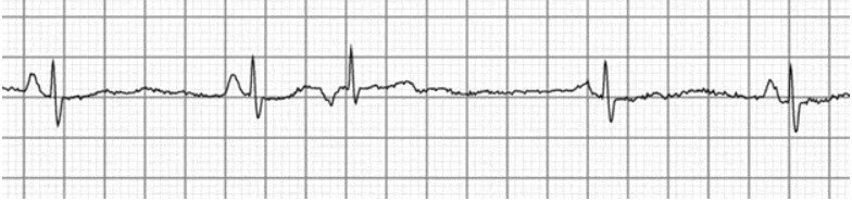
Your patient reports experiencing vomiting, nausea, and chest pain. You notice this rhythm after putting him on the monitor. What would you describe this rhythm as being?
A premature ventricular complex (PVC) occurs every other beat in this rhythm, which is known as a premature ventricular complex bigeminy. A premature ventricular beat follows each sinus beat in premature ventricular complex bigeminy. The premature beat and the prior QRS complex have a constant coupling interval (fixed coupling interval).
A patient of yours is complaining of weariness, dizziness, and chest pain. What is the beat of their hearts?
The patient has a complete electrical block at or below the AV node, which has led to a third-degree AV heart block (infranodal). The pacemaker for the atria is the SA node, whereas the pacemaker for the ventricles is an ectopic focus. The rhythmic occurrence of the P waves and QRS complexes is independent of one another.
This beat is known as .
A normal vertical P wave before each QRS complex and a ventricular rhythm of less than 60 beats per minute are indicators of sinus bradycardia (BPM). Sick sinus syndrome, hypoglycemia, AV blockers, elevated vagal tone seen in athletes, and hypothermia are some of the factors that contribute to sinus bradycardia.
This tracing is an illustration of
A kind of heart block known as Mobitz II second-degree AVB (AV block) causes sporadic loss of conduction below the level of the AV node, occasionally resulting in skipped beats without a progressive lengthening of the PR interval.
This beat is called a .
There won't be any discernible P waves and an extremely erratic QRS complex in atrial fibrillation. Unless the patient is taking medications that block AV nodes, such as beta blockers, the ventricular rate is frequently high.
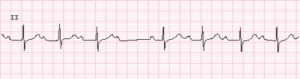
Following complaints of weakness and overall malaise, a patient is put on the heart monitor. What kind of rhythm is this?
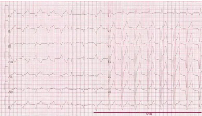
Accelerated idioventricular rhythm describes this beat. A regular beat with three or more ventricular complexes and a QRS complex longer than 120 milliseconds is referred to as an accelerated idioventricular rhythm. With a few capture or fusion beats here and there, the ventricular rate will range between 50 and 110 beats per minute.
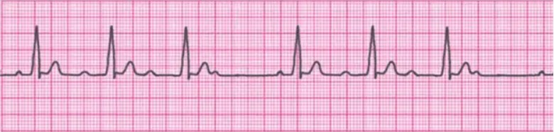
A continuously long PR interval defines this rhythm.
First-degree AV block is frequently asymptomatic and doesn't need to be treated. When someone has a healthy heart, it could be a benign finding. However, it could be a sign of underlying heart issues or prescription side effects that could impair the heart's electrical conduction system. Therefore, it's crucial to consider the clinical setting, together with any symptoms or risk factors that may be present, in order to decide whether more research or care is required. A healthcare expert should be consulted for an accurate assessment and recommendations.
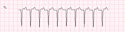
Typically, the rate of paroxysmal tachycardia is:
During paroxysmal tachycardia, the heart rate can vary greatly, often falling between 100 and 250 beats per minute. It's crucial to remember that a person's individual heart rate can differ from one another and occasionally even go above this range.
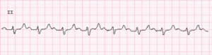
When your patient complains of chest pain, the cardiac monitor is activated. What would you say about this rhythm?
Premature atrial complex (PAC), a premature beat brought on by ectopic pacemaking tissue in the atria, is present in this rhythm. A PAC is characterized by an aberrant P wave that is typically followed by a typical QRS complex lasting less than 120 milliseconds.
What would you call this rhythm?

Please select 2 correct answers
It is junctional tachycardia in this rhythm. P waves will be absent from junctional rhythms, but the QRS complexes will still be present. A junction escape rhythm of 40 to 60 beats per minute will be present along with a regular junctional rhythm. Junctional tachycardia is any rate exceeding 100 beats per minute, while accelerated junctional rhythms range from 60 to 100 beats per minute.
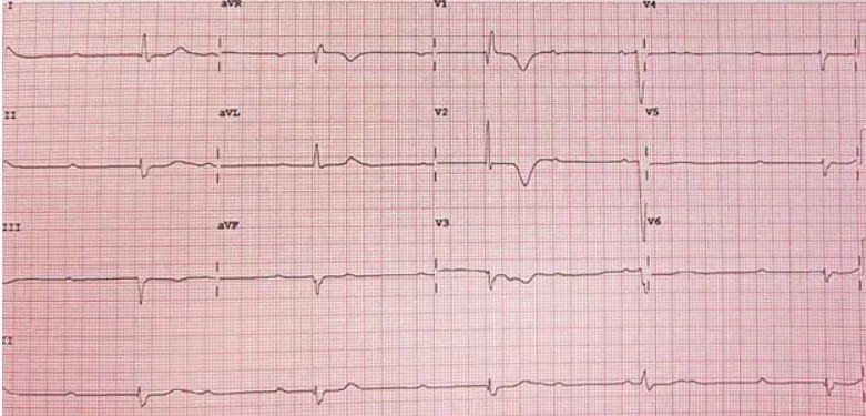
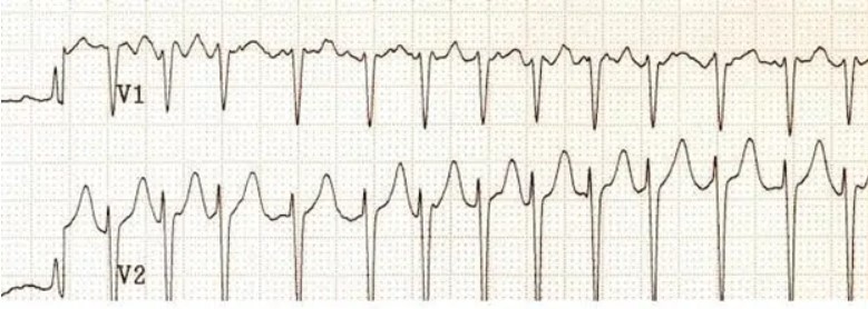
You observe this rhythm after placing your patient on the cardiac monitor. What kind of rhythm would you describe?
This rhythm is an intermittent second-degree AV heart block type 1 (Wenckebach), which typically occurs at the level of the SA node. From beat to beat, the conduction delay gets longer and longer until conduction to the ventricle is blocked. When Wenckebach is present, a distinctive cyclical pattern is created in which the PR interval gradually lengthens until a P wave without a QRS complex follows.
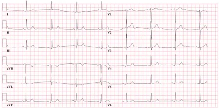
This tracing is an illustration of
A normal sinus rhythm with a protracted PR interval, which denotes a delay in the electrical transmission between the atria and ventricles, is referred to as a sinus rhythm with first-degree AVB (AV block).
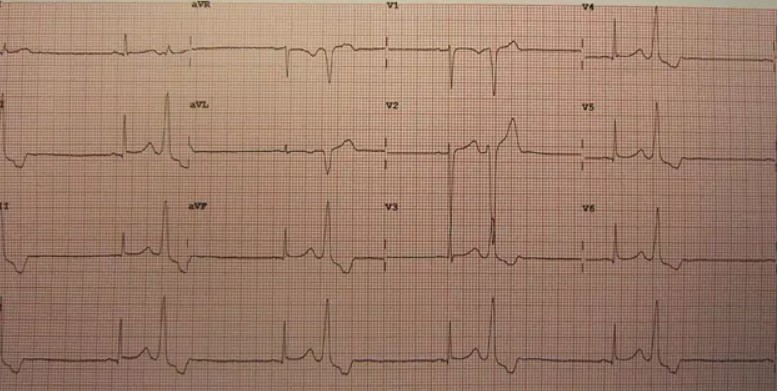
A patient is visiting her cardiologist for a routine examination. What does the monitor's rhythm look like?
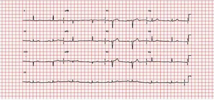
This rhythm is characterized by a PR interval that is regularly greater than 0.20 seconds, which represents a first-degree AV heart block. Increased vagal tone, inferior myocardial infarction, myocarditis, mitral valve surgery, and AV nodal blocking medications can all contribute to this disease, which is characterized by unusually slow conduction through the atrioventricular (AV) node.
The heart pumps the following amount of blood into the aorta each minute:
The cardiac output is the volume of blood that the heart pumps into the aorta in a minute. Heart rate (heartbeats per minute) and stroke volume together make up cardiac output (volume of blood pumped by the heart with each beat).
This tracing is an illustration of
The AV junction, which is the region between the atria and the ventricles, is the source of the fast heart rate known as junctional tachycardia. The electrical impulses that regulate the heartbeat in this disease come from the AV junction rather than the sinus node, circumventing the heart's usual electrical conduction system.
The principal blood vessel responsible for carrying blood from the head and upper extremities to the heart is the:
A significant vein in the human anatomy that returns deoxygenated blood from the upper body and upper extremities to the heart is called the superior vena cava (SVC). One of the two major veins, the other being the inferior vena cava, returns blood to the right atrium of the heart.