FREE EKG Certification Interpretation Question and Answers
In a normal sinus rhythm with PVC's, the heart's electrical impulses usually originate from the sinoatrial (SA) node and follow the normal conduction pathway through the atria, atrioventricular (AV) node, bundle of His, and its branches, resulting in a coordinated contraction of the ventricles. However, occasionally, an extra electrical impulse arises from an area outside the normal conduction pathway, often from the ventricles themselves. This premature impulse can disrupt the regular rhythm and cause a premature contraction of the ventricles, known as a premature ventricular contraction or PVC.
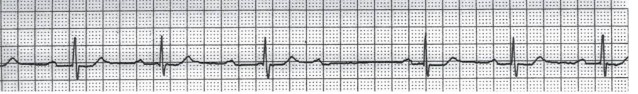
In idioventricular rhythm, the electrical impulses that initiate each heartbeat arise from within the ventricles instead of the normal conduction pathway originating from the SA node in the atria. These impulses are slower in rate compared to the normal sinus rhythm and typically result in a heart rate of 20 to 40 beats per minute, although it can vary. On an electrocardiogram (ECG), idioventricular rhythm appears as wide QRS complexes with a slow rate, often accompanied by an absence of P waves or dissociation between the atrial and ventricular activity. The QRS complexes can have a similar appearance to those seen in ventricular tachycardia.
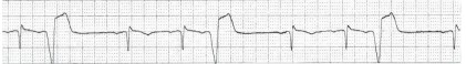
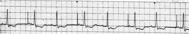
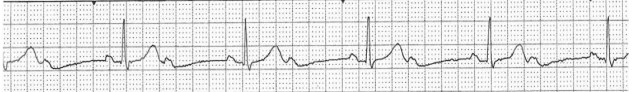
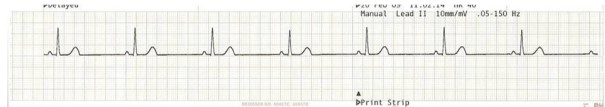
NSR with 1st degree AV block refers to a normal sinus rhythm (NSR) in which there is a delay in the conduction of electrical impulses between the atria and the ventricles. It is the mildest form of AV block, and it is characterized by a prolonged PR interval on an electrocardiogram (ECG). In NSR with 1st degree AV block, the electrical signals originating from the sinoatrial (SA) node, the heart's natural pacemaker, follow the normal pathway through the atria and the atrioventricular (AV) node. However, there is a delay in the conduction of the electrical impulse at the AV node before it reaches the ventricles. This results in a longer-than-normal PR interval on the ECG.
Atrial fibrillation (AF) is a common heart rhythm disorder characterized by rapid and irregular electrical activity in the atria, the upper chambers of the heart. During atrial fibrillation, the atria quiver or fibrillate instead of contracting normally, resulting in an irregular heartbeat. In atrial fibrillation, the normal electrical signals that initiate each heartbeat are disrupted, leading to chaotic electrical impulses firing from different locations in the atria. As a result, the atria do not contract effectively, which can lead to an irregular and often rapid ventricular heart rate.
Sinus rhythm with PAC (Premature Atrial Contraction) refers to a normal heart rhythm, known as sinus rhythm, that is occasionally interrupted by premature electrical impulses originating from the atria. In sinus rhythm, the electrical impulses that initiate each heartbeat originate from the sinoatrial (SA) node, the heart's natural pacemaker. These impulses travel through the atria, causing them to contract and pump blood into the ventricles. From there, the impulses reach the atrioventricular (AV) node and the bundle of His, stimulating the ventricles to contract and pump blood to the rest of the body.
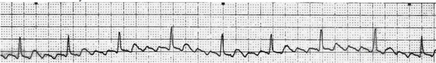
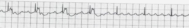
On an ECG, atrial flutter appears as a series of rapid, regular, and sawtooth-shaped waves, known as flutter waves, typically seen in the inferior leads (II, III, aVF) and sometimes in the V1 lead. The flutter waves have a characteristic "sawtooth" appearance due to the rapid and organized atrial activity. The ventricular response appears as a regular and often rapid heart rate, typically between 100 and 150 beats per minute, although it can vary.
A normal sinus rhythm on an electrocardiogram (ECG) will show certain characteristic features. These include a P wave preceding each QRS complex, a regular R-R interval (the time between consecutive R waves), a consistent PR interval (the time between the start of the P wave and the start of the QRS complex), and a normal QRS duration (the width of the QRS complex). The T wave represents the repolarization of the ventricles and should also follow a consistent pattern.
In an accelerated junctional rhythm, the electrical impulses that initiate each heartbeat arise from the AV junction rather than the normal pacemaker, the sinoatrial (SA) node. The heart rate in AJR is typically between 60 and 100 beats per minute, which is faster than the normal junctional rhythm but slower than the typical sinus rhythm.
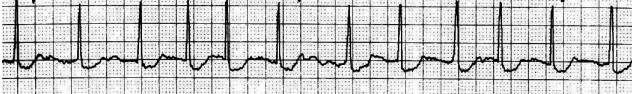
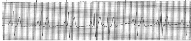
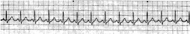
Supraventricular tachycardia (SVT) is a type of abnormal heart rhythm characterized by a rapid heart rate originating above the ventricles. It involves abnormal electrical circuits or pathways in the heart, leading to episodes of rapid and regular heartbeats. In SVT, the rapid heart rate typically exceeds 150 beats per minute, but it can range from 140 to 250 beats per minute. The fast heart rate originates from the atria (supraventricular), bypassing the normal conduction pathways or engaging in abnormal electrical circuits.
Second-degree atrioventricular (AV) block, Type I, also known as Mobitz Type I or Wenckebach block, is a specific type of abnormal heart rhythm characterized by progressively lengthening delays in electrical conduction between the atria and ventricles. In Type I AV block, some of the electrical signals originating from the atria are not transmitted to the ventricles in a regular and consistent manner. The electrical conduction through the AV node becomes progressively delayed with each heartbeat until a signal is completely blocked and fails to reach the ventricles. This results in a dropped beat or a skipped QRS complex on an electrocardiogram (ECG).
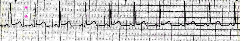
On an electrocardiogram (ECG), sinus rhythm with atrial pacing will typically show the characteristics of normal sinus rhythm. These include regular P waves preceding each QRS complex, a consistent PR interval, and a normal QRS duration. However, the presence of pacing spikes may also be observed, indicating the delivery of artificial pacing stimuli to the atria.
Second-degree atrioventricular (AV) block, Type II, is a specific type of abnormal heart rhythm characterized by intermittent or occasional blockage of electrical impulses between the atria and ventricles. In Type II AV block, not all atrial electrical signals are conducted to the ventricles. In Type II AV block, some of the electrical signals originating from the atria fail to be transmitted to the ventricles in a consistent and predictable manner. This results in intermittent "dropped beats" or skipped QRS complexes on an electrocardiogram (ECG). Unlike Type I AV block (Wenckebach block), Type II AV block does not show progressive lengthening of the PR interval before the dropped beat.
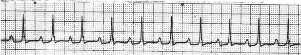
On an electrocardiogram (ECG), sinus tachycardia is characterized by a normal P wave preceding each QRS complex, a regular R-R interval (the time between consecutive R waves), and a consistent PR interval (the time between the start of the P wave and the start of the QRS complex). The QRS complex and T wave follow a normal pattern as well, but the overall heart rate is faster than the usual range of 60 to 100 beats per minute.
"V paced with failure to capture" refers to a situation where ventricular pacing (V paced) is attempted but fails to generate a ventricular contraction (capture). Ventricular pacing involves delivering electrical impulses to the ventricles through an artificial pacemaker to initiate a heartbeat when the heart's natural pacemaker is not functioning properly.
On an electrocardiogram (ECG), sinus bradycardia is characterized by a normal P wave preceding each QRS complex, a regular R-R interval (the time between consecutive R waves), and a consistent PR interval (the time between the start of the P wave and the start of the QRS complex). The QRS complex and T wave follow a normal pattern as well, but the overall heart rate is slower than the usual range of 60 to 100 beats per minute.
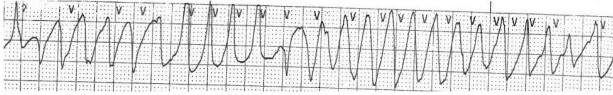
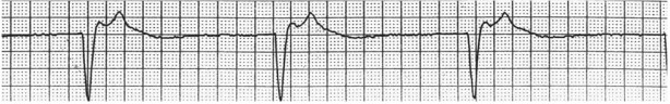
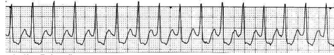
During Torsades de Pointes, the heart's electrical activity becomes chaotic, resulting in a rapid heart rate that ranges from 200 to 250 beats per minute or even higher. This arrhythmia is typically associated with a prolonged QT interval on the ECG, indicating an abnormality in the repolarization phase of the cardiac cycle. The prolonged QT interval increases the risk of developing Torsades de Pointes.